

Search Count: 31
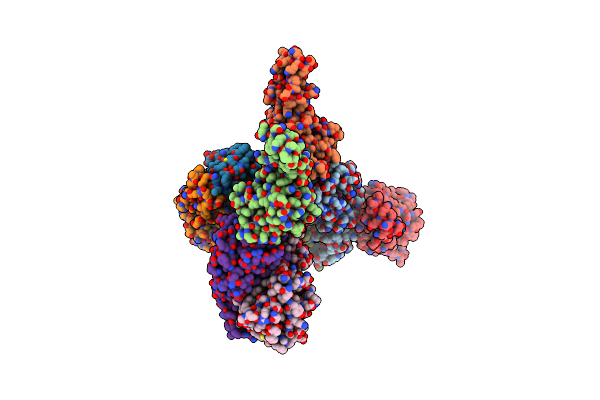 |
Structure Of S78C Human Peroxiredoxin 3 As Three Stacked Rings
Organism: Homo sapiens
Method: X-RAY DIFFRACTION Resolution:2.40 Å Release Date: 2018-01-10 Classification: OXIDOREDUCTASE |
Organism: Homo sapiens
Method: X-RAY DIFFRACTION
Release Date: 2018-01-10
 |
Structure Of Human Peroxiredoxin 3 As Three Stacked Rings
Organism: Homo sapiens
Method: X-RAY DIFFRACTION Resolution:2.80 Å Release Date: 2016-05-25 Classification: OXIDOREDUCTASE |
Organism: Homo sapiens
Method: X-RAY DIFFRACTION
Release Date: 2016-05-25
 |
0.89 Angstrom Resolution Crystal Structure Of (Gly-Pro-Hyp)10
Organism: Mammalian
Method: X-RAY DIFFRACTION Resolution:0.89 Å Release Date: 2015-03-04 Classification: STRUCTURAL PROTEIN |
Organism: Mammalian
Method: X-RAY DIFFRACTION
Release Date: 2015-03-04
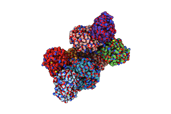 |
Dihydrodipicolinate Synthase From The Common Grapevine With Pyruvate And Lysine
Organism: Vitis vinifera
Method: X-RAY DIFFRACTION Resolution:2.40 Å Release Date: 2013-09-04 Classification: HYDROLASE Ligands: LYS |
Organism: Vitis vinifera
Method: X-RAY DIFFRACTION
Release Date: 2013-09-04
Ligands: LYS
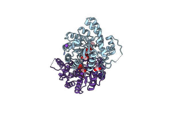 |
Structure, Function, Stability And Knockout Phenotype Of Dihydrodipicolinate Synthase From Streptococcus Pneumoniae
Organism: Streptococcus pneumoniae
Method: X-RAY DIFFRACTION Resolution:1.91 Å Release Date: 2013-06-12 Classification: LYASE Ligands: K, GOL, MES |
Organism: Streptococcus pneumoniae
Method: X-RAY DIFFRACTION
Release Date: 2013-06-12
Ligands: K, GOL, MES
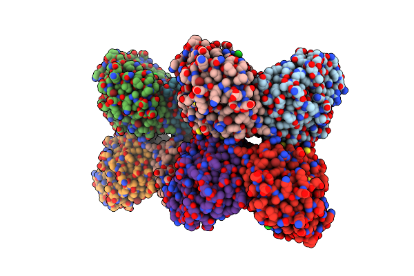 |
Structure Of Dihydrodipicolinate Synthase From The Common Grapevine
Organism: Vitis vinifera
Method: X-RAY DIFFRACTION Resolution:2.20 Å Release Date: 2012-07-25 Classification: LYASE Ligands: BR, CL |
Organism: Vitis vinifera
Method: X-RAY DIFFRACTION
Release Date: 2012-07-25
Ligands: BR, CL
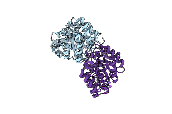 |
The Structure Of Dihydrodipicolinate Synthase 2 From Arabidopsis Thaliana
Organism: Arabidopsis thaliana
Method: X-RAY DIFFRACTION Resolution:2.00 Å Release Date: 2012-07-18 Classification: LYASE |
Organism: Arabidopsis thaliana
Method: X-RAY DIFFRACTION
Release Date: 2012-07-18
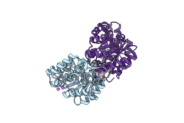 |
The Structure Of Dihydrodipicolinate Synthase 2 From Arabidopsis Thaliana In Complex With (S)-Lysine
Organism: Arabidopsis thaliana
Method: X-RAY DIFFRACTION Resolution:2.20 Å Release Date: 2012-07-18 Classification: LYASE |
Organism: Arabidopsis thaliana
Method: X-RAY DIFFRACTION
Release Date: 2012-07-18
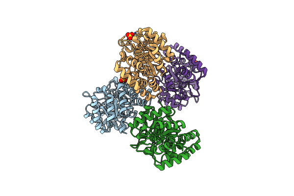 |
Characterisation Of The First Monomeric Dihydrodipicolinate Synthase Variant Reveals Evolutionary Insights
Organism: Thermotoga maritima
Method: X-RAY DIFFRACTION Resolution:2.00 Å Release Date: 2011-11-23 Classification: LYASE Ligands: SO4 |
Organism: Thermotoga maritima
Method: X-RAY DIFFRACTION
Release Date: 2011-11-23
Ligands: SO4
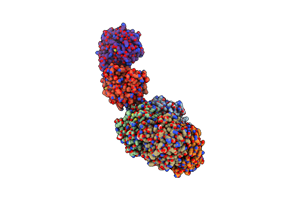 |
Characterisation Of The First Monomeric Dihydrodipicolinate Synthase Variant Reveals Evolutionary Insights
Organism: Thermotoga maritima
Method: X-RAY DIFFRACTION Resolution:1.90 Å Release Date: 2011-11-23 Classification: LYASE Ligands: GOL |
Organism: Thermotoga maritima
Method: X-RAY DIFFRACTION
Release Date: 2011-11-23
Ligands: GOL
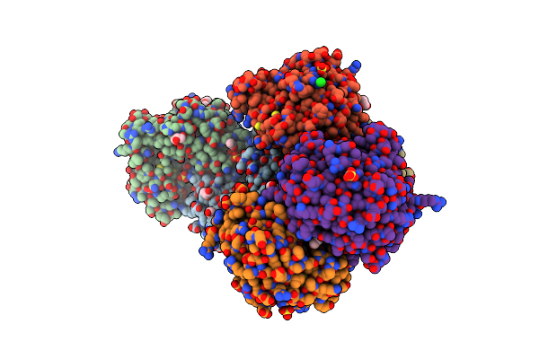 |
The Crystal Structure Of A Dimeric Mutant Of Dihydrodipicolinate Synthase (Dapa, Rv2753C) From Mycobacterium Tuberculosis - Dhdps-A204R
Organism: Mycobacterium tuberculosis
Method: X-RAY DIFFRACTION Resolution:2.10 Å Release Date: 2010-12-15 Classification: TRANSFERASE Ligands: SO4, GOL, ACT, BME, PYR, CL |
Organism: Mycobacterium tuberculosis
Method: X-RAY DIFFRACTION
Release Date: 2010-12-15
Ligands: SO4, GOL, ACT, BME, PYR, CL
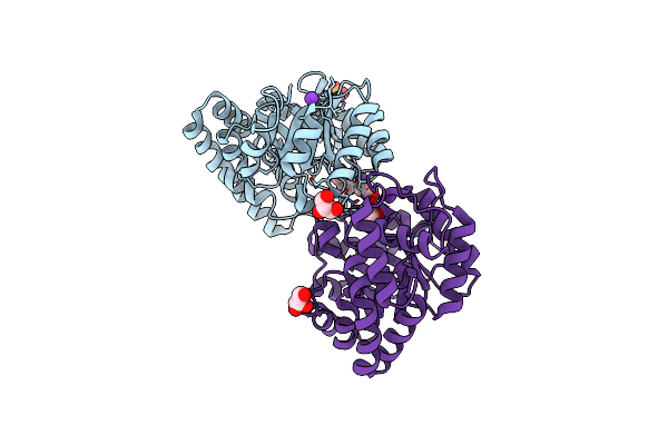 |
Dihydrodipicolinate Synthase Mutant - K161A
Organism: Escherichia coli
Method: X-RAY DIFFRACTION Resolution:2.00 Å Release Date: 2010-04-14 Classification: LYASE Ligands: GOL, K, PO4 |
Organism: Escherichia coli
Method: X-RAY DIFFRACTION
Release Date: 2010-04-14
Ligands: GOL, K, PO4
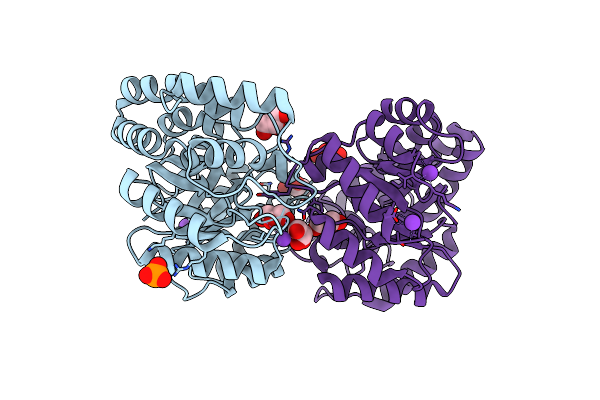 |
Dihydrodipicolinate Synthase - K161R
Organism: Escherichia coli k-12
Method: X-RAY DIFFRACTION Resolution:2.10 Å Release Date: 2010-04-14 Classification: LYASE Ligands: K, GOL, CL, PO4 |
Organism: Escherichia coli k-12
Method: X-RAY DIFFRACTION
Release Date: 2010-04-14
Ligands: K, GOL, CL, PO4
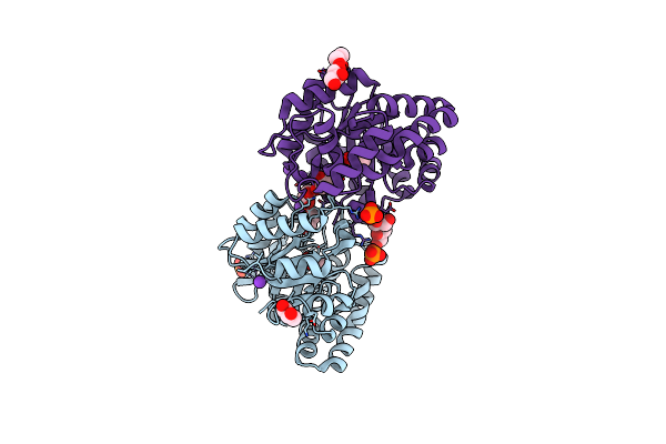 |
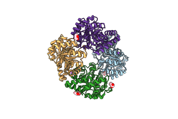
Dihydrodipicolinate Synthase Mutant - K161A - With The Substrate Pyruvate Bound In The Active Site.
Organism: Escherichia coli k-12
Method: X-RAY DIFFRACTION Resolution:2.30 Å Release Date: 2010-04-14 Classification: LYASE Ligands: GOL, K, PYR, PO4 |
Organism: Escherichia coli k-12
Method: X-RAY DIFFRACTION
Release Date: 2010-04-14
Ligands: GOL, K, PYR, PO4
 |
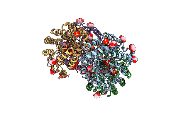
Crystal Structure Of Dihydrodipicolinate Synthase From Bacillus Anthracis In Complex With Its Substrate, Pyruvate
Organism: Bacillus anthracis
Method: X-RAY DIFFRACTION Resolution:2.15 Å Release Date: 2009-11-24 Classification: LYASE Ligands: GOL, NA |
Organism: Bacillus anthracis
Method: X-RAY DIFFRACTION
Release Date: 2009-11-24
Ligands: GOL, NA
 |
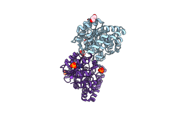
Crystal Structure Of Dihydrodipicolinate Synthase From The Pathogen Neisseria Meningitidis
Organism: Neisseria meningitidis serogroup b
Method: X-RAY DIFFRACTION Resolution:2.00 Å Release Date: 2009-06-23 Classification: LYASE Ligands: SO4, GOL |
Organism: Neisseria meningitidis serogroup b
Method: X-RAY DIFFRACTION
Release Date: 2009-06-23
Ligands: SO4, GOL
 |
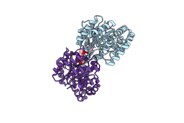
Structure Of E. Coli Dhdps Mutant Y107W
Organism: Escherichia coli k-12
Method: X-RAY DIFFRACTION Resolution:2.20 Å Release Date: 2008-11-25 Classification: LYASE Ligands: K, GOL, PO4 |
Organism: Escherichia coli k-12
Method: X-RAY DIFFRACTION
Release Date: 2008-11-25
Ligands: K, GOL, PO4
 |
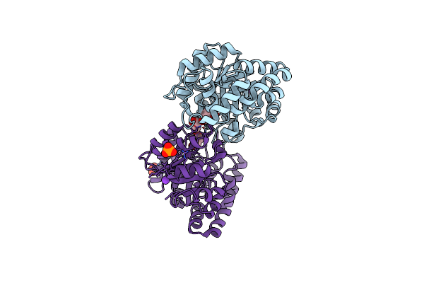
E. Coli Dihydrodipicolinate Synthase With First Substrate, Pyruvate, Bound In Active Site
Organism: Escherichia coli
Method: X-RAY DIFFRACTION Resolution:2.00 Å Release Date: 2008-11-18 Classification: LYASE Ligands: K, GOL, CL |
Organism: Escherichia coli
Method: X-RAY DIFFRACTION
Release Date: 2008-11-18
Ligands: K, GOL, CL
 |
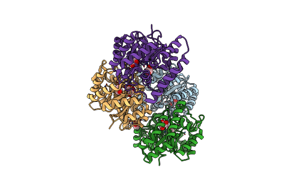
Structure Of E. Coli Dihydrodipicolinate Synthase Complexed With Hydroxypyruvate
Organism: Escherichia coli
Method: X-RAY DIFFRACTION Resolution:2.40 Å Release Date: 2008-09-23 Classification: LYASE Ligands: K, GOL, PO4 |
Organism: Escherichia coli
Method: X-RAY DIFFRACTION
Release Date: 2008-09-23
Ligands: K, GOL, PO4
 |
Crystal Structure Of Dihydrodipicolinate Synthase From Methicillin-Resistant Staphylococcus Aureus
Organism: Staphylococcus aureus
Method: X-RAY DIFFRACTION Resolution:1.45 Å Release Date: 2008-08-05 Classification: LYASE Ligands: CL, GOL |
Organism: Staphylococcus aureus
Method: X-RAY DIFFRACTION
Release Date: 2008-08-05
Ligands: CL, GOL