

Search Count: 15
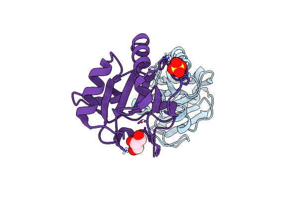 |
Membrane-Associated Thioredoxin Oxidoreductase Fete From Campylobacter Jejuni
Organism: Campylobacter jejuni subsp. jejuni
Method: X-RAY DIFFRACTION Resolution:1.50 Å Release Date: 2023-08-09 Classification: OXIDOREDUCTASE Ligands: SO4, GOL |
Organism: Campylobacter jejuni subsp. jejuni
Method: X-RAY DIFFRACTION
Release Date: 2023-08-09
Ligands: SO4, GOL
 |
Crystal Structure Of The Helical Cell Shape Determining Protein Pgp2 From Campylobacter Jejuni
Organism: Campylobacter jejuni subsp. jejuni
Method: X-RAY DIFFRACTION Resolution:1.50 Å Release Date: 2021-03-17 Classification: HYDROLASE |
Organism: Campylobacter jejuni subsp. jejuni
Method: X-RAY DIFFRACTION
Release Date: 2021-03-17
 |
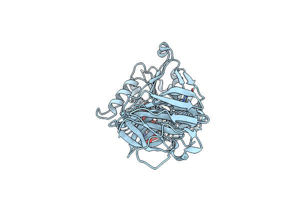
Crystal Structure Of The Helical Cell Shape Determining Protein Pgp2 (K307A Mutant) From Campylobacter Jejuni
Organism: Campylobacter jejuni subsp. jejuni
Method: X-RAY DIFFRACTION Resolution:1.85 Å Release Date: 2021-03-17 Classification: HYDROLASE |
Organism: Campylobacter jejuni subsp. jejuni
Method: X-RAY DIFFRACTION
Release Date: 2021-03-17
 |
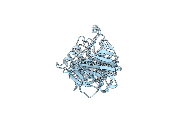
Inhibitor Bound Cell Shape Determinant Protein Csd4 From Helicobacter Pylori
Organism: Helicobacter pylori
Method: X-RAY DIFFRACTION Resolution:1.90 Å Release Date: 2016-01-20 Classification: hydrolase/hydrolase inhibitor Ligands: ZN, IOD, 56W |
Organism: Helicobacter pylori
Method: X-RAY DIFFRACTION
Release Date: 2016-01-20
Ligands: ZN, IOD, 56W
 |
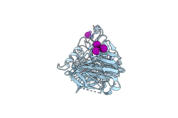
Crystal Structure Of Apo Cell Shape Determinant Protein Csd4 From Helicobacter Pylori
Organism: Helicobacter pylori
Method: X-RAY DIFFRACTION Resolution:1.40 Å Release Date: 2014-12-24 Classification: HYDROLASE Ligands: IOD, API |
Organism: Helicobacter pylori
Method: X-RAY DIFFRACTION
Release Date: 2014-12-24
Ligands: IOD, API
 |
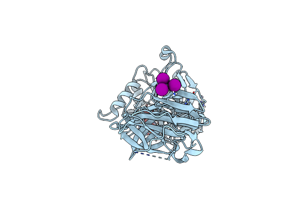
Crystal Structure Of Product Bound Cell Shape Determinant Protein Csd4 From Helicobacter Pylori
Organism: Helicobacter pylori
Method: X-RAY DIFFRACTION Resolution:1.85 Å Release Date: 2014-12-24 Classification: HYDROLASE Ligands: ZN, IOD, API, NA |
Organism: Helicobacter pylori
Method: X-RAY DIFFRACTION
Release Date: 2014-12-24
Ligands: ZN, IOD, API, NA
 |
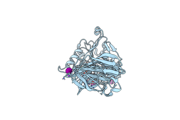
Crystal Structure Of Cell Shape Determinant Protein Csd4 Gln46His Variant From Helicobacter Pylori
Organism: Helicobacter pylori
Method: X-RAY DIFFRACTION Resolution:1.75 Å Release Date: 2014-12-24 Classification: HYDROLASE Ligands: IOD, ZN, PO4, API |
Organism: Helicobacter pylori
Method: X-RAY DIFFRACTION
Release Date: 2014-12-24
Ligands: IOD, ZN, PO4, API
 |
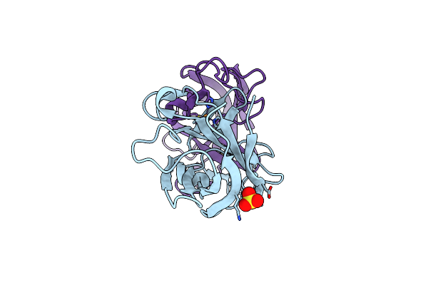
Crystal Structure Of Tripeptide Bound Cell Shape Determinant Csd4 Protein From Helicobacter Pylori
Organism: Helicobacter pylori
Method: X-RAY DIFFRACTION Resolution:1.75 Å Release Date: 2014-12-24 Classification: HYDROLASE Ligands: IOD, ZN, NA, 3KS |
Organism: Helicobacter pylori
Method: X-RAY DIFFRACTION
Release Date: 2014-12-24
Ligands: IOD, ZN, NA, 3KS
 |
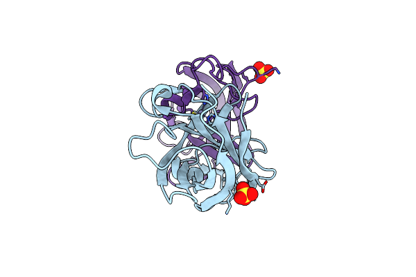
Crystal Structure Analysis Of The As-Solated P19 Protein From Campylobacter Jejuni At 1.45 A At Ph 9.0
Organism: Campylobacter jejuni
Method: X-RAY DIFFRACTION Resolution:1.45 Å Release Date: 2010-07-21 Classification: TRANSPORT PROTEIN Ligands: CU, SO4 |
Organism: Campylobacter jejuni
Method: X-RAY DIFFRACTION
Release Date: 2010-07-21
Ligands: CU, SO4
 |
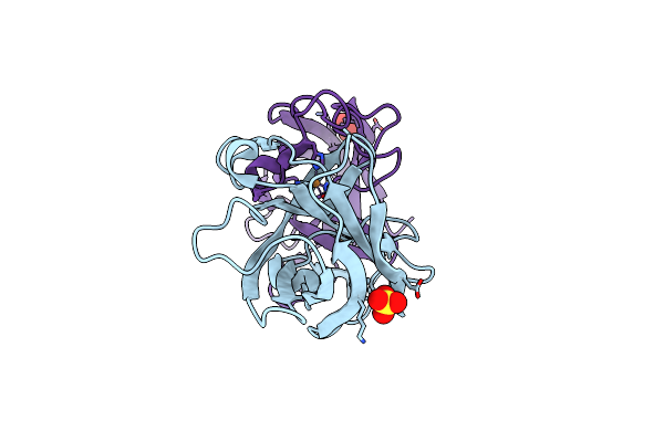
Crystal Structure Analysis Of The Apo P19 Protein From Campylobacter Jejuni At 1.59 A At Ph 9
Organism: Campylobacter jejuni
Method: X-RAY DIFFRACTION Resolution:1.59 Å Release Date: 2010-07-21 Classification: TRANSPORT PROTEIN Ligands: ZN, SO4 |
Organism: Campylobacter jejuni
Method: X-RAY DIFFRACTION
Release Date: 2010-07-21
Ligands: ZN, SO4
 |
Crystal Structure Analysis Of The Copper-Reconstituted P19 Protein From Campylobacter Jejuni At 1.65 A At Ph 10.0
Organism: Campylobacter jejuni
Method: X-RAY DIFFRACTION Resolution:1.65 Å Release Date: 2010-07-21 Classification: TRANSPORT PROTEIN Ligands: CU, SO4 |
Organism: Campylobacter jejuni
Method: X-RAY DIFFRACTION
Release Date: 2010-07-21
Ligands: CU, SO4
 |
Crystal Structure Analysis Of The 'As-Isolated' P19 Protein From Campylobacter Jejuni At 1.65 A At Ph 9.0
Organism: Campylobacter jejuni
Method: X-RAY DIFFRACTION Resolution:1.65 Å Release Date: 2010-07-21 Classification: TRANSPORT PROTEIN Ligands: CU, SO4 |
Organism: Campylobacter jejuni
Method: X-RAY DIFFRACTION
Release Date: 2010-07-21
Ligands: CU, SO4
 |
Crystal Structure Analysis Of Manganese Treated P19 Protein From Campylobacter Jejuni At 1.41 A At Ph 9
Organism: Campylobacter jejuni
Method: X-RAY DIFFRACTION Resolution:1.41 Å Release Date: 2010-07-21 Classification: TRANSPORT PROTEIN Ligands: CU, MN, SO4 |
Organism: Campylobacter jejuni
Method: X-RAY DIFFRACTION
Release Date: 2010-07-21
Ligands: CU, MN, SO4
 |
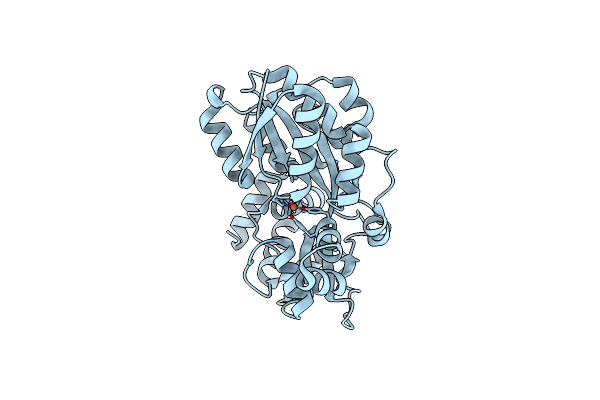
Crystal Structure Analysis Of Manganese Treated P19 Protein From Campylobacter Jejuni At 2.73 A At Ph 9 And Manganese Peak Wavelength (1.893 A)
Organism: Campylobacter jejuni
Method: X-RAY DIFFRACTION Resolution:2.73 Å Release Date: 2010-07-21 Classification: TRANSPORT PROTEIN Ligands: CU, MN, SO4 |
Organism: Campylobacter jejuni
Method: X-RAY DIFFRACTION
Release Date: 2010-07-21
Ligands: CU, MN, SO4
 |
Iron Reconstituted Ferric Binding Protein From Campylobacter Jejuni
Organism: Campylobacter jejuni
Method: X-RAY DIFFRACTION Resolution:1.60 Å Release Date: 2009-08-11 Classification: METAL BINDING PROTEIN Ligands: FE |
Organism: Campylobacter jejuni
Method: X-RAY DIFFRACTION
Release Date: 2009-08-11
Ligands: FE