

Search Count: 27
 |
Crystal Structure Of P53 Cancer Mutant Y220N In Complex With Rezatapopt
Organism: Homo sapiens
Method: X-RAY DIFFRACTION Resolution:1.87 Å Release Date: 2026-03-18 Classification: TRANSCRIPTION Ligands: ZN, A1JMR, EDO, PEG |
Organism: Homo sapiens
Method: X-RAY DIFFRACTION
Release Date: 2026-03-18
Ligands: ZN, A1JMR, EDO, PEG
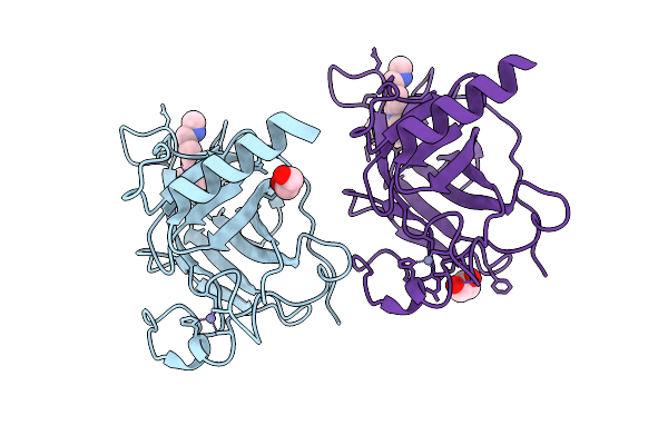 |
Crystal Structure Of P53 Cancer Mutant Y220C In Complex With Rezatapopt Precursor
Organism: Homo sapiens
Method: X-RAY DIFFRACTION Resolution:1.38 Å Release Date: 2026-03-11 Classification: TRANSCRIPTION Ligands: ZN, A1JMS, EDO, GOL |
Organism: Homo sapiens
Method: X-RAY DIFFRACTION
Release Date: 2026-03-11
Ligands: ZN, A1JMS, EDO, GOL
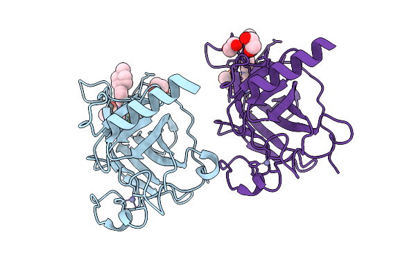 |
Crystal Structure Of P53 Cancer Mutant Y220C In Complex With Rezatapopt Analog 7
Organism: Homo sapiens
Method: X-RAY DIFFRACTION Resolution:1.83 Å Release Date: 2026-03-11 Classification: TRANSCRIPTION Ligands: A1JMO, EDO, ZN |
Organism: Homo sapiens
Method: X-RAY DIFFRACTION
Release Date: 2026-03-11
Ligands: A1JMO, EDO, ZN
 |
Crystal Structure Of P53 Cancer Mutant Y220C In Complex With Rezatapopt Analog 8
Organism: Homo sapiens
Method: X-RAY DIFFRACTION Resolution:1.72 Å Release Date: 2026-03-11 Classification: TRANSCRIPTION Ligands: A1JMP, EDO, ZN |
Organism: Homo sapiens
Method: X-RAY DIFFRACTION
Release Date: 2026-03-11
Ligands: A1JMP, EDO, ZN
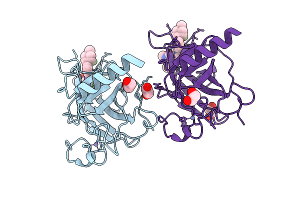 |
Crystal Structure Of P53 Cancer Mutant Y220C In Complex With Rezatapopt
Organism: Homo sapiens
Method: X-RAY DIFFRACTION Resolution:1.49 Å Release Date: 2026-03-11 Classification: TRANSCRIPTION Ligands: ZN, A1JMR, EDO |
Organism: Homo sapiens
Method: X-RAY DIFFRACTION
Release Date: 2026-03-11
Ligands: ZN, A1JMR, EDO
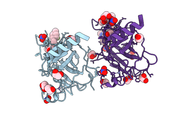 |
Crystal Structure Of P53 Cancer Mutant Y220S In Complex With Rezatapopt
Organism: Homo sapiens
Method: X-RAY DIFFRACTION Resolution:1.40 Å Release Date: 2026-03-11 Classification: TRANSCRIPTION Ligands: ZN, A1JMR, EDO, NO3 |
Organism: Homo sapiens
Method: X-RAY DIFFRACTION
Release Date: 2026-03-11
Ligands: ZN, A1JMR, EDO, NO3
 |
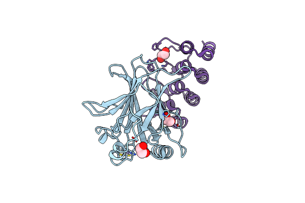
Crystal Structure Of P53 Cancer Mutant Y220N
Organism: Homo sapiens
Method: X-RAY DIFFRACTION Resolution:1.70 Å Release Date: 2026-03-11 Classification: TRANSCRIPTION Ligands: ZN, EDO |
Organism: Homo sapiens
Method: X-RAY DIFFRACTION
Release Date: 2026-03-11
Ligands: ZN, EDO
 |
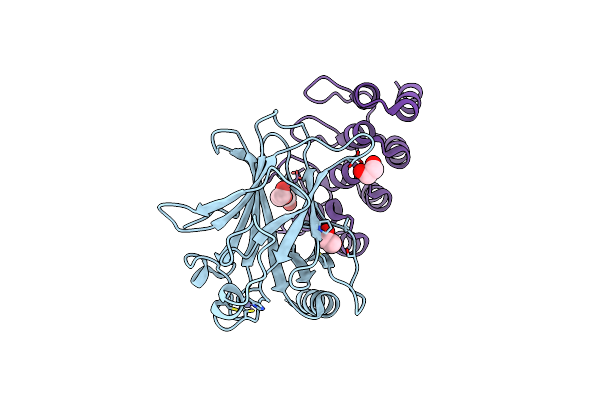
Human P53 Dna-Binding Domain Bound To Darpin C10-H82R
Organism: Homo sapiens, Synthetic construct
Method: X-RAY DIFFRACTION Resolution:1.44 Å Release Date: 2025-04-02 Classification: DNA BINDING PROTEIN Ligands: EDO, ZN |
Organism: Homo sapiens, Synthetic construct
Method: X-RAY DIFFRACTION
Release Date: 2025-04-02
Ligands: EDO, ZN
 |
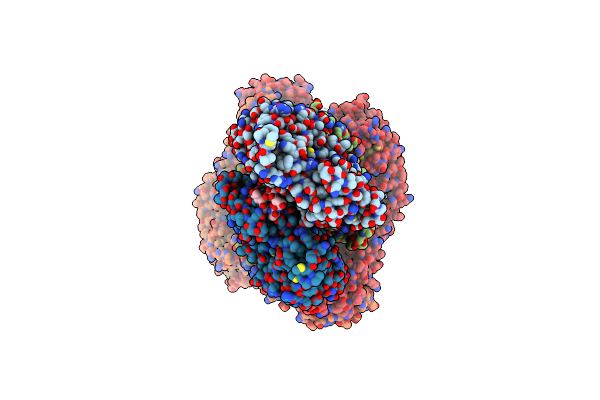
Human P53 Dna-Binding Domain Bound To Darpin C10
Organism: Homo sapiens, Synthetic construct
Method: X-RAY DIFFRACTION Resolution:1.50 Å Release Date: 2024-12-18 Classification: DNA BINDING PROTEIN Ligands: EDO, ZN |
Organism: Homo sapiens, Synthetic construct
Method: X-RAY DIFFRACTION
Release Date: 2024-12-18
Ligands: EDO, ZN
 |
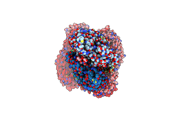
Structure Of The Formin Cdc12 Bound To The Barbed End Of Phalloidin-Stabilized F-Actin.
Organism: Homo sapiens, Schizosaccharomyces pombe, Amanita phalloides
Method: ELECTRON MICROSCOPY Release Date: 2024-04-10 Classification: STRUCTURAL PROTEIN Ligands: ADP, MG, PO4 |
Organism: Homo sapiens, Schizosaccharomyces pombe, Amanita phalloides
Method: ELECTRON MICROSCOPY
Release Date: 2024-04-10
Ligands: ADP, MG, PO4
 |
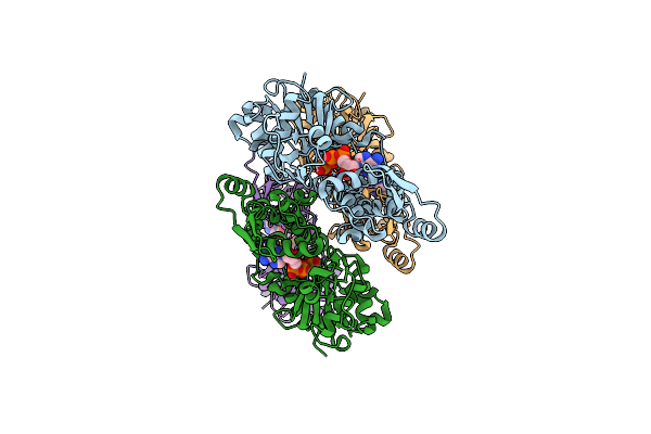
Structure Of The F-Actin Barbed End Bound By Cdc12 And Profilin (Ring Complex) At A Resolution Of 6.3 Angstrom
Organism: Homo sapiens, Schizosaccharomyces pombe, Amanita phalloides
Method: ELECTRON MICROSCOPY Release Date: 2024-04-10 Classification: STRUCTURAL PROTEIN Ligands: ADP, MG, PO4 |
Organism: Homo sapiens, Schizosaccharomyces pombe, Amanita phalloides
Method: ELECTRON MICROSCOPY
Release Date: 2024-04-10
Ligands: ADP, MG, PO4
 |
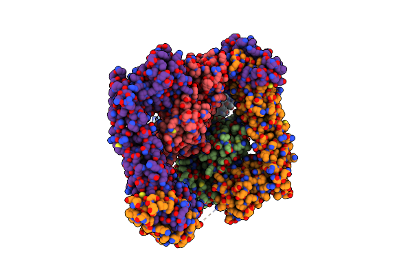
Structure Of The Undecorated Barbed End Of F-Actin.
Organism: Oryctolagus cuniculus
Method: ELECTRON MICROSCOPY Release Date: 2024-04-10 Classification: STRUCTURAL PROTEIN Ligands: ADP, MG, PO4, ATP |
Organism: Oryctolagus cuniculus
Method: ELECTRON MICROSCOPY
Release Date: 2024-04-10
Ligands: ADP, MG, PO4, ATP
 |
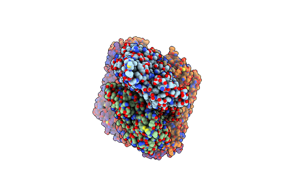
Structure Of The F-Actin Barbed End Bound By Formin Mdia1
Organism: Homo sapiens, Mus musculus
Method: ELECTRON MICROSCOPY Release Date: 2024-04-10 Classification: STRUCTURAL PROTEIN Ligands: ADP, MG |
Organism: Homo sapiens, Mus musculus
Method: ELECTRON MICROSCOPY
Release Date: 2024-04-10
Ligands: ADP, MG
 |
Structure Of The Formin Inf2 Bound To The Barbed End Of F-Actin.
Organism: Homo sapiens, Oryctolagus cuniculus
Method: ELECTRON MICROSCOPY Release Date: 2024-04-10 Classification: STRUCTURAL PROTEIN Ligands: ADP, MG, PO4, ATP |
Organism: Homo sapiens, Oryctolagus cuniculus
Method: ELECTRON MICROSCOPY
Release Date: 2024-04-10
Ligands: ADP, MG, PO4, ATP
 |
Structure Of P. Luminescens Tccc3-F-Actin Complex
Organism: Photorhabdus luminescens, Oryctolagus cuniculus
Method: ELECTRON MICROSCOPY Release Date: 2022-06-29 Classification: TOXIN Ligands: ADP, MG, APR, NCA |
Organism: Photorhabdus luminescens, Oryctolagus cuniculus
Method: ELECTRON MICROSCOPY
Release Date: 2022-06-29
Ligands: ADP, MG, APR, NCA
 |
Structure Of Adp-Ribosylated F-Actin
Organism: Oryctolagus cuniculus
Method: ELECTRON MICROSCOPY Release Date: 2022-06-29 Classification: CYTOSOLIC PROTEIN Ligands: ADP, MG, APR |
Organism: Oryctolagus cuniculus
Method: ELECTRON MICROSCOPY
Release Date: 2022-06-29
Ligands: ADP, MG, APR
 |
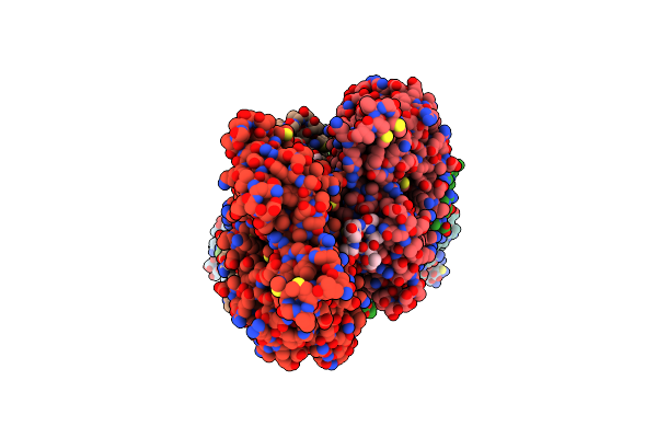
Structure Of The Adp-Ribosyltransferase Tccc3Hvr From Photorhabdus Luminescens
Organism: Photorhabdus luminescens
Method: SOLUTION NMR Release Date: 2022-06-29 Classification: TOXIN |
Organism: Photorhabdus luminescens
Method: SOLUTION NMR
Release Date: 2022-06-29
 |
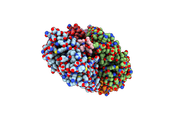
Structure Of Capping Protein Bound To The Barbed End Of A Cytoplasmic Actin Filament
Organism: Mus musculus, Synthetic construct, Bos taurus
Method: ELECTRON MICROSCOPY Release Date: 2021-09-01 Classification: STRUCTURAL PROTEIN Ligands: ADP, MG, PO4 |
Organism: Mus musculus, Synthetic construct, Bos taurus
Method: ELECTRON MICROSCOPY
Release Date: 2021-09-01
Ligands: ADP, MG, PO4
 |
Cryo-Em Structure Of F-Actin Stabilized By Cis-Optojasp-8
Organism: Oryctolagus cuniculus
Method: ELECTRON MICROSCOPY Release Date: 2021-01-27 Classification: STRUCTURAL PROTEIN Ligands: ADP, MG, PO4, RLZ |
Organism: Oryctolagus cuniculus
Method: ELECTRON MICROSCOPY
Release Date: 2021-01-27
Ligands: ADP, MG, PO4, RLZ
 |
Cryo-Em Structure Of F-Actin Stabilized By Trans-Optojasp-8
Organism: Oryctolagus cuniculus
Method: ELECTRON MICROSCOPY Release Date: 2021-01-27 Classification: STRUCTURAL PROTEIN Ligands: ADP, PO4, MG, RLZ |
Organism: Oryctolagus cuniculus
Method: ELECTRON MICROSCOPY
Release Date: 2021-01-27
Ligands: ADP, PO4, MG, RLZ