

Search Count: 78
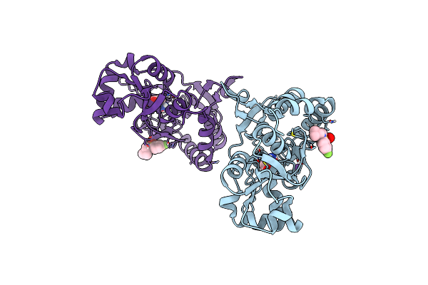 |
Crystal Structure Of The Gluk2 Ligand-Binding Domain In Complex With L-Glutamate And Bpam344 At 1.60 A Resolution
Organism: Rattus norvegicus
Method: X-RAY DIFFRACTION Resolution:1.60 Å Release Date: 2024-09-18 Classification: MEMBRANE PROTEIN Ligands: GLU, 2J9, CL, NA |
Organism: Rattus norvegicus
Method: X-RAY DIFFRACTION
Release Date: 2024-09-18
Ligands: GLU, 2J9, CL, NA
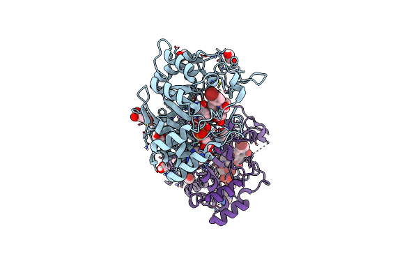 |
Structure Of Aldo-Keto Reductase 1C3 (Akr1C3) In Complex With An Inhibitor Meds765
Organism: Homo sapiens
Method: X-RAY DIFFRACTION Resolution:1.75 Å Release Date: 2024-08-21 Classification: OXIDOREDUCTASE Ligands: NAP, A1ICG, EDO, MES |
Organism: Homo sapiens
Method: X-RAY DIFFRACTION
Release Date: 2024-08-21
Ligands: NAP, A1ICG, EDO, MES
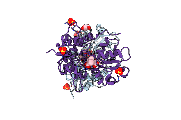 |
Crystal Structure Of The Gluk1 Ligand-Binding Domain In Complex With Kainate And Bpam538 At 1.90 A Resolution
Organism: Rattus norvegicus
Method: X-RAY DIFFRACTION Resolution:1.90 Å Release Date: 2024-08-14 Classification: MEMBRANE PROTEIN Ligands: KAI, SO4, CL, GOL, 9TE |
Organism: Rattus norvegicus
Method: X-RAY DIFFRACTION
Release Date: 2024-08-14
Ligands: KAI, SO4, CL, GOL, 9TE
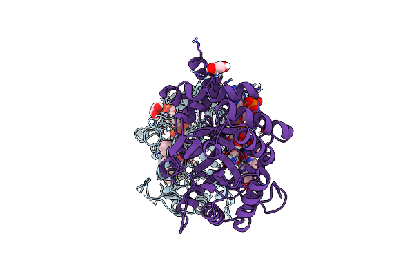 |
Structure Of Aldo-Keto Reductase 1C3 (Akr1C3) In Complex With An Inhibitor M689, With The 3-Hydroxy-Benzoisoxazole Moiety. Resolution 2.0A
Organism: Homo sapiens
Method: X-RAY DIFFRACTION Resolution:2.00 Å Release Date: 2024-03-06 Classification: OXIDOREDUCTASE Ligands: NAP, YMC, EDO |
Organism: Homo sapiens
Method: X-RAY DIFFRACTION
Release Date: 2024-03-06
Ligands: NAP, YMC, EDO
 |
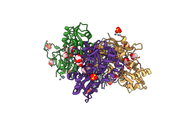
Crystal Structure Of The Ampa Receptor Glua2-L504Y-N775S Ligand Binding Domain In Complex With L-Glutamate And Positive Allosteric Modulator Bpam395 At 1.55A Resolution
Organism: Rattus norvegicus
Method: X-RAY DIFFRACTION Resolution:1.55 Å Release Date: 2023-12-27 Classification: MEMBRANE PROTEIN Ligands: GLU, ZN, ACT, UF5, CL, GOL, CAC |
Organism: Rattus norvegicus
Method: X-RAY DIFFRACTION
Release Date: 2023-12-27
Ligands: GLU, ZN, ACT, UF5, CL, GOL, CAC
 |
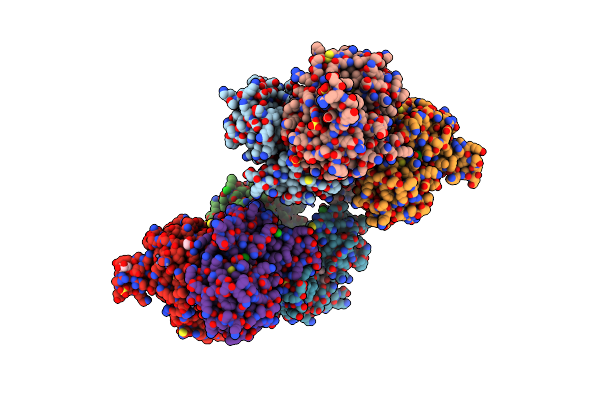
Crystal Structure Of The Kainate Receptor Gluk3-H523A Ligand Binding Domain In Complex With Kainate At 2.7A Resolution
Organism: Rattus norvegicus
Method: X-RAY DIFFRACTION Resolution:2.70 Å Release Date: 2023-12-13 Classification: MEMBRANE PROTEIN Ligands: ACT, ZN, CL, SO4, PEG, KAI, GOL |
Organism: Rattus norvegicus
Method: X-RAY DIFFRACTION
Release Date: 2023-12-13
Ligands: ACT, ZN, CL, SO4, PEG, KAI, GOL
 |
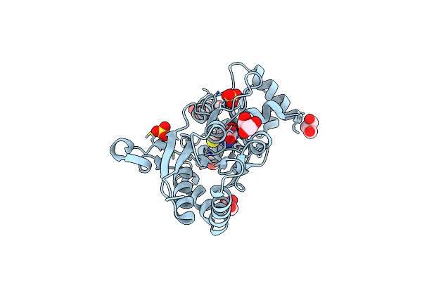
Crystal Structure Of The Kainate Receptor Gluk3-H523A Ligand Binding Domain In Complex With Kainate And The Positive Allosteric Modulator Bpam344 At 2.9A Resolution
Organism: Rattus norvegicus
Method: X-RAY DIFFRACTION Resolution:2.90 Å Release Date: 2023-12-13 Classification: MEMBRANE PROTEIN Ligands: 2J9, ZN, GOL, CL, SO4, KAI, ACT |
Organism: Rattus norvegicus
Method: X-RAY DIFFRACTION
Release Date: 2023-12-13
Ligands: 2J9, ZN, GOL, CL, SO4, KAI, ACT
 |
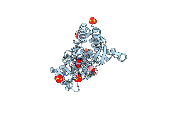
Structure Of The Ampa Receptor Glua2O Ligand-Binding Domain (S1S2J) In Complex With The Compound (S)-1-[2'-Amino-2'-Carboxyethyl]-5,7-Dihydrothieno[3,4-D]Pyrimidin- 2,4(1H,3H)-Dione At Resolution 1.60A
Organism: Rattus norvegicus
Method: X-RAY DIFFRACTION Resolution:1.61 Å Release Date: 2020-06-03 Classification: MEMBRANE PROTEIN Ligands: SO4, GOL, CGW, CL |
Organism: Rattus norvegicus
Method: X-RAY DIFFRACTION
Release Date: 2020-06-03
Ligands: SO4, GOL, CGW, CL
 |
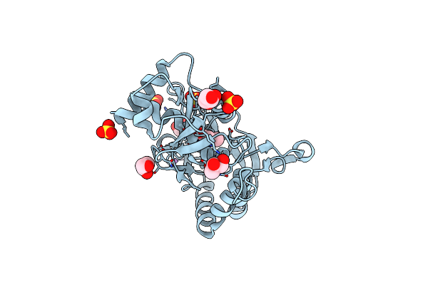
Structure Of The Ampa Receptor Glua2O Ligand-Binding Domain (S1S2J) In Complex With The Compound ( S) - 1- [2'-Amino-2'-Carboxyethyl]-5 ,7- Dihydropyrrolo[3,4-D]Pyrimidin-2,4(1H,3H)-Dione At Resolution 1.20A
Organism: Rattus norvegicus
Method: X-RAY DIFFRACTION Resolution:1.20 Å Release Date: 2020-06-03 Classification: MEMBRANE PROTEIN Ligands: GOL, SO4, PVQ, NH4, CL |
Organism: Rattus norvegicus
Method: X-RAY DIFFRACTION
Release Date: 2020-06-03
Ligands: GOL, SO4, PVQ, NH4, CL
 |
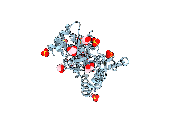
Structure Of The Ampa Receptor Glua2O Ligand-Binding Domain (S1S2J) In Complex With The Compound ( S) - 1- [2'-Amino-2'-Carboxyethyl]-6-Methyl-5 ,7- Dihydropyrrolo[3,4-D]Pyrimidin-2,4(1H,3H)-Dione At Resolution 1.00A
Organism: Rattus norvegicus
Method: X-RAY DIFFRACTION Resolution:1.00 Å Release Date: 2020-06-03 Classification: MEMBRANE PROTEIN Ligands: SO4, GOL, LI, CG8, CL |
Organism: Rattus norvegicus
Method: X-RAY DIFFRACTION
Release Date: 2020-06-03
Ligands: SO4, GOL, LI, CG8, CL
 |
Structure Of The Ampa Receptor Glua2O Ligand-Binding Domain (S1S2J) In Complex With The Compound (S)-1-(2'-Amino-2'-Carboxyethyl)-5,7-Dihydrofuro[3,4-D]- Pyrimidine-2,4(1H,3H)-Dione At Resolution 1.15A
Organism: Rattus norvegicus
Method: X-RAY DIFFRACTION Resolution:1.15 Å Release Date: 2020-06-03 Classification: MEMBRANE PROTEIN Ligands: GOL, SO4, LI, PVK, CL |
Organism: Rattus norvegicus
Method: X-RAY DIFFRACTION
Release Date: 2020-06-03
Ligands: GOL, SO4, LI, PVK, CL
 |
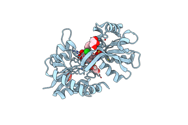
Structure Of The Ampa Receptor Glua2O Ligand-Binding Domain (S1S2J) In Complex With The Compound (S)-1-(2'-Amino-2'-Carboxyethyl)Furo[3,4-D]Pyrimidin-2,4-Dione At Resolution 1.47A
Organism: Rattus norvegicus
Method: X-RAY DIFFRACTION Resolution:1.47 Å Release Date: 2020-06-03 Classification: MEMBRANE PROTEIN Ligands: SO4, GOL, LI, CL, OUB |
Organism: Rattus norvegicus
Method: X-RAY DIFFRACTION
Release Date: 2020-06-03
Ligands: SO4, GOL, LI, CL, OUB
 |
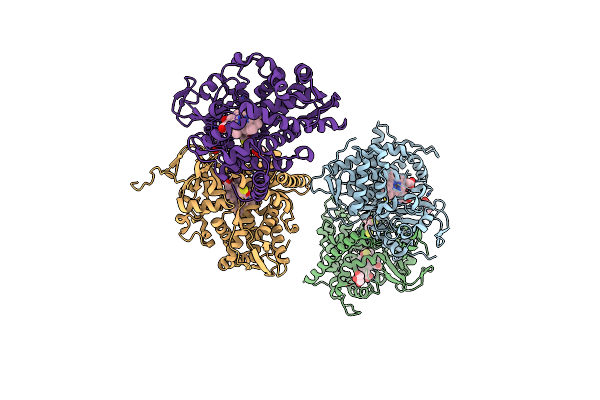
Structure Of Gluk1 Ligand-Binding Domain (S1S2) In Complex With N-(7-(1H-Imidazol-1-Yl)-2,3-Dioxo-6-(Trifluoromethyl)-3,4-Dihydroquinoxalin-1(2H)-Yl Benzamide At 2.3 A Resolution
Organism: Rattus norvegicus
Method: X-RAY DIFFRACTION Resolution:2.30 Å Release Date: 2019-10-30 Classification: MEMBRANE PROTEIN Ligands: SO4, L5H, CL, GOL |
Organism: Rattus norvegicus
Method: X-RAY DIFFRACTION
Release Date: 2019-10-30
Ligands: SO4, L5H, CL, GOL
 |
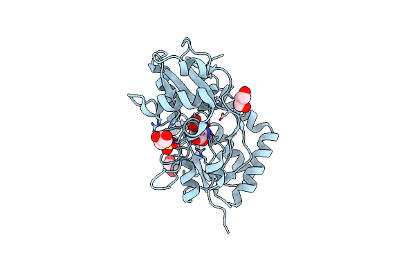
Structure Of Cytochrome P450 Bm3 M11 Mutant In Complex With Dtt At Resolution 2.16A
Organism: Bacillus megaterium
Method: X-RAY DIFFRACTION Resolution:2.16 Å Release Date: 2019-06-05 Classification: OXIDOREDUCTASE Ligands: HEM, DTT, CL, GOL, PEG |
Organism: Bacillus megaterium
Method: X-RAY DIFFRACTION
Release Date: 2019-06-05
Ligands: HEM, DTT, CL, GOL, PEG
 |
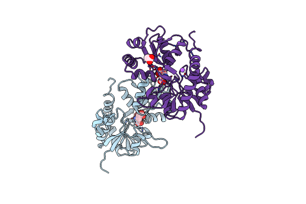
Structure Of Glua2-N775S Ligand-Binding Domain (S1S2J) In Complex With Glutamate And Rubidium Bromide At 1.75 A Resolution
Organism: Rattus norvegicus
Method: X-RAY DIFFRACTION Resolution:1.75 Å Release Date: 2019-05-15 Classification: MEMBRANE PROTEIN Ligands: GLU, SO4, GOL, BR |
Organism: Rattus norvegicus
Method: X-RAY DIFFRACTION
Release Date: 2019-05-15
Ligands: GLU, SO4, GOL, BR
 |
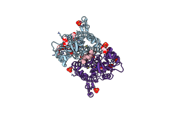
Structure Of Glua2O Ligand-Binding Domain (S1S2J) In Complex With Glutamate And Sodium Bromide At 1.95 A Resolution
Organism: Rattus norvegicus
Method: X-RAY DIFFRACTION Resolution:1.95 Å Release Date: 2019-05-15 Classification: MEMBRANE PROTEIN Ligands: GLU, BR, GOL, NA, ACT |
Organism: Rattus norvegicus
Method: X-RAY DIFFRACTION
Release Date: 2019-05-15
Ligands: GLU, BR, GOL, NA, ACT
 |
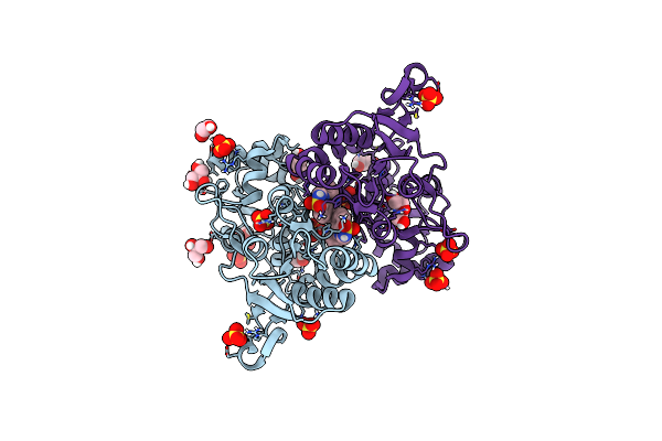
Structure Of Glua2 Ligand-Binding Domain (S1S2J-L504Y-N775S) In Complex With Glutamate And Tdpam02 At 2.4 A Resolution.
Organism: Rattus norvegicus
Method: X-RAY DIFFRACTION Resolution:2.40 Å Release Date: 2019-04-03 Classification: MEMBRANE PROTEIN Ligands: GOL, CL, GLU, SO4, PEG, PGE, FXW |
Organism: Rattus norvegicus
Method: X-RAY DIFFRACTION
Release Date: 2019-04-03
Ligands: GOL, CL, GLU, SO4, PEG, PGE, FXW
 |
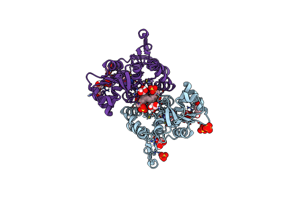
Structure Of Glua2 Ligand-Binding Domain (S1S1J) In Complex With Positive Allosteric Modulator Tdpam02 At 1.8 A Resolution
Organism: Rattus norvegicus
Method: X-RAY DIFFRACTION Resolution:1.88 Å Release Date: 2019-04-03 Classification: MEMBRANE PROTEIN Ligands: SO4, GOL, CL, FXW, PEG |
Organism: Rattus norvegicus
Method: X-RAY DIFFRACTION
Release Date: 2019-04-03
Ligands: SO4, GOL, CL, FXW, PEG
 |
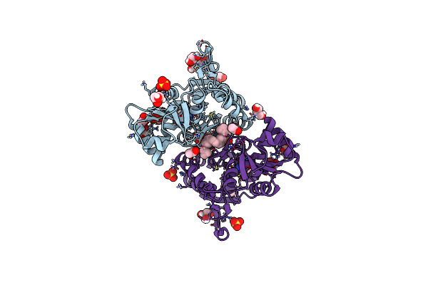
Structure Of Glua2 Ligand-Binding Domain (S1S2J-N775S) In Complex With Glutamate And Tdpam01 At 1.9 A Resolution.
Organism: Rattus norvegicus
Method: X-RAY DIFFRACTION Resolution:1.90 Å Release Date: 2019-04-03 Classification: MEMBRANE PROTEIN Ligands: SO4, GLU, GOL, CL, PEG, D45 |
Organism: Rattus norvegicus
Method: X-RAY DIFFRACTION
Release Date: 2019-04-03
Ligands: SO4, GLU, GOL, CL, PEG, D45
 |
Structure Of Glua2 Ligand-Binding Domain (S1S2J-N775S) In Complex With Glutamate And Tdpam02 At 1.6 A Resolution.
Organism: Rattus norvegicus
Method: X-RAY DIFFRACTION Resolution:1.62 Å Release Date: 2019-04-03 Classification: MEMBRANE PROTEIN Ligands: FXW, GLU, SO4, GOL, PEG, CL, ACT |
Organism: Rattus norvegicus
Method: X-RAY DIFFRACTION
Release Date: 2019-04-03
Ligands: FXW, GLU, SO4, GOL, PEG, CL, ACT