

Search Count: 21
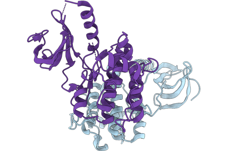 |
Streptococcus Pneumoniae Stkp Catalytic Domain T167A/T169A Double Mutant
Organism: Streptococcus pneumoniae r6
Method: X-RAY DIFFRACTION Resolution:2.80 Å Release Date: 2026-06-10 Classification: CELL CYCLE |
Organism: Streptococcus pneumoniae r6
Method: X-RAY DIFFRACTION
Release Date: 2026-06-10
 |
Streptococcus Pneumoniae Stkp Catalytic Domain T167E/T169E Double Mutant In Complex With Amp-Pnp And Mn2+
Organism: Streptococcus pneumoniae r6
Method: X-RAY DIFFRACTION Resolution:1.60 Å Release Date: 2026-06-10 Classification: CELL CYCLE Ligands: ANP, MN |
Organism: Streptococcus pneumoniae r6
Method: X-RAY DIFFRACTION
Release Date: 2026-06-10
Ligands: ANP, MN
 |
Streptococcus Pneumoniae Stkp Catalytic Domain T167A/T169A Double Mutant In Complex With Amp-Pnp And Mn2+
Organism: Streptococcus pneumoniae r6
Method: X-RAY DIFFRACTION Resolution:1.88 Å Release Date: 2026-06-10 Classification: CELL CYCLE Ligands: ANP, MN |
Organism: Streptococcus pneumoniae r6
Method: X-RAY DIFFRACTION
Release Date: 2026-06-10
Ligands: ANP, MN
 |
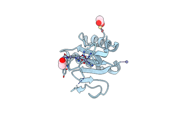
Streptococcus Pneumoniae Stkp Catalytic Domain T167A/T169A Double Mutant In Complex With Amp-Pnp And Mg2+
Organism: Streptococcus pneumoniae r6
Method: X-RAY DIFFRACTION Resolution:1.80 Å Release Date: 2026-06-10 Classification: CELL CYCLE Ligands: ANP, MG, MPD, GOL |
Organism: Streptococcus pneumoniae r6
Method: X-RAY DIFFRACTION
Release Date: 2026-06-10
Ligands: ANP, MG, MPD, GOL
 |
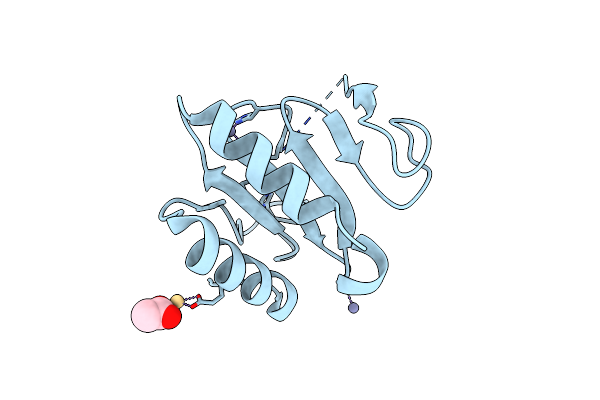
Crystal Structure Of The C-Terminal Domain Of Vlde From Streptococcus Pneumoniae Containing Four Zinc Atoms At The Binding Site
Organism: Streptococcus pneumoniae r6
Method: X-RAY DIFFRACTION Resolution:1.50 Å Release Date: 2025-01-22 Classification: METAL BINDING PROTEIN Ligands: ZN, CD, ACT |
Organism: Streptococcus pneumoniae r6
Method: X-RAY DIFFRACTION
Release Date: 2025-01-22
Ligands: ZN, CD, ACT
 |
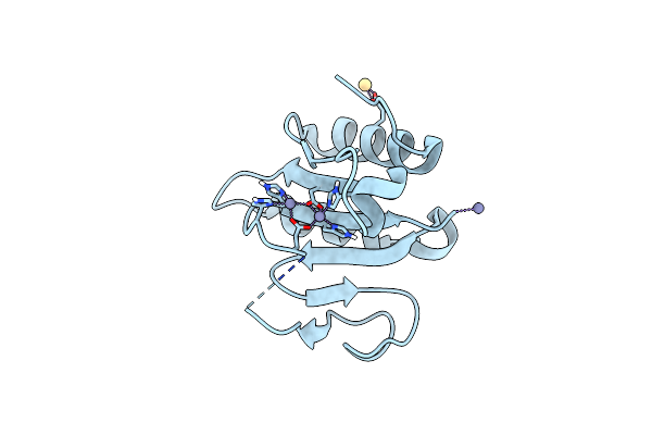
Crystal Structure Of The C-Terminal Domain Of Vlde From Streptococcus Pneumoniae Containing Three Zinc Atoms At The Binding Site
Organism: Streptococcus pneumoniae r6
Method: X-RAY DIFFRACTION Resolution:1.60 Å Release Date: 2025-01-22 Classification: METAL BINDING PROTEIN Ligands: ACT, ZN, CD |
Organism: Streptococcus pneumoniae r6
Method: X-RAY DIFFRACTION
Release Date: 2025-01-22
Ligands: ACT, ZN, CD
 |
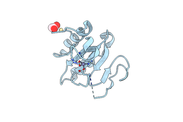
Crystal Structure Of The C-Terminal Domain Of Vlde From Streptococcus Pneumoniae Containing Two Zinc Atoms At The Binding Site
Organism: Streptococcus pneumoniae r6
Method: X-RAY DIFFRACTION Resolution:1.85 Å Release Date: 2025-01-22 Classification: METAL BINDING PROTEIN Ligands: ZN, CD |
Organism: Streptococcus pneumoniae r6
Method: X-RAY DIFFRACTION
Release Date: 2025-01-22
Ligands: ZN, CD
 |
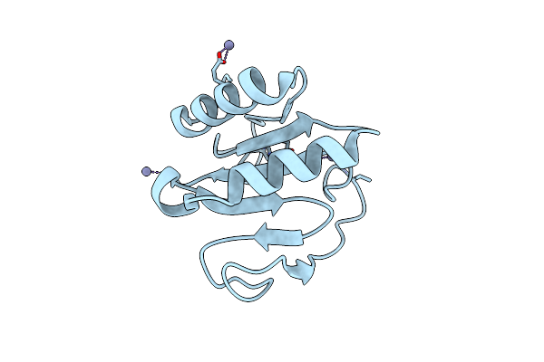
Crystal Structure Of The C-Terminal Domain Of Vlde From Streptococcus Pneumoniae Containing A Zinc Atom At The Binding Site
Organism: Streptococcus pneumoniae r6
Method: X-RAY DIFFRACTION Resolution:2.80 Å Release Date: 2025-01-22 Classification: METAL BINDING PROTEIN Ligands: ACT, CD, ZN |
Organism: Streptococcus pneumoniae r6
Method: X-RAY DIFFRACTION
Release Date: 2025-01-22
Ligands: ACT, CD, ZN
 |
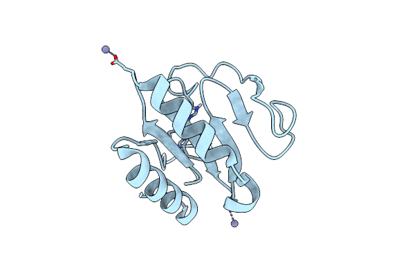
Crystal Structure Of The C-Terminal Domain Of Vlde From Streptococcus Pneumoniae In A Catalytically Competent Conformation
Organism: Streptococcus pneumoniae r6
Method: X-RAY DIFFRACTION Resolution:1.50 Å Release Date: 2025-01-22 Classification: METAL BINDING PROTEIN Ligands: ZN |
Organism: Streptococcus pneumoniae r6
Method: X-RAY DIFFRACTION
Release Date: 2025-01-22
Ligands: ZN
 |
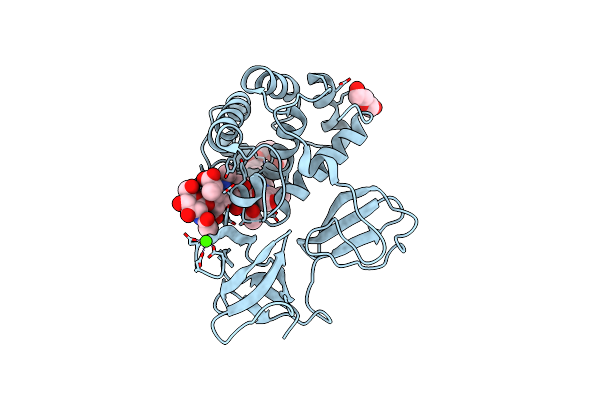
Crystal Structure Of The C-Terminal Domain Of Vlde H373A From Streptococcus Pneumoniae
Organism: Streptococcus pneumoniae r6
Method: X-RAY DIFFRACTION Resolution:1.14 Å Release Date: 2025-01-22 Classification: METAL BINDING PROTEIN Ligands: ZN |
Organism: Streptococcus pneumoniae r6
Method: X-RAY DIFFRACTION
Release Date: 2025-01-22
Ligands: ZN
 |
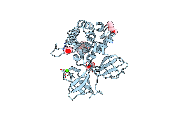
Crystal Structure Of Catalytic Domain Of Lytb (E585Q) From Streptococcus Pneumoniae In Complex With Nag-Nam-Nag-Nam Tetrasaccharide
Organism: Streptococcus pneumoniae (strain atcc baa-255 / r6)
Method: X-RAY DIFFRACTION Resolution:1.50 Å Release Date: 2022-09-21 Classification: HYDROLASE Ligands: 1PE, PEG, CA |
Organism: Streptococcus pneumoniae (strain atcc baa-255 / r6)
Method: X-RAY DIFFRACTION
Release Date: 2022-09-21
Ligands: 1PE, PEG, CA
 |
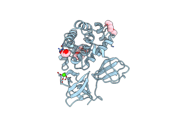
Crystal Structure Of Catalytic Domain In Open Conformation Of Lytb From Streptococcus Pneumoniae
Organism: Streptococcus pneumoniae (strain atcc baa-255 / r6)
Method: X-RAY DIFFRACTION Resolution:1.43 Å Release Date: 2022-09-07 Classification: HYDROLASE Ligands: 1PE, PGE, PEG, CA |
Organism: Streptococcus pneumoniae (strain atcc baa-255 / r6)
Method: X-RAY DIFFRACTION
Release Date: 2022-09-07
Ligands: 1PE, PGE, PEG, CA
 |
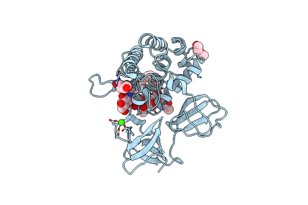
Crystal Structure Of Catalytic Domain In Closed Conformation Of Lytb (E585Q)From Streptococcus Pneumoniae
Organism: Streptococcus pneumoniae (strain atcc baa-255 / r6)
Method: X-RAY DIFFRACTION Resolution:1.25 Å Release Date: 2022-09-07 Classification: HYDROLASE Ligands: 1PE, PGE, PEG, ACT, CA |
Organism: Streptococcus pneumoniae (strain atcc baa-255 / r6)
Method: X-RAY DIFFRACTION
Release Date: 2022-09-07
Ligands: 1PE, PGE, PEG, ACT, CA
 |
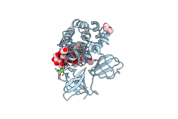
Crystal Structure Of Catalytic Domain Of Lytb From Streptococcus Pneumoniae In Complex With Nag-Nag-Nag-Nag Tetrasaccharide
Organism: Streptococcus pneumoniae (strain atcc baa-255 / r6)
Method: X-RAY DIFFRACTION Resolution:1.55 Å Release Date: 2022-09-07 Classification: HYDROLASE Ligands: 1PE, PEG, CA |
Organism: Streptococcus pneumoniae (strain atcc baa-255 / r6)
Method: X-RAY DIFFRACTION
Release Date: 2022-09-07
Ligands: 1PE, PEG, CA
 |
Crystal Structure Of Catalytic Domain Of Lytb (E585Q) From Streptococcus Pneumoniae In Complex With Nag-Nam-Nag-Nam-Nag Peptidolycan Analogue
Organism: Streptococcus pneumoniae (strain atcc baa-255 / r6)
Method: X-RAY DIFFRACTION Resolution:1.30 Å Release Date: 2022-09-07 Classification: HYDROLASE Ligands: 1PE, PGE, PEG, CA |
Organism: Streptococcus pneumoniae (strain atcc baa-255 / r6)
Method: X-RAY DIFFRACTION
Release Date: 2022-09-07
Ligands: 1PE, PGE, PEG, CA
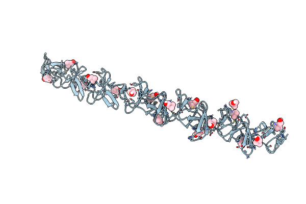 |
Crystal Structure Of Choline-Binding Module Of Lytb From Streptococcus Pneumoniae
Organism: Streptococcus pneumoniae (strain atcc baa-255 / r6)
Method: X-RAY DIFFRACTION Resolution:2.98 Å Release Date: 2022-09-07 Classification: HYDROLASE Ligands: CHT |
Organism: Streptococcus pneumoniae (strain atcc baa-255 / r6)
Method: X-RAY DIFFRACTION
Release Date: 2022-09-07
Ligands: CHT
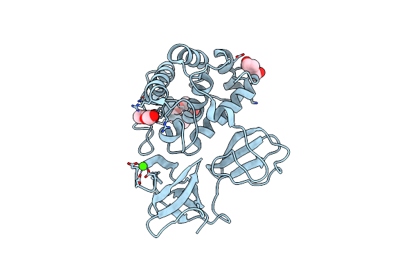 |
Crystal Structure Of Catalytic Domain In Closed Conformation Of Lytb From Streptococcus Pneumoniae
Organism: Streptococcus pneumoniae r6
Method: X-RAY DIFFRACTION Resolution:1.80 Å Release Date: 2022-09-07 Classification: HYDROLASE Ligands: 1PE, PEG, CA |
Organism: Streptococcus pneumoniae r6
Method: X-RAY DIFFRACTION
Release Date: 2022-09-07
Ligands: 1PE, PEG, CA
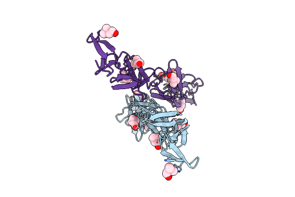 |
Crystal Structure Of Choline-Binding Module (R1-R9) Of Lytb From Streptococcus Pneumoniae
Organism: Streptococcus pneumoniae (strain atcc baa-255 / r6)
Method: X-RAY DIFFRACTION Resolution:1.99 Å Release Date: 2022-09-07 Classification: HYDROLASE Ligands: CHT, ZN, PGE |
Organism: Streptococcus pneumoniae (strain atcc baa-255 / r6)
Method: X-RAY DIFFRACTION
Release Date: 2022-09-07
Ligands: CHT, ZN, PGE
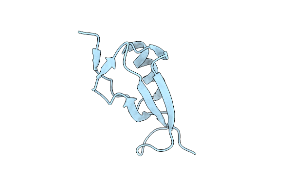 |
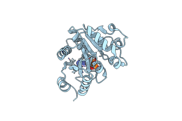
Pasta Subunit 4 Of Streptococcus Pneumoniae Stkp Crystallized With Peg And Succinate
Organism: Streptococcus pneumoniae (strain atcc baa-255 / r6)
Method: X-RAY DIFFRACTION Resolution:1.90 Å Release Date: 2017-11-08 Classification: TRANSFERASE |
Organism: Streptococcus pneumoniae (strain atcc baa-255 / r6)
Method: X-RAY DIFFRACTION
Release Date: 2017-11-08
 |
Crystal Structure Of The Chimerical Protein Capa1B1 In Complex With Adp-Mg
Organism: Staphylococcus aureus
Method: X-RAY DIFFRACTION Resolution:2.20 Å Release Date: 2014-02-05 Classification: TRANSFERASE Ligands: ADP, MG |
Organism: Staphylococcus aureus
Method: X-RAY DIFFRACTION
Release Date: 2014-02-05
Ligands: ADP, MG