

Search Count: 28
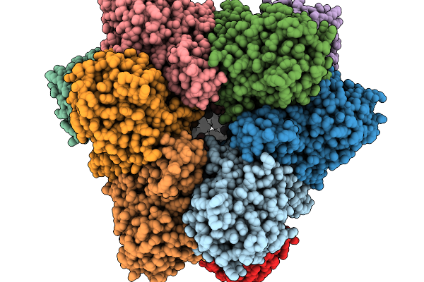 |
Cryoem Structure Of The Tetrahedral M42 Aminopeptidase From M. Jannaschii
Organism: Methanocaldococcus jannaschii
Method: ELECTRON MICROSCOPY Release Date: 2026-01-14 Classification: CYTOSOLIC PROTEIN |
Organism: Methanocaldococcus jannaschii
Method: ELECTRON MICROSCOPY
Release Date: 2026-01-14
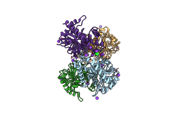 |
Crystal Structure Of Malate Dehydrogenase From Haloarcula Marismortui With Potassium And Chloride Ions
Organism: Haloarcula marismortui
Method: X-RAY DIFFRACTION Resolution:1.95 Å Release Date: 2022-02-16 Classification: OXIDOREDUCTASE Ligands: CL, K |
Organism: Haloarcula marismortui
Method: X-RAY DIFFRACTION
Release Date: 2022-02-16
Ligands: CL, K
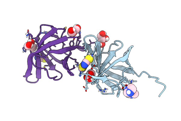 |
Crystal Structure Of Pyrococcus Abyssi Pbp11
Organism: Pyrococcus abyssi (strain ge5 / orsay)
Method: X-RAY DIFFRACTION Resolution:1.64 Å Release Date: 2021-11-03 Classification: PROTEIN BINDING Ligands: NI, GOL, IMD, SCN |
Organism: Pyrococcus abyssi (strain ge5 / orsay)
Method: X-RAY DIFFRACTION
Release Date: 2021-11-03
Ligands: NI, GOL, IMD, SCN
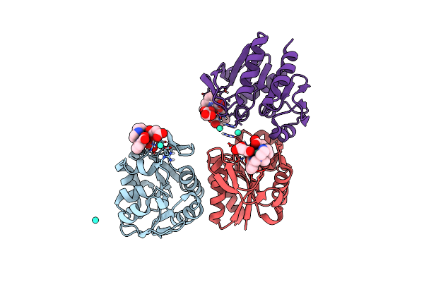 |
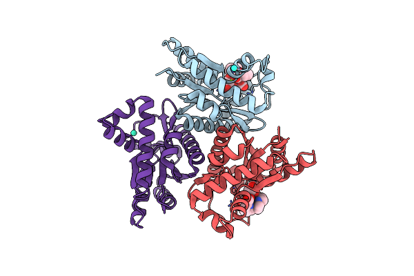
Crystal Structure Of The Protease 1 (E29A,E60A,E80A) From Pyrococcus Horikoshii Co-Crystallized With Tb-Xo4.
Organism: Pyrococcus horikoshii (strain atcc 700860 / dsm 12428 / jcm 9974 / nbrc 100139 / ot-3)
Method: X-RAY DIFFRACTION Resolution:2.00 Å Release Date: 2019-06-19 Classification: HYDROLASE Ligands: 7MT, MLI, TB |
Organism: Pyrococcus horikoshii (strain atcc 700860 / dsm 12428 / jcm 9974 / nbrc 100139 / ot-3)
Method: X-RAY DIFFRACTION
Release Date: 2019-06-19
Ligands: 7MT, MLI, TB
 |
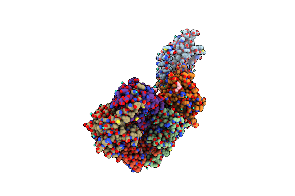
Crystal Structure Of The Adenylate Kinase From Methanothermococcus Thermolithotrophicus Co-Crystallized With Tb-Xo4
Organism: Methanothermococcus thermolithotrophicus
Method: X-RAY DIFFRACTION Resolution:1.96 Å Release Date: 2019-06-19 Classification: TRANSFERASE Ligands: 7MT, TB, MG, GOL |
Organism: Methanothermococcus thermolithotrophicus
Method: X-RAY DIFFRACTION
Release Date: 2019-06-19
Ligands: 7MT, TB, MG, GOL
 |
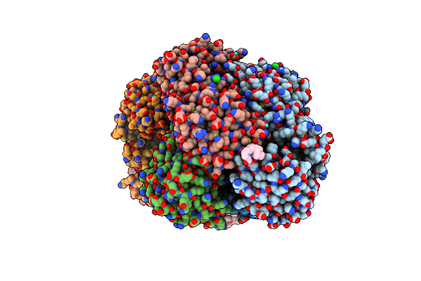
Crystal Structure Of The Thiazole Synthase From Methanothermococcus Thermolithotrophicus Co-Crystallized With Tb-Xo4
Organism: Methanothermococcus thermolithotrophicus
Method: X-RAY DIFFRACTION Resolution:2.55 Å Release Date: 2019-06-19 Classification: BIOSYNTHETIC PROTEIN Ligands: TB, 48F, GOL, PGE, PEG, NA |
Organism: Methanothermococcus thermolithotrophicus
Method: X-RAY DIFFRACTION
Release Date: 2019-06-19
Ligands: TB, 48F, GOL, PGE, PEG, NA
 |
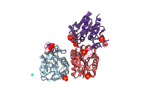
Crystal Structure Of Coenzyme F420H2 Oxidase (Fpra) Co-Crystallized With 10 Mm Tb-Xo4
Organism: Methanothermococcus thermolithotrophicus
Method: X-RAY DIFFRACTION Resolution:2.20 Å Release Date: 2018-10-31 Classification: OXIDOREDUCTASE Ligands: FMN, 7MT, TB, CL, FE |
Organism: Methanothermococcus thermolithotrophicus
Method: X-RAY DIFFRACTION
Release Date: 2018-10-31
Ligands: FMN, 7MT, TB, CL, FE
 |
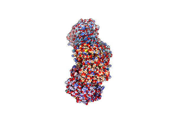
Crystal Structure Of Protease 1 From Pyrococcus Horikoshii Co-Cystallized In Presence Of 10 Mm Tb-Xo4 And Ammonium Sulfate.
Organism: Pyrococcus horikoshii
Method: X-RAY DIFFRACTION Resolution:1.65 Å Release Date: 2018-10-03 Classification: CELL CYCLE Ligands: 7MT, SO4, TB, 2HA |
Organism: Pyrococcus horikoshii
Method: X-RAY DIFFRACTION
Release Date: 2018-10-03
Ligands: 7MT, SO4, TB, 2HA
 |
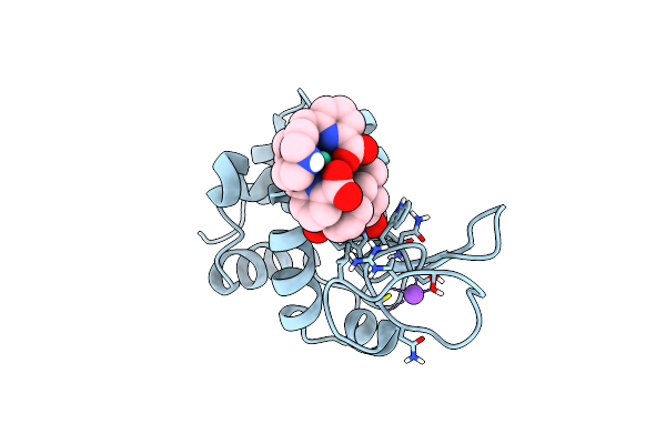
Structure Of Protease 1 From Pyrococcus Horikoshii Co-Crystallized In Presence Of 10 Mm Tb-Xo4 And Potassium Iodide.
Organism: Pyrococcus horikoshii
Method: X-RAY DIFFRACTION Resolution:2.19 Å Release Date: 2018-10-03 Classification: HYDROLASE Ligands: 7MT, IOD, TB |
Organism: Pyrococcus horikoshii
Method: X-RAY DIFFRACTION
Release Date: 2018-10-03
Ligands: 7MT, IOD, TB
 |
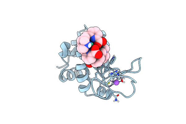
Crystal Structure Of Hen Egg-White Lysozyme Co-Crystallized In Presence Of 100 Mm Tb-Xo4
Organism: Gallus gallus
Method: X-RAY DIFFRACTION Resolution:1.20 Å Release Date: 2018-10-03 Classification: HYDROLASE Ligands: TB, 7MT, CL, NA, ACT |
Organism: Gallus gallus
Method: X-RAY DIFFRACTION
Release Date: 2018-10-03
Ligands: TB, 7MT, CL, NA, ACT
 |
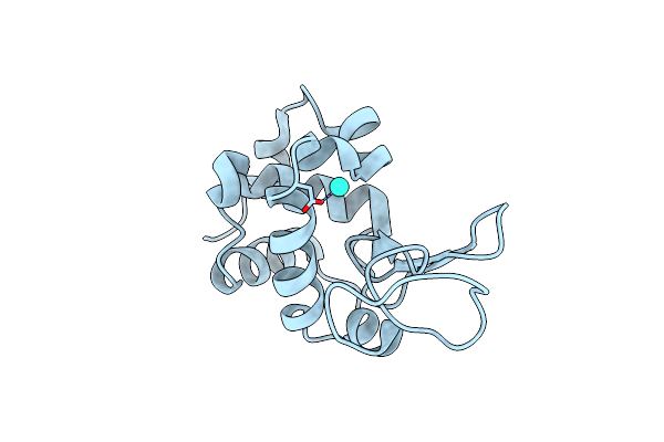
Crystal Structure Of Hen Egg-White Lysozyme Co-Crystallized In Presence Of 100 Mm Tb-Xo4 And 100 Mm Sodium Sulfate
Organism: Gallus gallus
Method: X-RAY DIFFRACTION Resolution:1.30 Å Release Date: 2018-10-03 Classification: HYDROLASE Ligands: TB, CL, NA, 7MT |
Organism: Gallus gallus
Method: X-RAY DIFFRACTION
Release Date: 2018-10-03
Ligands: TB, CL, NA, 7MT
 |
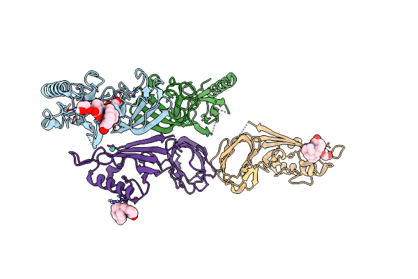
Crystal Structure Of Hen Egg-White Lysozyme Co-Crystallized In Presence Of 100 Mm Tb-Xo4 And 100 Mm Potassium Phosphate Monobasic.
Organism: Gallus gallus
Method: X-RAY DIFFRACTION Resolution:1.50 Å Release Date: 2018-10-03 Classification: HYDROLASE Ligands: TB, CL |
Organism: Gallus gallus
Method: X-RAY DIFFRACTION
Release Date: 2018-10-03
Ligands: TB, CL
 |
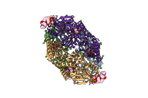
Structure Of The Bacteriophage T5 Distal Tail Protein Pb9 Co-Crystallized With 10Mm Tb-Xo4
Organism: Escherichia phage t5
Method: X-RAY DIFFRACTION Resolution:1.80 Å Release Date: 2018-10-03 Classification: VIRAL PROTEIN Ligands: 7MT, EDO, CL, NA, TB |
Organism: Escherichia phage t5
Method: X-RAY DIFFRACTION
Release Date: 2018-10-03
Ligands: 7MT, EDO, CL, NA, TB
 |
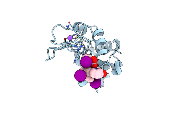
Structure Of F420H2 Oxidase (Fpra) Co-Crystallized With 10Mm Tb-Xo4 And Calcium Chloride
Organism: Methanothermococcus thermolithotrophicus
Method: X-RAY DIFFRACTION Resolution:1.74 Å Release Date: 2018-10-03 Classification: OXIDOREDUCTASE Ligands: FMN, CA, TB, GOL, PE4, 7MT, NA |
Organism: Methanothermococcus thermolithotrophicus
Method: X-RAY DIFFRACTION
Release Date: 2018-10-03
Ligands: FMN, CA, TB, GOL, PE4, 7MT, NA
 |
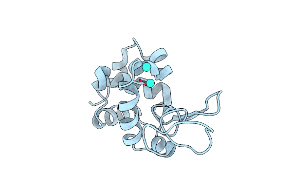
Crystal Structure Of Hen Egg-White Lysozyme Co-Crystallized In Presence Of 100 Mm Tb-Xo4 And 100 Mm Potassium Iodide.
Organism: Gallus gallus
Method: X-RAY DIFFRACTION Resolution:1.42 Å Release Date: 2018-10-03 Classification: HYDROLASE Ligands: TB, IOD, NA, 7MT |
Organism: Gallus gallus
Method: X-RAY DIFFRACTION
Release Date: 2018-10-03
Ligands: TB, IOD, NA, 7MT
 |
Structure Of Tetragonal Hen Egg-White Lysozyme Co-Crystallized In Presence Of 100 Mm Tb-Xo4 And 100 Mm Potassium Sodium Tartrate Tetrahydrate.
Organism: Gallus gallus
Method: X-RAY DIFFRACTION Resolution:1.69 Å Release Date: 2018-10-03 Classification: HYDROLASE Ligands: TB, CL |
Organism: Gallus gallus
Method: X-RAY DIFFRACTION
Release Date: 2018-10-03
Ligands: TB, CL
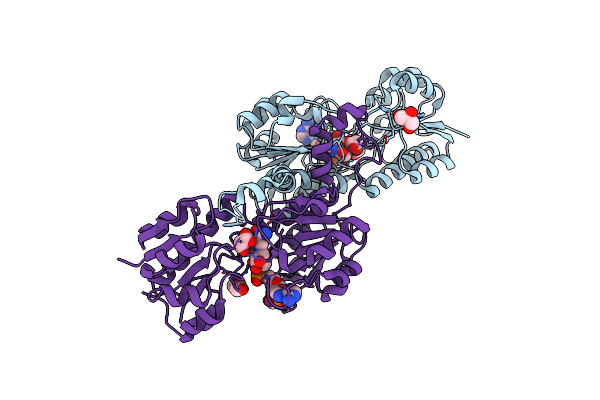 |
Crystal Structure Of Pyrococcus Yayanosii Glyoxylate Hydroxypyruvate Reductase In Complex With Nadp And Malonate (Re-Refinement Of 5Aow)
Organism: Pyrococcus yayanosii (strain ch1 / jcm 16557)
Method: X-RAY DIFFRACTION Resolution:2.00 Å Release Date: 2018-01-17 Classification: OXIDOREDUCTASE Ligands: NAP, MLI, GOL |
Organism: Pyrococcus yayanosii (strain ch1 / jcm 16557)
Method: X-RAY DIFFRACTION
Release Date: 2018-01-17
Ligands: NAP, MLI, GOL
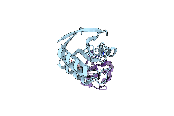 |
Structure Of Deubiquitinating Enzyme Homolog (Pyrococcus Furiosus Jamm1) In Complex With Ubiquitin-Like Samp2.
Organism: Pyrococcus furiosus, Pyrococcus furiosus (strain atcc 43587 / dsm 3638 / jcm 8422 / vc1)
Method: X-RAY DIFFRACTION Resolution:1.90 Å Release Date: 2017-06-21 Classification: CELL CYCLE Ligands: ZN, GOL |
Organism: Pyrococcus furiosus, Pyrococcus furiosus (strain atcc 43587 / dsm 3638 / jcm 8422 / vc1)
Method: X-RAY DIFFRACTION
Release Date: 2017-06-21
Ligands: ZN, GOL
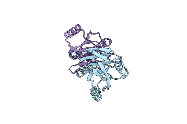 |
Structure Of Deubiquitinating Enzyme Homolog, Pyrococcus Furiosus Jamm1.
Organism: Pyrococcus furiosus (strain atcc 43587 / dsm 3638 / jcm 8422 / vc1)
Method: X-RAY DIFFRACTION Resolution:1.73 Å Release Date: 2017-05-17 Classification: HYDROLASE Ligands: ZN, CL |
Organism: Pyrococcus furiosus (strain atcc 43587 / dsm 3638 / jcm 8422 / vc1)
Method: X-RAY DIFFRACTION
Release Date: 2017-05-17
Ligands: ZN, CL
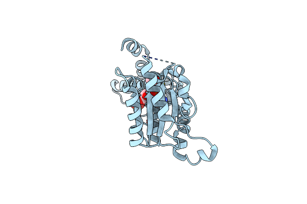 |
Crystal Structure Of The Atpase Domain Of Pan From Pyrococcus Horikoshii
Organism: Pyrococcus horikoshii
Method: X-RAY DIFFRACTION Resolution:1.94 Å Release Date: 2016-11-23 Classification: HYDROLASE Ligands: ADP, ACT, SO4 |
Organism: Pyrococcus horikoshii
Method: X-RAY DIFFRACTION
Release Date: 2016-11-23
Ligands: ADP, ACT, SO4