

Search Count: 11
 |
Spinach 19S Proteasome
Organism: Spinacia oleracea
Method: ELECTRON MICROSCOPY Release Date: 2022-08-24 Classification: PLANT PROTEIN Ligands: ATP, ADP |
Organism: Spinacia oleracea
Method: ELECTRON MICROSCOPY
Release Date: 2022-08-24
Ligands: ATP, ADP
 |
Spinach 20S Proteasome
Organism: Spinacia oleracea
Method: ELECTRON MICROSCOPY Release Date: 2022-05-25 Classification: PLANT PROTEIN |
Organism: Spinacia oleracea
Method: ELECTRON MICROSCOPY
Release Date: 2022-05-25
 |
Sars-Cov-2 Spike In Prefusion State
Organism: Severe acute respiratory syndrome coronavirus 2
Method: ELECTRON MICROSCOPY Release Date: 2020-07-29 Classification: VIRAL PROTEIN Ligands: NAG, MAN, DMS |
Organism: Severe acute respiratory syndrome coronavirus 2
Method: ELECTRON MICROSCOPY
Release Date: 2020-07-29
Ligands: NAG, MAN, DMS
 |
Sars-Cov-2 Spike In Prefusion State (Flexibility Analysis, 1-Up Closed Conformation)
Organism: Severe acute respiratory syndrome coronavirus 2
Method: ELECTRON MICROSCOPY Release Date: 2020-07-29 Classification: VIRAL PROTEIN Ligands: NAG, MAN, DMS |
Organism: Severe acute respiratory syndrome coronavirus 2
Method: ELECTRON MICROSCOPY
Release Date: 2020-07-29
Ligands: NAG, MAN, DMS
 |
Sars-Cov-2 Spike In Prefusion State (Flexibility Analysis, 1-Up Open Conformation)
Organism: Severe acute respiratory syndrome coronavirus 2
Method: ELECTRON MICROSCOPY Release Date: 2020-07-29 Classification: VIRAL PROTEIN Ligands: NAG, MAN, DMS |
Organism: Severe acute respiratory syndrome coronavirus 2
Method: ELECTRON MICROSCOPY
Release Date: 2020-07-29
Ligands: NAG, MAN, DMS
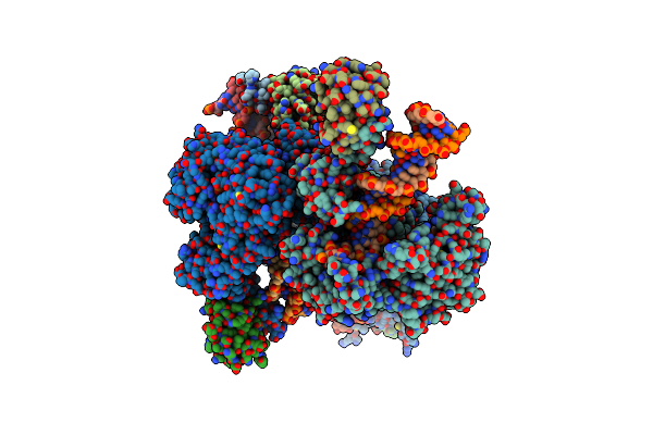 |
Structure Of T7 Dna Polymerase Bound To A Primer/Template Dna And A Peptide That Mimics The C-Terminal Tail Of The Primase-Helicase
Organism: Enterobacteria phage t7, Escherichia coli
Method: X-RAY DIFFRACTION Resolution:3.00 Å Release Date: 2020-03-04 Classification: TRANSFERASE/DNA BINDING PROTEIN/DNA Ligands: TTP, MG |
Organism: Enterobacteria phage t7, Escherichia coli
Method: X-RAY DIFFRACTION
Release Date: 2020-03-04
Ligands: TTP, MG
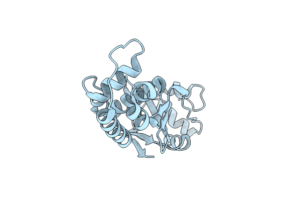 |
The Crystal Structure Of Sats C-Terminal Domain
Organism: Mycobacterium tuberculosis h37rv
Method: X-RAY DIFFRACTION Resolution:1.40 Å Release Date: 2019-01-23 Classification: CHAPERONE |
Organism: Mycobacterium tuberculosis h37rv
Method: X-RAY DIFFRACTION
Release Date: 2019-01-23
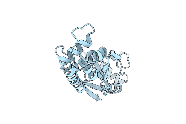 |
The Crystal Structure Of Sats C-Terminal Domain In Complex With Bromine
Organism: Mycobacterium tuberculosis
Method: X-RAY DIFFRACTION Resolution:2.30 Å Release Date: 2019-01-23 Classification: CHAPERONE Ligands: BR |
Organism: Mycobacterium tuberculosis
Method: X-RAY DIFFRACTION
Release Date: 2019-01-23
Ligands: BR
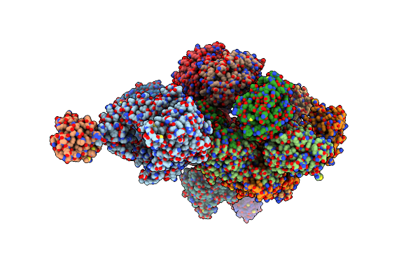 |
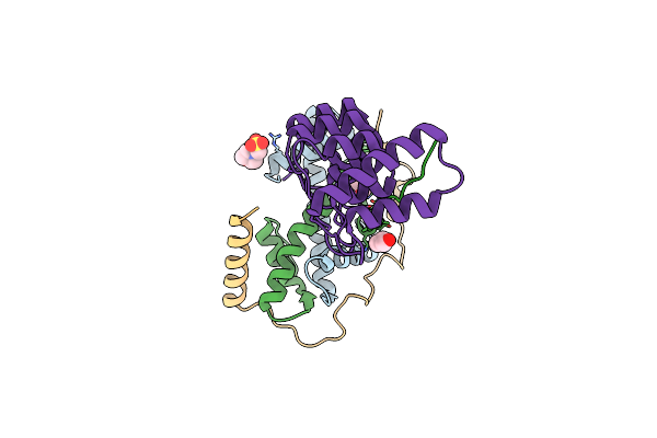
Crystal Structure Of The T7 Replisome In The Absence Of Dna
Organism: Enterobacteria phage t7, Escherichia coli o157:h7
Method: X-RAY DIFFRACTION Resolution:4.80 Å Release Date: 2016-12-07 Classification: TRANSFERASE |
Organism: Enterobacteria phage t7, Escherichia coli o157:h7
Method: X-RAY DIFFRACTION
Release Date: 2016-12-07
 |
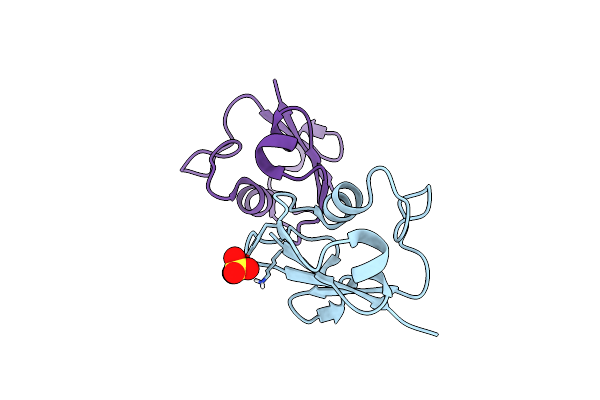
Crystal Structure Of Human Tonsl And Mcm2 Hbds Binding To A Histone H3-H4 Tetramer
Organism: Homo sapiens
Method: X-RAY DIFFRACTION Resolution:2.42 Å Release Date: 2016-06-15 Classification: CHAPERONE Ligands: MES, GOL |
Organism: Homo sapiens
Method: X-RAY DIFFRACTION
Release Date: 2016-06-15
Ligands: MES, GOL
 |
X-Ray Crystal Structure Of Sulfolobus Solfataricus Urm1
Organism: Sulfolobus solfataricus
Method: X-RAY DIFFRACTION Resolution:2.20 Å Release Date: 2015-09-23 Classification: SIGNALING PROTEIN Ligands: SO4 |
Organism: Sulfolobus solfataricus
Method: X-RAY DIFFRACTION
Release Date: 2015-09-23
Ligands: SO4