

Search Count: 19
 |
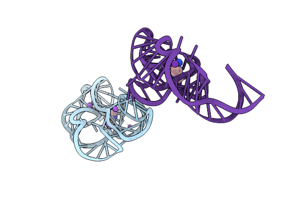
Crystal Structure Of The E. Coli Thim Riboswitch Bound To N-Methyl-1-(Quinoxalin-6-Yl)Methanamine (Compound 16)
Organism: Escherichia coli
Method: X-RAY DIFFRACTION Resolution:2.70 Å Release Date: 2022-05-25 Classification: RNA Ligands: KWU, MG, K, NA |
Organism: Escherichia coli
Method: X-RAY DIFFRACTION
Release Date: 2022-05-25
Ligands: KWU, MG, K, NA
 |
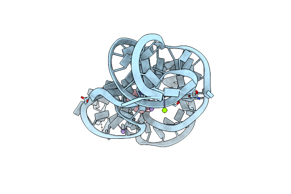
Crystal Structure Of The E. Coli Thim Riboswitch In Complex With Quinoxalin-6-Ylmethanamine (Compound 17)
Organism: Escherichia coli
Method: X-RAY DIFFRACTION Resolution:2.21 Å Release Date: 2022-05-25 Classification: RNA Ligands: KX3, MG, K, NA |
Organism: Escherichia coli
Method: X-RAY DIFFRACTION
Release Date: 2022-05-25
Ligands: KX3, MG, K, NA
 |
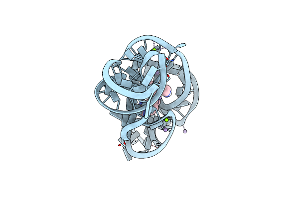
Crystal Structure Of The E. Coli Thim Riboswitch In Complex With N1,N1-Dimethyl-N2-(Quinoxalin-6-Ylmethyl)Ethane-1,2-Diamine (Linked Compound 37)
Organism: Escherichia coli
Method: X-RAY DIFFRACTION Resolution:2.96 Å Release Date: 2022-05-25 Classification: RNA Ligands: MN, MG, KXC |
Organism: Escherichia coli
Method: X-RAY DIFFRACTION
Release Date: 2022-05-25
Ligands: MN, MG, KXC
 |
Crystal Structure Of The E. Coli Thim Riboswitch Bound To 1-(4-(Piperazin-1-Yl)Pyridin-3-Yl)-N-(Quinoxalin-6-Ylmethyl)Methanamine (Linked Compound 38)
Organism: Escherichia coli
Method: X-RAY DIFFRACTION Resolution:2.87 Å Release Date: 2022-05-25 Classification: RNA Ligands: KWL, MG, MN |
Organism: Escherichia coli
Method: X-RAY DIFFRACTION
Release Date: 2022-05-25
Ligands: KWL, MG, MN
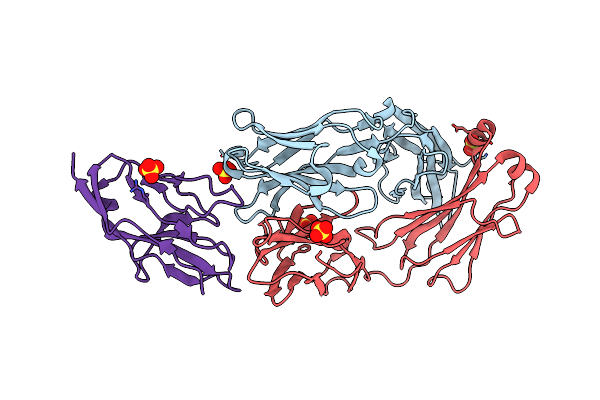 |
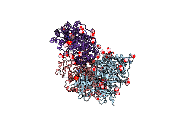
Blocking Fab 25 Anti-Sirp-Alpha Antibody In Complex With Sirp-Alpha Variant 1
Organism: Homo sapiens
Method: X-RAY DIFFRACTION Resolution:2.27 Å Release Date: 2020-12-16 Classification: SIGNALING PROTEIN Ligands: SO4 |
Organism: Homo sapiens
Method: X-RAY DIFFRACTION
Release Date: 2020-12-16
Ligands: SO4
 |
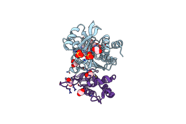
Crystal Structure Of Photinus Pyralis Luciferase Pps6 Mutant In Complex With Dlsa
Organism: Photinus pyralis
Method: X-RAY DIFFRACTION Resolution:2.75 Å Release Date: 2019-10-16 Classification: LIGASE Ligands: SO4, DYD, EDO, SLU |
Organism: Photinus pyralis
Method: X-RAY DIFFRACTION
Release Date: 2019-10-16
Ligands: SO4, DYD, EDO, SLU
 |
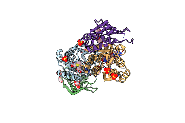
Crystal Structure Of Human Mettl5-Trmt112 Complex, The 18S Rrna M6A1832 Methyltransferase At 1.6A Resolution
Organism: Homo sapiens
Method: X-RAY DIFFRACTION Resolution:1.60 Å Release Date: 2019-07-31 Classification: TRANSFERASE Ligands: SAM, EDO, SO4 |
Organism: Homo sapiens
Method: X-RAY DIFFRACTION
Release Date: 2019-07-31
Ligands: SAM, EDO, SO4
 |
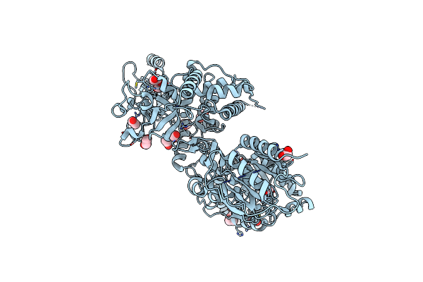
Crystal Structure Of Human Mettl5-Trmt112 Complex, The 18S Rrna M6A1832 Methyltransferase At 2.5A Resolution
Organism: Homo sapiens
Method: X-RAY DIFFRACTION Resolution:2.49 Å Release Date: 2019-07-31 Classification: TRANSFERASE Ligands: SAM, SO4, PEG, EDO, PG0 |
Organism: Homo sapiens
Method: X-RAY DIFFRACTION
Release Date: 2019-07-31
Ligands: SAM, SO4, PEG, EDO, PG0
 |
Crystal Structure Of S. Cerevisiae Deah-Box Rna Helicase Dhr1, Essential For Small Ribosomal Subunit Biogenesis
Organism: Saccharomyces cerevisiae (strain atcc 204508 / s288c)
Method: X-RAY DIFFRACTION Resolution:2.30 Å Release Date: 2019-06-19 Classification: HYDROLASE Ligands: EDO, MG, CL |
Organism: Saccharomyces cerevisiae (strain atcc 204508 / s288c)
Method: X-RAY DIFFRACTION
Release Date: 2019-06-19
Ligands: EDO, MG, CL
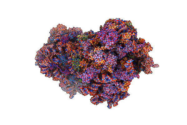 |
Crystal Structure Of Haemanthamine Bound To The 80S Ribosome
Organism: Saccharomyces cerevisiae (strain atcc 204508 / s288c)
Method: X-RAY DIFFRACTION Resolution:3.10 Å Release Date: 2018-02-28 Classification: RIBOSOME Ligands: OHX, MG, HN8, GOL, ZN |
Organism: Saccharomyces cerevisiae (strain atcc 204508 / s288c)
Method: X-RAY DIFFRACTION
Release Date: 2018-02-28
Ligands: OHX, MG, HN8, GOL, ZN
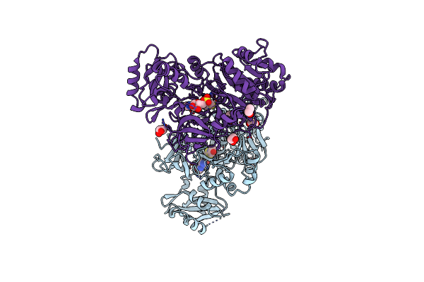 |
Structure Of Photinus Pyralis Luciferase Red Light Emitting Variant
Organism: Photinus pyralis
Method: X-RAY DIFFRACTION Resolution:2.00 Å Release Date: 2016-12-21 Classification: OXIDOREDUCTASE Ligands: SLU, EDO |
Organism: Photinus pyralis
Method: X-RAY DIFFRACTION
Release Date: 2016-12-21
Ligands: SLU, EDO
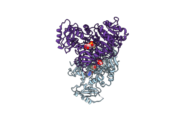 |
Structure Of Photinus Pyralis Luciferase Green Shifted Light Emitting Variant
Organism: Photinus pyralis
Method: X-RAY DIFFRACTION Resolution:2.50 Å Release Date: 2016-12-21 Classification: OXIDOREDUCTASE Ligands: SLU, TLA |
Organism: Photinus pyralis
Method: X-RAY DIFFRACTION
Release Date: 2016-12-21
Ligands: SLU, TLA
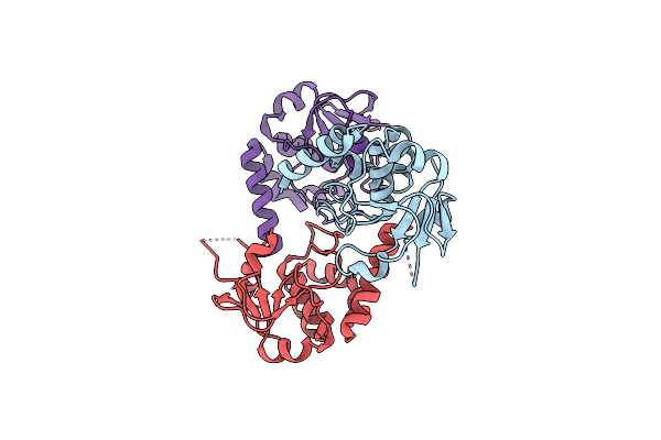 |
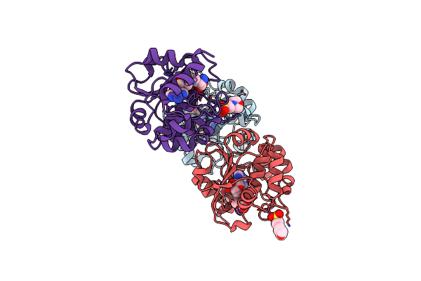
Structure Of The Sam-Dependent Rrna:Acp-Transferase Tsr3 From S. Solfataricus
Organism: Sulfolobus solfataricus
Method: X-RAY DIFFRACTION Resolution:2.25 Å Release Date: 2016-04-27 Classification: TRANSFERASE |
Organism: Sulfolobus solfataricus
Method: X-RAY DIFFRACTION
Release Date: 2016-04-27
 |
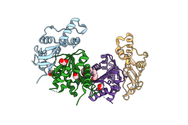
Structure Of The Sam-Dependent Rrna:Acp-Transferase Tsr3 From Vulcanisaeta Distributa
Organism: Vulcanisaeta distributa
Method: X-RAY DIFFRACTION Resolution:1.60 Å Release Date: 2016-04-27 Classification: TRANSFERASE Ligands: EEM, MES |
Organism: Vulcanisaeta distributa
Method: X-RAY DIFFRACTION
Release Date: 2016-04-27
Ligands: EEM, MES
 |
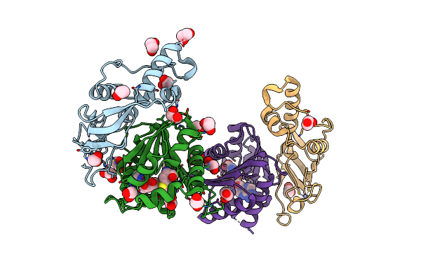
Structure Of S. Cerevisiae Bud23-Trm112 Complex Involved In Formation Of M7G1575 On 18S Rrna (Apo-Form)
Organism: Saccharomyces cerevisiae s288c
Method: X-RAY DIFFRACTION Resolution:2.00 Å Release Date: 2014-12-24 Classification: TRANSFERASE Ligands: ZN, EDO, 12P |
Organism: Saccharomyces cerevisiae s288c
Method: X-RAY DIFFRACTION
Release Date: 2014-12-24
Ligands: ZN, EDO, 12P
 |
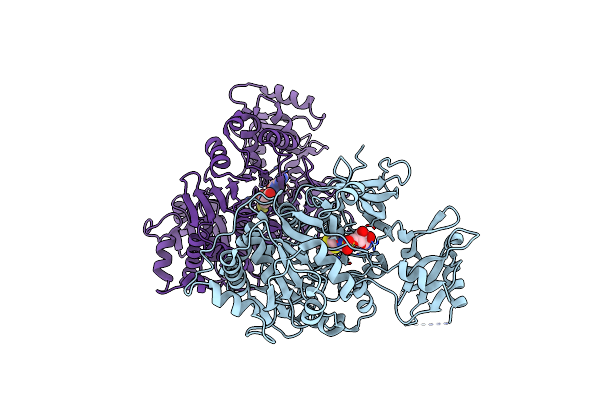
Structure Of S. Cerevisiae Bud23-Trm112 Complex Involved In Formation Of M7G1575 On 18S Rrna (Sam Bound Form)
Organism: Saccharomyces cerevisiae s288c
Method: X-RAY DIFFRACTION Resolution:2.12 Å Release Date: 2014-12-24 Classification: TRANSFERASE Ligands: ZN, EDO, SAM |
Organism: Saccharomyces cerevisiae s288c
Method: X-RAY DIFFRACTION
Release Date: 2014-12-24
Ligands: ZN, EDO, SAM
 |
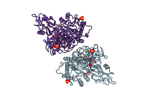
Photinus Pyralis Luciferase In The Adenylate-Forming Conformation Bound To Dlsa
Organism: Photinus pyralis
Method: X-RAY DIFFRACTION Resolution:2.62 Å Release Date: 2012-08-15 Classification: LIGASE Ligands: SLU |
Organism: Photinus pyralis
Method: X-RAY DIFFRACTION
Release Date: 2012-08-15
Ligands: SLU
 |
Structure Of Cross-Linked Firefly Luciferase In Second Catalytic Conformation
Organism: Photinus pyralis
Method: X-RAY DIFFRACTION Resolution:2.40 Å Release Date: 2012-08-15 Classification: LIGASE Ligands: SLU, XLX, SO4, CL |
Organism: Photinus pyralis
Method: X-RAY DIFFRACTION
Release Date: 2012-08-15
Ligands: SLU, XLX, SO4, CL
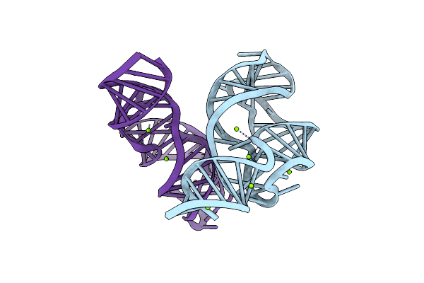 |
Structure Of The U65C Mutant A-Riboswitch Aptamer From The Bacillus Subtilis Pbue Operon
Organism: Bacillus subtilis
Method: X-RAY DIFFRACTION Resolution:2.80 Å Release Date: 2010-01-19 Classification: RNA Ligands: MG, BR |
Organism: Bacillus subtilis
Method: X-RAY DIFFRACTION
Release Date: 2010-01-19
Ligands: MG, BR