

Search Count: 9
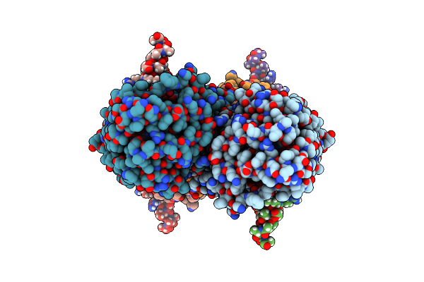 |
Partial Structure Of Tyrosine Hydroxylase Lacking The First 35 Residues In Complex With Dopamine.
Organism: Homo sapiens
Method: ELECTRON MICROSCOPY Release Date: 2021-12-22 Classification: OXIDOREDUCTASE Ligands: LDP, FE |
Organism: Homo sapiens
Method: ELECTRON MICROSCOPY
Release Date: 2021-12-22
Ligands: LDP, FE
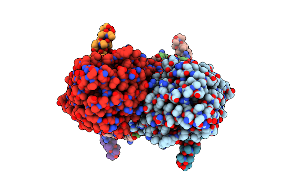 |
Partial Structure Of Tyrosine Hydroxylase In Complex With Dopamine Showing The Catalytic Domain And An Alpha-Helix From The Regulatory Domain Involved In Dopamine Binding.
Organism: Homo sapiens
Method: ELECTRON MICROSCOPY Release Date: 2021-12-08 Classification: OXIDOREDUCTASE Ligands: LDP, FE |
Organism: Homo sapiens
Method: ELECTRON MICROSCOPY
Release Date: 2021-12-08
Ligands: LDP, FE
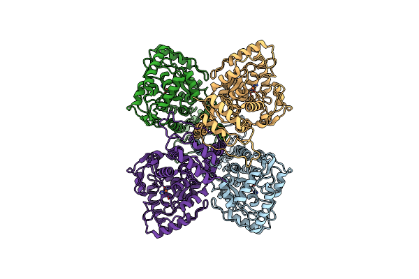 |
Full-Length Structure Of The Substrate-Free Tyrosine Hydroxylase (Apo-Th).
Organism: Homo sapiens
Method: ELECTRON MICROSCOPY Release Date: 2021-12-01 Classification: OXIDOREDUCTASE Ligands: FE |
Organism: Homo sapiens
Method: ELECTRON MICROSCOPY
Release Date: 2021-12-01
Ligands: FE
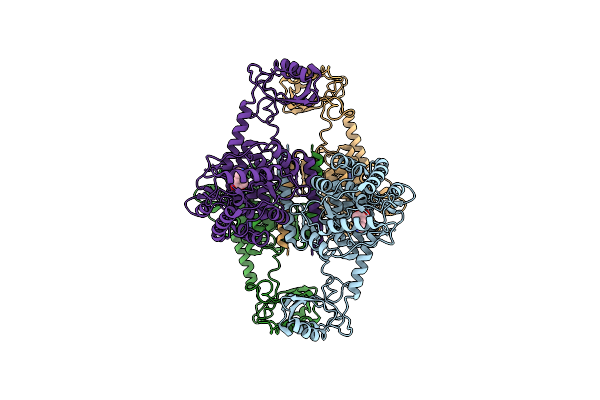 |
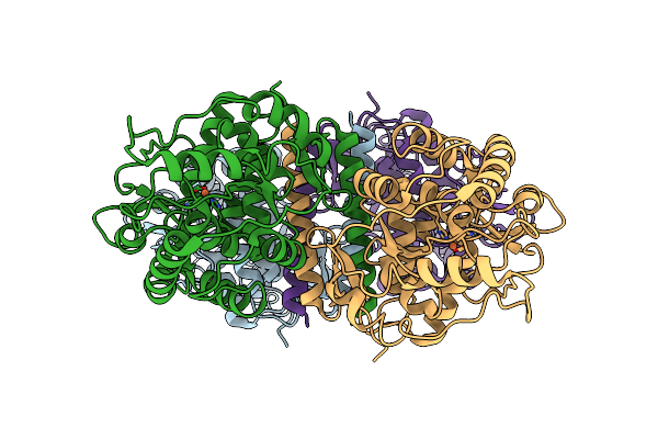
Atomic Model Of The Em-Based Structure Of The Full-Length Tyrosine Hydroxylase In Complex With Dopamine (Residues 40-497) In Which The Regulatory Domain (Residues 40-165) Has Been Included Only With The Backbone Atoms
Organism: Homo sapiens
Method: ELECTRON MICROSCOPY Release Date: 2021-11-17 Classification: OXIDOREDUCTASE Ligands: FE, LDP |
Organism: Homo sapiens
Method: ELECTRON MICROSCOPY
Release Date: 2021-11-17
Ligands: FE, LDP
 |
Partial Structure Of The Substrate-Free Tyrosine Hydroxylase (Apo-Th).
Organism: Homo sapiens
Method: ELECTRON MICROSCOPY Release Date: 2021-11-17 Classification: OXIDOREDUCTASE Ligands: FE |
Organism: Homo sapiens
Method: ELECTRON MICROSCOPY
Release Date: 2021-11-17
Ligands: FE
 |
Atomic-Resolution Structure Of The Coiled-Coil Dimerisation Domain Of Human Arc
Organism: Homo sapiens
Method: X-RAY DIFFRACTION Resolution:0.95 Å Release Date: 2021-03-03 Classification: SIGNALING PROTEIN Ligands: MPD, CL |
Organism: Homo sapiens
Method: X-RAY DIFFRACTION
Release Date: 2021-03-03
Ligands: MPD, CL
 |
Crystallographic Structure Of The Catalytic Domain Of Human Phenylalanine Hydroxylase (Hpah Cd) In Complex With Iron At 1.6 Angstrom
Organism: Homo sapiens
Method: X-RAY DIFFRACTION Resolution:1.67 Å Release Date: 2019-06-05 Classification: OXIDOREDUCTASE Ligands: FE |
Organism: Homo sapiens
Method: X-RAY DIFFRACTION
Release Date: 2019-06-05
Ligands: FE
 |
The Structure Of Full-Length Human Phenylalanine Hydroxylase In Complex With The Cofactor And Negative Regulator Tetrahydrobiopterin
Organism: Homo sapiens
Method: X-RAY DIFFRACTION Resolution:3.18 Å Release Date: 2019-06-05 Classification: OXIDOREDUCTASE Ligands: H4B |
Organism: Homo sapiens
Method: X-RAY DIFFRACTION
Release Date: 2019-06-05
Ligands: H4B
 |
Structural And Thermodynamic Insight Into Phenylalanine Hydroxylase From The Human Pathogen Legionella Pneumophila
Organism: Legionella pneumophila
Method: X-RAY DIFFRACTION Resolution:2.50 Å Release Date: 2013-08-21 Classification: OXIDOREDUCTASE Ligands: PEG |
Organism: Legionella pneumophila
Method: X-RAY DIFFRACTION
Release Date: 2013-08-21
Ligands: PEG