

Search Count: 118
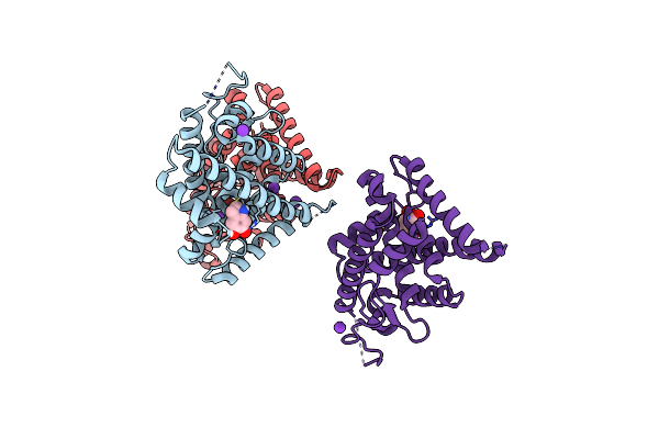 |
Nurr1 Covalently Modified By A Dopamine Metabolite
Organism: Homo sapiens
Method: X-RAY DIFFRACTION Resolution:3.20 Å Release Date: 2019-03-20 Classification: TRANSCRIPTION Ligands: K, G7J, BR |
Organism: Homo sapiens
Method: X-RAY DIFFRACTION
Release Date: 2019-03-20
Ligands: K, G7J, BR
 |
Crystal Structure Of A Nanog Homeobox (Nanog) From Homo Sapiens At 3.30 A Resolution
Organism: Homo sapiens
Method: X-RAY DIFFRACTION Resolution:3.30 Å Release Date: 2014-10-01 Classification: DNA BINDING PROTEIN, TRANSCRIPTION/DNA |
Organism: Homo sapiens
Method: X-RAY DIFFRACTION
Release Date: 2014-10-01
 |
Crystal Structure Of Human Nuclear Receptor Sf-1 (Nr5A1) Bound To Its Hormone Pip3 At 2.4 A Resolution
Organism: Homo sapiens
Method: X-RAY DIFFRACTION Resolution:2.40 Å Release Date: 2014-07-30 Classification: TRANSCRIPTION FACTOR/HORMONE Ligands: PIZ, ACT |
Organism: Homo sapiens
Method: X-RAY DIFFRACTION
Release Date: 2014-07-30
Ligands: PIZ, ACT
 |
Crystal Structure Of Human Nuclear Receptor Sf-1 (Nr5A1) Bound To Pip2 At 2.8 A Resolution
Organism: Homo sapiens
Method: X-RAY DIFFRACTION Resolution:2.81 Å Release Date: 2014-07-30 Classification: TRANSCRIPTION FACTOR/HORMONE Ligands: PIK, EDO |
Organism: Homo sapiens
Method: X-RAY DIFFRACTION
Release Date: 2014-07-30
Ligands: PIK, EDO
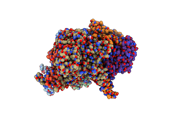 |
Crystal Structure Of Taf1-Taf7, A Tfiid Subcomplex
Organism: Saccharomyces cerevisiae
Method: X-RAY DIFFRACTION Resolution:2.90 Å Release Date: 2014-06-25 Classification: TRANSCRIPTION Ligands: ZN, TRS |
Organism: Saccharomyces cerevisiae
Method: X-RAY DIFFRACTION
Release Date: 2014-06-25
Ligands: ZN, TRS
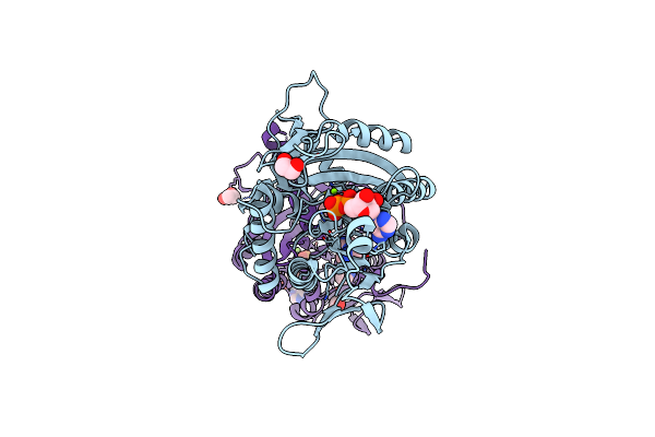 |
Arabidopsis Kcbp Motor Domain Dimerized Via Regulatory Domain
Organism: Arabidopsis thaliana
Method: X-RAY DIFFRACTION Resolution:2.40 Å Release Date: 2013-07-10 Classification: MOTOR PROTEIN Ligands: MG, ADP, IMD, EDO |
Organism: Arabidopsis thaliana
Method: X-RAY DIFFRACTION
Release Date: 2013-07-10
Ligands: MG, ADP, IMD, EDO
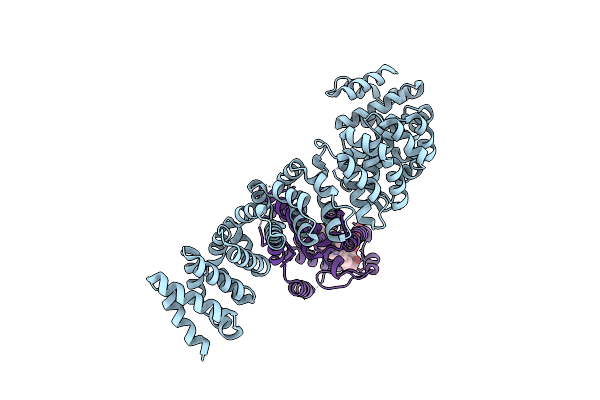 |
Crystal Structure Of Lrh-1/Beta-Catenin Complex
Organism: Homo sapiens
Method: X-RAY DIFFRACTION Resolution:2.76 Å Release Date: 2011-12-14 Classification: PROTEIN BINDING Ligands: P6L |
Organism: Homo sapiens
Method: X-RAY DIFFRACTION
Release Date: 2011-12-14
Ligands: P6L
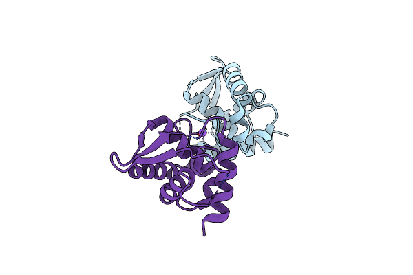 |
Crystal Structure Of A Fragment Of Nuclear Factor Related To Kappa-B-Binding Protein (Residues 370-495) (Nfrkb) From Homo Sapiens At 2.18 A Resolution
Organism: Homo sapiens
Method: X-RAY DIFFRACTION Resolution:2.18 Å Release Date: 2011-11-02 Classification: Transcription regulation, DNA BINDING |
Organism: Homo sapiens
Method: X-RAY DIFFRACTION
Release Date: 2011-11-02
 |
Crystal Structure Of A Clathrin Heavy Chain And Clathrin Light Chain Complex
Organism: Bos taurus
Method: X-RAY DIFFRACTION Resolution:7.94 Å Release Date: 2010-06-09 Classification: STRUCTURAL PROTEIN |
Organism: Bos taurus
Method: X-RAY DIFFRACTION
Release Date: 2010-06-09
 |
Crystal Structure Of A Clathrin Heavy Chain And Clathrin Light Chain Complex
Organism: Bos taurus
Method: X-RAY DIFFRACTION Resolution:9.00 Å Release Date: 2010-06-09 Classification: STRUCTURAL PROTEIN |
Organism: Bos taurus
Method: X-RAY DIFFRACTION
Release Date: 2010-06-09
 |
Crystal Structure Of The Huntingtin Interacting Protein 1 Coiled Coil Domain
Organism: Homo sapiens
Method: X-RAY DIFFRACTION Resolution:2.30 Å Release Date: 2010-02-23 Classification: TRANSCRIPTION |
Organism: Homo sapiens
Method: X-RAY DIFFRACTION
Release Date: 2010-02-23
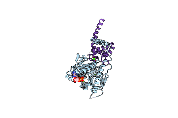 |
Structure Of The Complex Of A Mitotic Kinesin With Its Calcium Binding Regulator
Organism: Arabidopsis thaliana
Method: X-RAY DIFFRACTION Resolution:2.40 Å Release Date: 2009-05-19 Classification: motor protein/Calcium binding protein Ligands: ADP, MG, CA |
Organism: Arabidopsis thaliana
Method: X-RAY DIFFRACTION
Release Date: 2009-05-19
Ligands: ADP, MG, CA
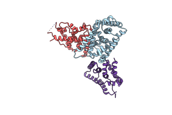 |
Structure Of Dax-1:Lrh-1 Complex
Organism: Mus musculus
Method: X-RAY DIFFRACTION Resolution:3.00 Å Release Date: 2008-12-23 Classification: TRANSCRIPTION |
Organism: Mus musculus
Method: X-RAY DIFFRACTION
Release Date: 2008-12-23
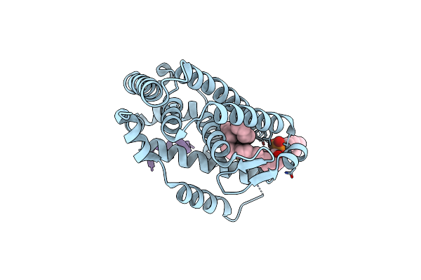 |
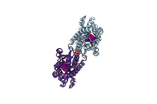
Sf-1 Lbd Bound By Phosphatidylcholine
Organism: Mus musculus
Method: X-RAY DIFFRACTION Resolution:2.20 Å Release Date: 2008-12-09 Classification: TRANSCRIPTION Ligands: P42 |
Organism: Mus musculus
Method: X-RAY DIFFRACTION
Release Date: 2008-12-09
Ligands: P42
 |
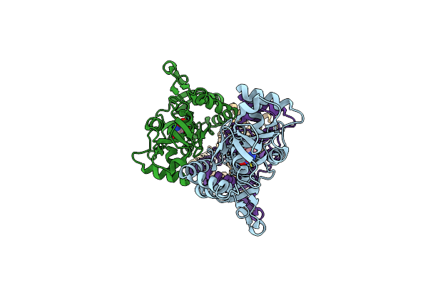
Tr Variant D355R
Organism: Homo sapiens
Method: X-RAY DIFFRACTION Resolution:2.20 Å Release Date: 2008-10-14 Classification: HORMONE, TRANSCRIPTION RECEPTOR Ligands: 4HY, SO4 |
Organism: Homo sapiens
Method: X-RAY DIFFRACTION
Release Date: 2008-10-14
Ligands: 4HY, SO4
 |
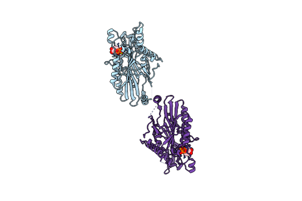
Crystal Structure Of The Glur2 Ligand Binding Core (S1S2J) In Complex With Fqx At 1.87 Angstroms
Organism: Rattus norvegicus
Method: X-RAY DIFFRACTION Resolution:1.87 Å Release Date: 2008-09-16 Classification: TRANSPORT PROTEIN Ligands: FQX |
Organism: Rattus norvegicus
Method: X-RAY DIFFRACTION
Release Date: 2008-09-16
Ligands: FQX
 |
Structural Dynamics Of The Microtubule Binding And Regulatory Elements In The Kinesin-Like Calmodulin Binding Protein
Organism: Solanum tuberosum
Method: X-RAY DIFFRACTION Resolution:2.20 Å Release Date: 2008-06-24 Classification: MOTOR PROTEIN Ligands: MG, ADP |
Organism: Solanum tuberosum
Method: X-RAY DIFFRACTION
Release Date: 2008-06-24
Ligands: MG, ADP
 |
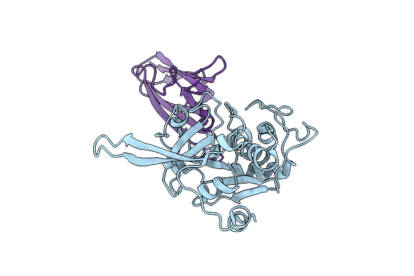
Structural Dynamics Of The Microtubule Binding And Regulatory Elements In The Kinesin-Like Calmodulin Binding Protein
Organism: Solanum tuberosum
Method: X-RAY DIFFRACTION Resolution:2.90 Å Release Date: 2008-06-17 Classification: MOTOR PROTEIN Ligands: MG, ADP |
Organism: Solanum tuberosum
Method: X-RAY DIFFRACTION
Release Date: 2008-06-17
Ligands: MG, ADP
 |
The Structure Of Chagasin In Complex With A Cysteine Protease Clarifies The Binding Mode And Evolution Of A New Inhibitor Family
Organism: Plasmodium falciparum, Trypanosoma cruzi
Method: X-RAY DIFFRACTION Resolution:2.20 Å Release Date: 2008-02-26 Classification: HYDROLASE/hydrolase inhibitor |
Organism: Plasmodium falciparum, Trypanosoma cruzi
Method: X-RAY DIFFRACTION
Release Date: 2008-02-26
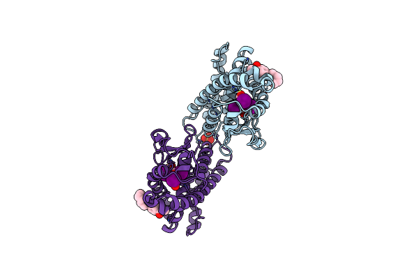 |
Thyroid Receptor Beta In Complex With Inhibitor
Organism: Homo sapiens
Method: X-RAY DIFFRACTION Resolution:2.30 Å Release Date: 2008-02-26 Classification: HORMONE RECEPTOR Ligands: 4HY, LEG, SO4 |
Organism: Homo sapiens
Method: X-RAY DIFFRACTION
Release Date: 2008-02-26
Ligands: 4HY, LEG, SO4