

Search Count: 24
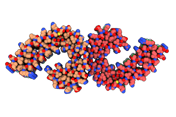 |
Tau Paired Helical Filament From Extracellular Vesicles From Alzheimer'S Disease Brain
Organism: Homo sapiens
Method: ELECTRON MICROSCOPY Resolution:3.16 Å Release Date: 2023-05-24 Classification: PROTEIN FIBRIL |
Organism: Homo sapiens
Method: ELECTRON MICROSCOPY
Release Date: 2023-05-24
 |
Tau Paired Helical Filament From Cellular Fraction Of Alzheimer'S Disease Brain
Organism: Homo sapiens
Method: ELECTRON MICROSCOPY Resolution:3.27 Å Release Date: 2023-05-24 Classification: PROTEIN FIBRIL |
Organism: Homo sapiens
Method: ELECTRON MICROSCOPY
Release Date: 2023-05-24
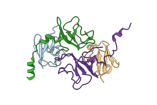 |
Structure Of Cyld Cap-Gly3 (467-565) Bound To Ub; Orthorhobic Space Group
Organism: Homo sapiens
Method: X-RAY DIFFRACTION Resolution:1.85 Å Release Date: 2021-10-20 Classification: IMMUNE SYSTEM |
Organism: Homo sapiens
Method: X-RAY DIFFRACTION
Release Date: 2021-10-20
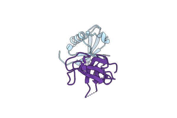 |
Structure Of Cyld Cap-Gly3 (467-552) Bound To Ub; Tetragonal Space Group
Organism: Homo sapiens
Method: X-RAY DIFFRACTION Resolution:1.71 Å Release Date: 2021-10-20 Classification: IMMUNE SYSTEM |
Organism: Homo sapiens
Method: X-RAY DIFFRACTION
Release Date: 2021-10-20
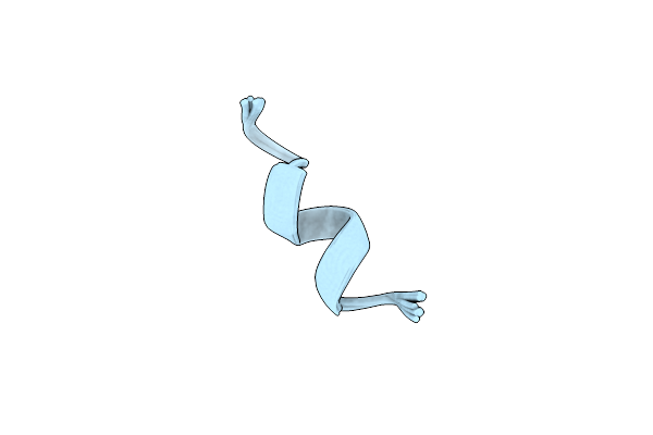 |
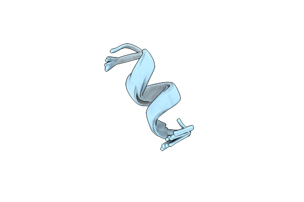
Nmr Solution Structure Of Harzianin Hk-Vi In Sds Micelles
Organism: Trichoderma pseudokoningii
Method: SOLUTION NMR Release Date: 2018-06-13 Classification: ANTIBIOTIC |
Organism: Trichoderma pseudokoningii
Method: SOLUTION NMR
Release Date: 2018-06-13
 |
Nmr Solution Structure Of Harzianin Hk-Vi In Trifluoroethanol
Organism: Trichoderma pseudokoningii
Method: SOLUTION NMR Release Date: 2018-06-13 Classification: ANTIBIOTIC |
Organism: Trichoderma pseudokoningii
Method: SOLUTION NMR
Release Date: 2018-06-13
 |
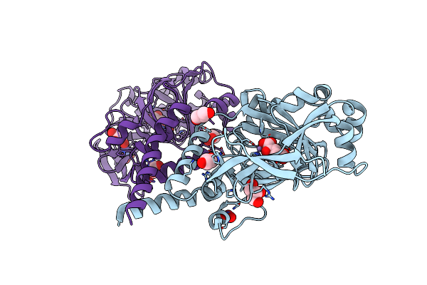
Jmjd7 In Complex With Mn And 2Og In The H32 Form
Organism: Homo sapiens
Method: X-RAY DIFFRACTION Resolution:2.98 Å Release Date: 2018-03-14 Classification: OXIDOREDUCTASE Ligands: MN, AKG, NO3, GOL |
Organism: Homo sapiens
Method: X-RAY DIFFRACTION
Release Date: 2018-03-14
Ligands: MN, AKG, NO3, GOL
 |
Human Jmjd7 In Complex With Mn And 2Og In The P21212 Form
Organism: Homo sapiens
Method: X-RAY DIFFRACTION Resolution:2.17 Å Release Date: 2018-03-14 Classification: OXIDOREDUCTASE Ligands: MN, AKG, GOL, NO3 |
Organism: Homo sapiens
Method: X-RAY DIFFRACTION
Release Date: 2018-03-14
Ligands: MN, AKG, GOL, NO3
 |
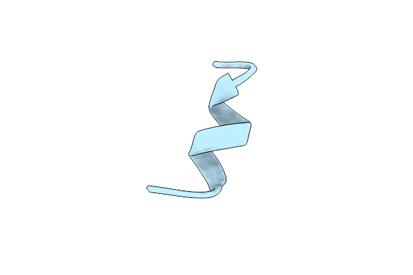
Crystal Structure Of Middle-East Respiratory Syndrome (Mers) Coronavirus Neutralizing Antibody Jc57-14 Isolated From A Vaccinated Rhesus Macaque In Complex With Mers Receptor Binding Domain
Organism: Macaca mulatta, Betacoronavirus england 1
Method: X-RAY DIFFRACTION Resolution:3.32 Å Release Date: 2018-03-07 Classification: IMMUNE SYSTEM Ligands: SO4 |
Organism: Macaca mulatta, Betacoronavirus england 1
Method: X-RAY DIFFRACTION
Release Date: 2018-03-07
Ligands: SO4
 |
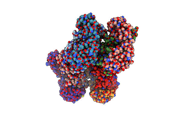
Nmr Solution Structure Of Harzianin Hk-Vi In Dpc Micelles
Organism: Trichoderma pseudokoningii
Method: SOLUTION NMR Release Date: 2018-02-28 Classification: ANTIBIOTIC |
Organism: Trichoderma pseudokoningii
Method: SOLUTION NMR
Release Date: 2018-02-28
 |
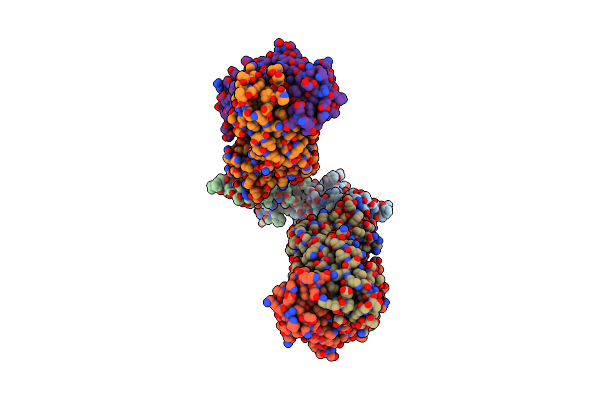
Crystal Structure Of Middle-East Respiratory Syndrome (Mers) Coronavirus Neutralizing Antibody Jc57-14 Isolated From A Vaccinated Rhesus Macaque.
Organism: Macaca mulatta
Method: X-RAY DIFFRACTION Resolution:1.99 Å Release Date: 2018-02-28 Classification: IMMUNE SYSTEM |
Organism: Macaca mulatta
Method: X-RAY DIFFRACTION
Release Date: 2018-02-28
 |
Crystal Structure Of Potent Neutralizing Antibody Cdc2-C2 In Complex With Mers-Cov S1 Rbd
Organism: Middle east respiratory syndrome-related coronavirus, Homo sapiens
Method: X-RAY DIFFRACTION Resolution:2.10 Å Release Date: 2018-01-31 Classification: IMMUNE SYSTEM Ligands: NAG |
Organism: Middle east respiratory syndrome-related coronavirus, Homo sapiens
Method: X-RAY DIFFRACTION
Release Date: 2018-01-31
Ligands: NAG
 |
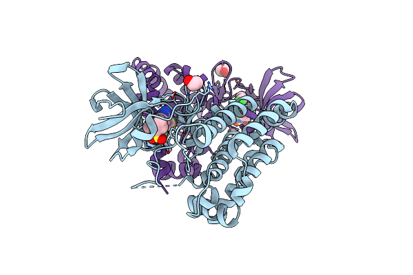
Crystal Structure Of Pcna Encoding The Hypomorphic Mutation S228I
Organism: Homo sapiens
Method: X-RAY DIFFRACTION Resolution:2.27 Å Release Date: 2017-01-25 Classification: DNA BINDING PROTEIN |
Organism: Homo sapiens
Method: X-RAY DIFFRACTION
Release Date: 2017-01-25
 |
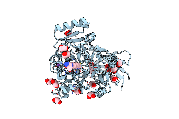
Human Leucine Zipper- And Sterile Alpha Motif-Containing Kinase (Zak, Mlt, Hccs-4, Mrk, Azk, Mltk) In Complex With Vemurafenib
Organism: Homo sapiens
Method: X-RAY DIFFRACTION Resolution:2.14 Å Release Date: 2016-03-30 Classification: TRANSFERASE Ligands: 032, EDO |
Organism: Homo sapiens
Method: X-RAY DIFFRACTION
Release Date: 2016-03-30
Ligands: 032, EDO
 |
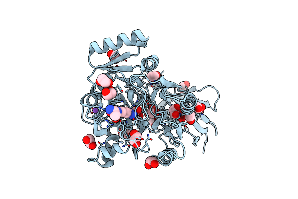
Structure Of Wt Porphyromonas Gingivalis Peptidylarginine Deiminase
Organism: Porphyromonas gingivalis
Method: X-RAY DIFFRACTION Resolution:1.46 Å Release Date: 2015-07-22 Classification: HYDROLASE Ligands: ALA, ARG, EDO, NA |
Organism: Porphyromonas gingivalis
Method: X-RAY DIFFRACTION
Release Date: 2015-07-22
Ligands: ALA, ARG, EDO, NA
 |
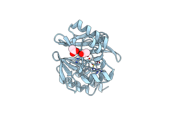
Structure Of C351A Mutant Of Porphyromonas Gingivalis Peptidylarginine Deiminase
Organism: Porphyromonas gingivalis
Method: X-RAY DIFFRACTION Resolution:1.48 Å Release Date: 2015-07-22 Classification: HYDROLASE Ligands: ALA, ARG, NA, EDO |
Organism: Porphyromonas gingivalis
Method: X-RAY DIFFRACTION
Release Date: 2015-07-22
Ligands: ALA, ARG, NA, EDO
 |
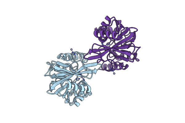
Crystal Structure Of The Metallo-Beta-Lactamase Vim-31 In Its Reduced Form At 1.61 A
Organism: Enterobacter cloacae
Method: X-RAY DIFFRACTION Resolution:1.61 Å Release Date: 2013-06-26 Classification: HYDROLASE Ligands: ZN, ACT, CL |
Organism: Enterobacter cloacae
Method: X-RAY DIFFRACTION
Release Date: 2013-06-26
Ligands: ZN, ACT, CL
 |
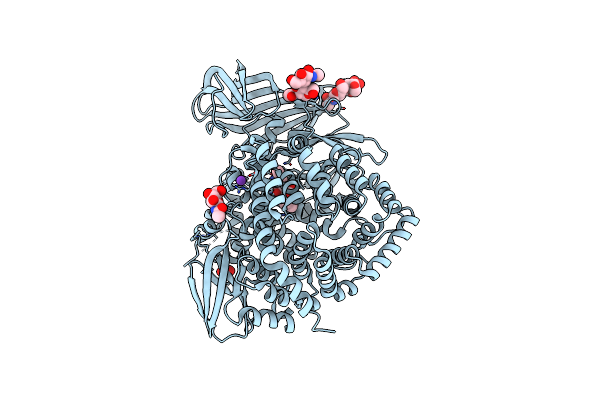
Crystal Structure Of The Metallo-Beta-Lactamase Vim-31 In Its Oxidized Form At 1.88 A
Organism: Enterobacter cloacae
Method: X-RAY DIFFRACTION Resolution:1.88 Å Release Date: 2013-06-26 Classification: HYDROLASE Ligands: ZN, CL |
Organism: Enterobacter cloacae
Method: X-RAY DIFFRACTION
Release Date: 2013-06-26
Ligands: ZN, CL
 |
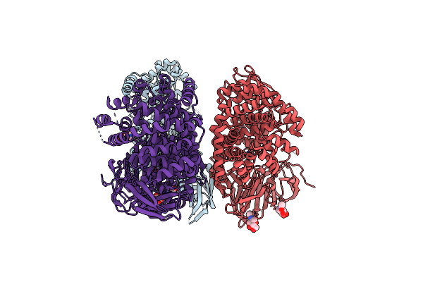
Crystal Structure Of The Soluble Domain Of Human Endoplasmic Reticulum Aminopeptidase 1 Erap1
Organism: Homo sapiens
Method: X-RAY DIFFRACTION Resolution:2.70 Å Release Date: 2011-04-13 Classification: HYDROLASE Ligands: NAG, ZN, K, EDO, BES |
Organism: Homo sapiens
Method: X-RAY DIFFRACTION
Release Date: 2011-04-13
Ligands: NAG, ZN, K, EDO, BES
 |
Crystal Structure Of The Open State Of Human Endoplasmic Reticulum Aminopeptidase 1 Erap1
Organism: Homo sapiens
Method: X-RAY DIFFRACTION Resolution:3.00 Å Release Date: 2011-02-23 Classification: HYDROLASE Ligands: ZN, NAG |
Organism: Homo sapiens
Method: X-RAY DIFFRACTION
Release Date: 2011-02-23
Ligands: ZN, NAG