

Search Count: 37
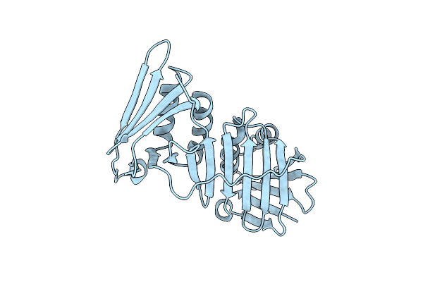 |
Crystal Structure Of Yeast Proliferating Cell Nuclear Antigen
Organism: Saccharomyces cerevisiae (strain atcc 204508 / s288c)
Method: X-RAY DIFFRACTION Resolution:2.95 Å Release Date: 2015-03-18 Classification: DNA BINDING PROTEIN |
Organism: Saccharomyces cerevisiae (strain atcc 204508 / s288c)
Method: X-RAY DIFFRACTION
Release Date: 2015-03-18
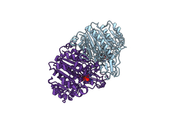 |
E. Coli Alkaline Phosphatase Mutant S102G/R166S In Complex With Inorganic Phosphate
Organism: Escherichia coli
Method: X-RAY DIFFRACTION Resolution:2.80 Å Release Date: 2013-07-24 Classification: HYDROLASE Ligands: PO4, ZN |
Organism: Escherichia coli
Method: X-RAY DIFFRACTION
Release Date: 2013-07-24
Ligands: PO4, ZN
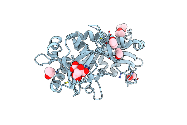 |
Crystal Structure Of Atg7 Ntd K14A F16A D18A Mutant
Organism: Saccharomyces cerevisiae
Method: X-RAY DIFFRACTION Resolution:1.70 Å Release Date: 2012-11-14 Classification: PROTEIN TRANSPORT Ligands: CIT, IPA |
Organism: Saccharomyces cerevisiae
Method: X-RAY DIFFRACTION
Release Date: 2012-11-14
Ligands: CIT, IPA
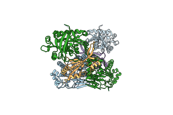 |
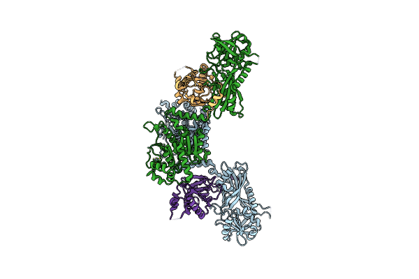
Crystal Structure Of An Atg7-Atg10 Crosslinked Complex
Organism: Saccharomyces cerevisiae
Method: X-RAY DIFFRACTION Resolution:2.90 Å Release Date: 2012-11-14 Classification: PROTEIN TRANSPORT/LIGASE Ligands: ZN |
Organism: Saccharomyces cerevisiae
Method: X-RAY DIFFRACTION
Release Date: 2012-11-14
Ligands: ZN
 |
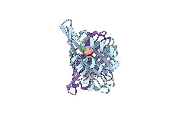
Crystal Structure Of An Atg7-Atg3 Crosslinked Complex
Organism: Saccharomyces cerevisiae
Method: X-RAY DIFFRACTION Resolution:2.70 Å Release Date: 2012-11-14 Classification: PROTEIN TRANSPORT Ligands: ZN |
Organism: Saccharomyces cerevisiae
Method: X-RAY DIFFRACTION
Release Date: 2012-11-14
Ligands: ZN
 |
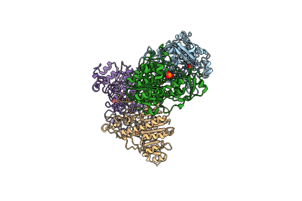
Crystal Structure Of Ketosteroid Isomerase D40N From Pseudomonas Putida (Pksi) With Bound 3-Fluoro-4-Nitrophenol
Organism: Pseudomonas putida
Method: X-RAY DIFFRACTION Resolution:1.30 Å Release Date: 2012-08-22 Classification: ISOMERASE Ligands: FNN |
Organism: Pseudomonas putida
Method: X-RAY DIFFRACTION
Release Date: 2012-08-22
Ligands: FNN
 |
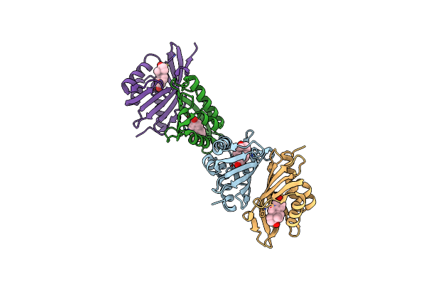
E. Coli Alkaline Phosphatase With Bound Inorganic Phosphate
Organism: Escherichia coli
Method: X-RAY DIFFRACTION Resolution:1.20 Å Release Date: 2011-11-09 Classification: HYDROLASE Ligands: ZN, MG, PO4 |
Organism: Escherichia coli
Method: X-RAY DIFFRACTION
Release Date: 2011-11-09
Ligands: ZN, MG, PO4
 |
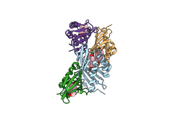
Crystal Structure Of Ketosteroid Isomerase D40N/C69S/C81S/C97S/M116C-Cn From P. Putida With Bound Equilenin
Organism: Pseudomonas putida
Method: X-RAY DIFFRACTION Resolution:1.71 Å Release Date: 2011-09-21 Classification: ISOMERASE Ligands: EQU |
Organism: Pseudomonas putida
Method: X-RAY DIFFRACTION
Release Date: 2011-09-21
Ligands: EQU
 |
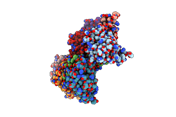
Crystal Structure Of Ketosteroid Isomerase D40N/C69S/C81S/C97S/F86C-Cn From P. Putida With Bound Equilenin
Organism: Pseudomonas putida
Method: X-RAY DIFFRACTION Resolution:1.70 Å Release Date: 2011-09-21 Classification: ISOMERASE Ligands: EQU |
Organism: Pseudomonas putida
Method: X-RAY DIFFRACTION
Release Date: 2011-09-21
Ligands: EQU
 |
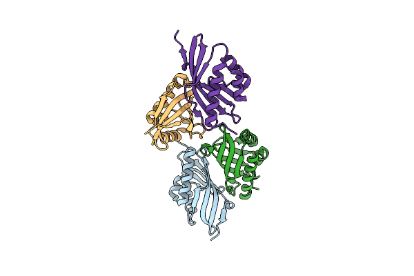
Crystal Structure Of Ketosteroid Isomerase D40N/C69S/C81S/C97S/M105C-Cn From P. Putida With Bound Equilenin
Organism: Pseudomonas putida
Method: X-RAY DIFFRACTION Resolution:2.30 Å Release Date: 2011-09-21 Classification: ISOMERASE Ligands: EQU |
Organism: Pseudomonas putida
Method: X-RAY DIFFRACTION
Release Date: 2011-09-21
Ligands: EQU
 |
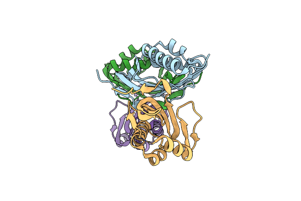
Crystal Structure Of Ketosteroid Isomerase D40N/C69S/C81S/C97S/F86C-Cn From P. Putida
Organism: Pseudomonas putida
Method: X-RAY DIFFRACTION Resolution:2.00 Å Release Date: 2011-09-21 Classification: ISOMERASE |
Organism: Pseudomonas putida
Method: X-RAY DIFFRACTION
Release Date: 2011-09-21
 |
Crystal Structure Of Ketosteroid Isomerase D40N/C69S/C81S/C97S/M116C-Cn From P. Putida
Organism: Pseudomonas putida
Method: X-RAY DIFFRACTION Resolution:1.89 Å Release Date: 2011-09-21 Classification: ISOMERASE |
Organism: Pseudomonas putida
Method: X-RAY DIFFRACTION
Release Date: 2011-09-21
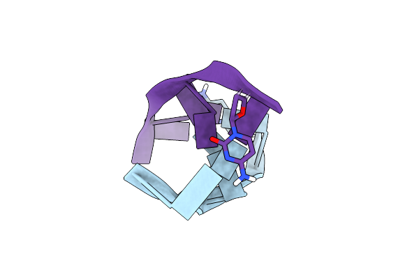 |
Reintroducing Electrostatics Into Macromolecular Crystallographic Refinement: Z-Dna (X-Ray)
Method: X-RAY DIFFRACTION, NEUTRON DIFFRACTION
Resolution:1.53 Å, 1.4 Å Release Date: 2011-03-02 Classification: DNA Ligands: DOD |
Method: X-RAY DIFFRACTION, NEUTRON DIFFRACTION
Release Date: 2011-03-02
Ligands: DOD
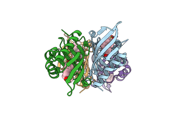 |
Crystal Structure Of Ketosteroid Isomerase Y16S/D40N From Pseudomonas Putida With Bound Equilenin
Organism: Pseudomonas putida
Method: X-RAY DIFFRACTION Resolution:1.63 Å Release Date: 2010-01-12 Classification: ISOMERASE Ligands: EQU |
Organism: Pseudomonas putida
Method: X-RAY DIFFRACTION
Release Date: 2010-01-12
Ligands: EQU
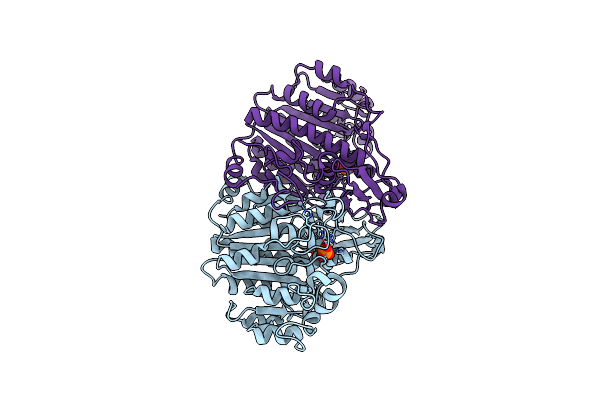 |
Structure Of E322Y Alkaline Phosphatase In Complex With Inorganic Phosphate
Organism: Escherichia coli
Method: X-RAY DIFFRACTION Resolution:2.30 Å Release Date: 2008-10-21 Classification: HYDROLASE Ligands: ZN, PO4 |
Organism: Escherichia coli
Method: X-RAY DIFFRACTION
Release Date: 2008-10-21
Ligands: ZN, PO4
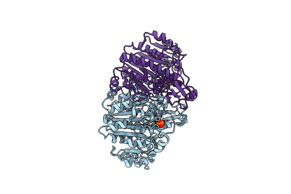 |
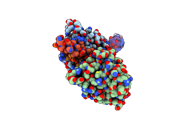
E. Coli Alkaline Phosphatase Mutant R166S In Complex With Phosphate
Organism: Escherichia coli
Method: X-RAY DIFFRACTION Resolution:2.05 Å Release Date: 2008-07-29 Classification: HYDROLASE Ligands: ZN, PO4, MG |
Organism: Escherichia coli
Method: X-RAY DIFFRACTION
Release Date: 2008-07-29
Ligands: ZN, PO4, MG
 |
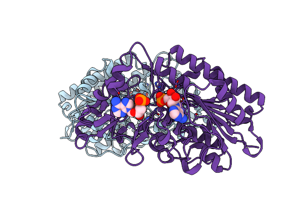
Rab6-Gtp:Gcc185 Rab Binding Domain Complex
Organism: Homo sapiens
Method: X-RAY DIFFRACTION Resolution:3.00 Å Release Date: 2008-02-05 Classification: Protein Transport/Splicing Ligands: MG, GTP |
Organism: Homo sapiens
Method: X-RAY DIFFRACTION
Release Date: 2008-02-05
Ligands: MG, GTP
 |
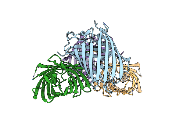
Structure Of Xac Npp For Evaluation Of Refinement Methodology
Organism: Xanthomonas axonopodis pv. citri
Method: X-RAY DIFFRACTION Resolution:1.45 Å Release Date: 2007-10-30 Classification: HYDROLASE Ligands: ZN, AMP |
Organism: Xanthomonas axonopodis pv. citri
Method: X-RAY DIFFRACTION
Release Date: 2007-10-30
Ligands: ZN, AMP
 |
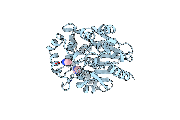
Crystal Structures Of The Luciferase And Green Fluorescent Protein From Renilla Reniformis
Organism: Renilla reniformis
Method: X-RAY DIFFRACTION Resolution:1.50 Å Release Date: 2007-10-23 Classification: FLUORESCENT PROTEIN |
Organism: Renilla reniformis
Method: X-RAY DIFFRACTION
Release Date: 2007-10-23
 |
Crystal Structures Of The Luciferase And Green Fluorescent Protein From Renilla Reniformis
Organism: Renilla reniformis
Method: X-RAY DIFFRACTION Resolution:1.40 Å Release Date: 2007-06-05 Classification: OXIDOREDUCTASE Ligands: IMD |
Organism: Renilla reniformis
Method: X-RAY DIFFRACTION
Release Date: 2007-06-05
Ligands: IMD