
Search Count: 84
 |
Organism: Mus musculus
Method: ELECTRON MICROSCOPY Resolution:3.13 Å Release Date: 2026-04-29 Classification: SIGNALING PROTEIN Ligands: PLM, OLC, POV |
 |
Organism: Mus musculus
Method: ELECTRON MICROSCOPY Resolution:2.80 Å Release Date: 2026-04-29 Classification: SIGNALING PROTEIN Ligands: PLM, OLC, POV |
 |
Organism: Mus musculus
Method: ELECTRON MICROSCOPY Resolution:3.04 Å Release Date: 2026-04-29 Classification: SIGNALING PROTEIN Ligands: PLM, OLC, POV |
 |
Organism: Mus musculus
Method: ELECTRON MICROSCOPY Resolution:2.87 Å Release Date: 2026-04-29 Classification: SIGNALING PROTEIN Ligands: PLM, OLC, POV |
 |
Organism: Mus musculus
Method: ELECTRON MICROSCOPY Resolution:3.36 Å Release Date: 2026-04-29 Classification: SIGNALING PROTEIN Ligands: PLM, OLC, POV |
 |
Organism: Mus musculus
Method: ELECTRON MICROSCOPY Resolution:3.08 Å Release Date: 2026-04-29 Classification: SIGNALING PROTEIN Ligands: PLM, OLC, POV |
 |
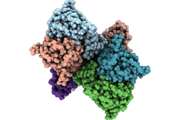
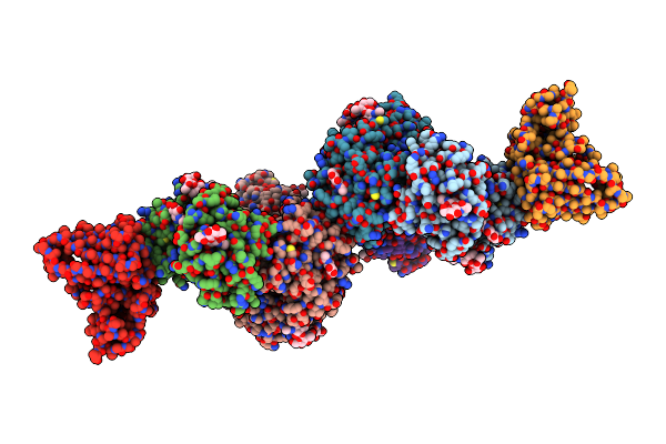
The Lbd-Tmd Structure Of Native Mouse Ampar With 2 Tarps 2 Cnihs And Prrt1/Syndig4
Organism: Mus musculus
Method: ELECTRON MICROSCOPY Resolution:3.36 Å Release Date: 2026-04-29 Classification: SIGNALING PROTEIN Ligands: PLM, OLC, POV |
 |
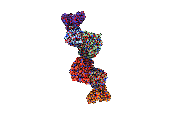
The Tmd Structure Of Native Mouse Ampar With 2 Tarps 2 Cnihs And Prrt1/Syndig4
Organism: Mus musculus
Method: ELECTRON MICROSCOPY Resolution:3.04 Å Release Date: 2026-04-29 Classification: SIGNALING PROTEIN Ligands: PLM, OLC, POV |
 |
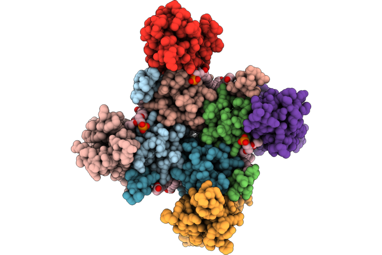
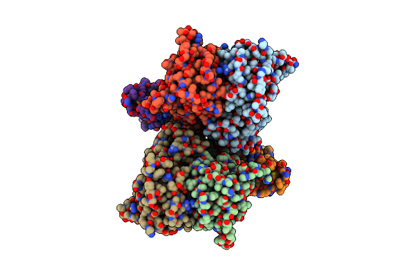
The Lbd-Tmd Structure Of Native Mouse Ampar With 3 Tarps 1 Cnih And Prrt1/Syndig4
Organism: Mus musculus
Method: ELECTRON MICROSCOPY Resolution:3.75 Å Release Date: 2026-04-29 Classification: SIGNALING PROTEIN Ligands: PLM, OLC, POV |
 |
Organism: Mus musculus
Method: ELECTRON MICROSCOPY Resolution:3.45 Å Release Date: 2026-04-29 Classification: SIGNALING PROTEIN Ligands: PLM, OLC, POV |
 |
Organism: Mus musculus
Method: ELECTRON MICROSCOPY Resolution:3.33 Å Release Date: 2026-04-29 Classification: SIGNALING PROTEIN Ligands: PLM, OLC, POV |
 |
Organism: Homo sapiens
Method: X-RAY DIFFRACTION Resolution:1.40 Å Release Date: 2025-11-19 Classification: OXIDOREDUCTASE Ligands: EDO, A1IDQ |
 |
Organism: Actinoalloteichus caeruleus
Method: X-RAY DIFFRACTION Resolution:2.70 Å Release Date: 2025-10-22 Classification: BIOSYNTHETIC PROTEIN Ligands: SAH |
 |
Organism: Mus musculus, Rattus norvegicus
Method: ELECTRON MICROSCOPY Release Date: 2025-07-02 Classification: MEMBRANE PROTEIN Ligands: NAG, CA |
 |
Organism: Rattus norvegicus
Method: ELECTRON MICROSCOPY Release Date: 2025-07-02 Classification: MEMBRANE PROTEIN Ligands: ZK1 |
 |
Organism: Mus musculus, Rattus norvegicus
Method: ELECTRON MICROSCOPY Resolution:3.53 Å Release Date: 2025-07-02 Classification: MEMBRANE PROTEIN Ligands: NAG |
 |
Organism: Rattus norvegicus
Method: ELECTRON MICROSCOPY Resolution:4.22 Å Release Date: 2025-07-02 Classification: MEMBRANE PROTEIN Ligands: ZK1 |
 |
Organism: Rattus norvegicus
Method: ELECTRON MICROSCOPY Release Date: 2025-07-02 Classification: MEMBRANE PROTEIN Ligands: ZK1 |
 |
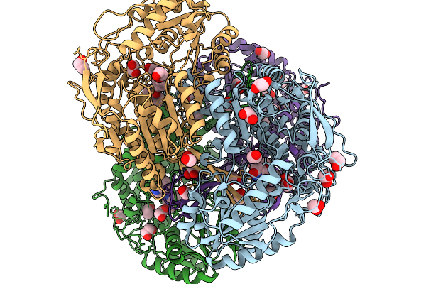
Cryoem Structure Of M. Tuberculosis Clpc1P1P2 Complex Bound To Bortezomib, Conformation 2
Organism: Mycobacterium tuberculosis h37rv, Bos grunniens
Method: ELECTRON MICROSCOPY Release Date: 2025-03-19 Classification: HYDROLASE Ligands: ADP, ATP, MG, BO2 |
 |
Organism: Mycobacterium tuberculosis h37rv
Method: ELECTRON MICROSCOPY Release Date: 2025-03-19 Classification: HYDROLASE Ligands: MG, ATP, ADP, BO2 |