

Search Count: 22
 |
Crystal Structure Of Mettl9 In Complex With Compound 15K
Organism: Homo sapiens
Method: X-RAY DIFFRACTION Resolution:2.28 Å Release Date: 2026-04-22 Classification: TRANSFERASE Ligands: A1ES7 |
Organism: Homo sapiens
Method: X-RAY DIFFRACTION
Release Date: 2026-04-22
Ligands: A1ES7
 |
Crystal Structure Of The Dhr2 Domain Of Dock10 In Complex With Cdc42
Organism: Homo sapiens
Method: X-RAY DIFFRACTION Resolution:2.55 Å Release Date: 2020-01-22 Classification: SIGNALING PROTEIN Ligands: GOL |
Organism: Homo sapiens
Method: X-RAY DIFFRACTION
Release Date: 2020-01-22
Ligands: GOL
 |
Crystal Structure Of The Dhr2 Domain Of Dock10 In Complex With Cdc42
Organism: Homo sapiens
Method: X-RAY DIFFRACTION Resolution:2.64 Å Release Date: 2020-01-22 Classification: SIGNALING PROTEIN Ligands: GDP, GOL |
Organism: Homo sapiens
Method: X-RAY DIFFRACTION
Release Date: 2020-01-22
Ligands: GDP, GOL
 |
Crystal Structure Of The Dhr2 Domain Of Dock10 In Complex With Rac3
Organism: Homo sapiens
Method: X-RAY DIFFRACTION Resolution:3.71 Å Release Date: 2020-01-22 Classification: SIGNALING PROTEIN |
Organism: Homo sapiens
Method: X-RAY DIFFRACTION
Release Date: 2020-01-22
 |
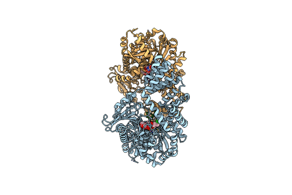
O-Glcnac Transferase In Complex With Covalent Inhibitor
Organism: Homo sapiens
Method: X-RAY DIFFRACTION Resolution:2.53 Å Release Date: 2019-11-06 Classification: TRANSFERASE/Inhibitor Ligands: HQV |
Organism: Homo sapiens
Method: X-RAY DIFFRACTION
Release Date: 2019-11-06
Ligands: HQV
 |
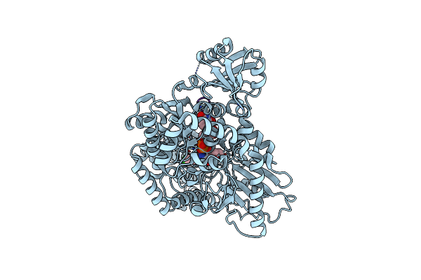
Electrophilic Probes For Deciphering Substrate Recognition By O-Glcnac Transferase
Organism: Homo sapiens
Method: X-RAY DIFFRACTION Resolution:2.60 Å Release Date: 2017-10-18 Classification: TRANSFERASE Ligands: UDP, 9C1, 9CD |
Organism: Homo sapiens
Method: X-RAY DIFFRACTION
Release Date: 2017-10-18
Ligands: UDP, 9C1, 9CD
 |
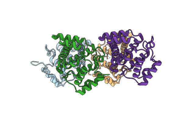
Electrophilic Probes For Deciphering Substrate Recognition By O-Glcnac Transferase
Organism: Homo sapiens
Method: X-RAY DIFFRACTION Resolution:2.25 Å Release Date: 2017-10-18 Classification: TRANSFERASE Ligands: UDP, 9C1 |
Organism: Homo sapiens
Method: X-RAY DIFFRACTION
Release Date: 2017-10-18
Ligands: UDP, 9C1
 |
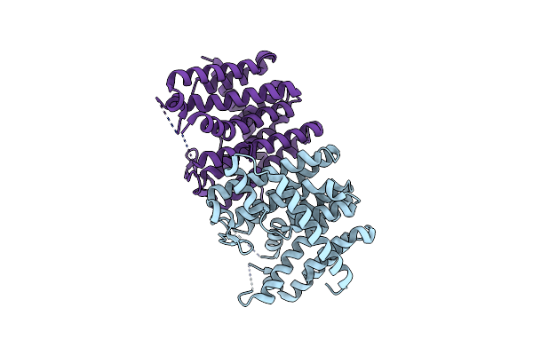
Crystal Structure Of Human Myosin 9B Rhogap Domain At 2.2 Angstrom
Organism: Homo sapiens
Method: X-RAY DIFFRACTION Resolution:2.20 Å Release Date: 2015-11-25 Classification: SIGNALING PROTEIN |
Organism: Homo sapiens
Method: X-RAY DIFFRACTION
Release Date: 2015-11-25
 |
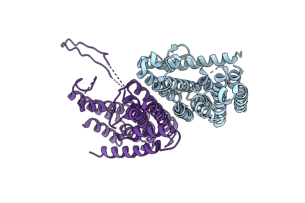
Crystal Structure Of Geranylfarnesyl Pyrophosphate Synthases 2 From Arabidopsis Thaliana
Organism: Arabidopsis thaliana
Method: X-RAY DIFFRACTION Resolution:2.30 Å Release Date: 2015-11-11 Classification: TRANSFERASE |
Organism: Arabidopsis thaliana
Method: X-RAY DIFFRACTION
Release Date: 2015-11-11
 |
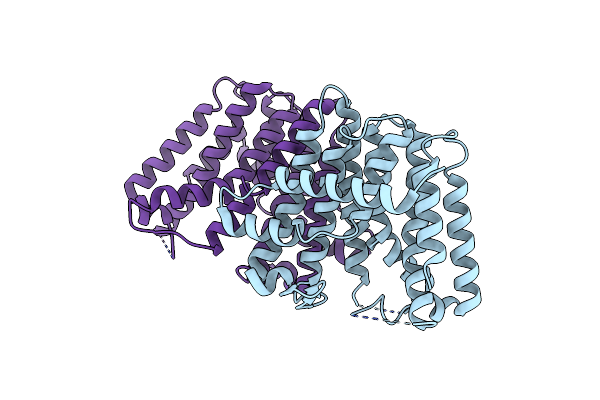
Crystal Structure Of Polyprenyl Pyrophosphate Synthase 2 From Arabidopsis Thaliana
Organism: Arabidopsis thaliana
Method: X-RAY DIFFRACTION Resolution:3.03 Å Release Date: 2015-11-11 Classification: TRANSFERASE |
Organism: Arabidopsis thaliana
Method: X-RAY DIFFRACTION
Release Date: 2015-11-11
 |
Crystal Structure Of Geranylgeranyl Pyrophosphate Synthase 11 From Arabidopsis Thaliana
Organism: Arabidopsis thaliana
Method: X-RAY DIFFRACTION Resolution:2.81 Å Release Date: 2015-11-11 Classification: TRANSFERASE |
Organism: Arabidopsis thaliana
Method: X-RAY DIFFRACTION
Release Date: 2015-11-11
 |
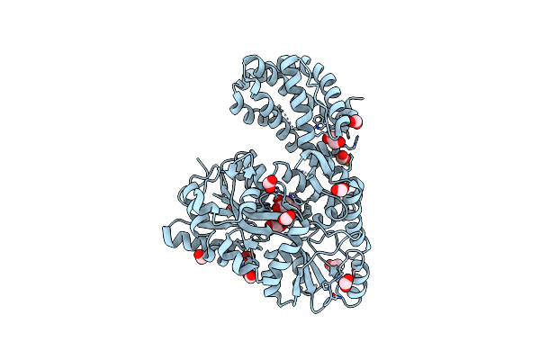
Structure Of Mcl1 Bound To Brd Inhibitor Ligand 1 At 1.7A
Organism: Homo sapiens
Method: X-RAY DIFFRACTION Resolution:1.70 Å Release Date: 2015-05-06 Classification: Apoptosis/Inhibitor Ligands: ZN, 865, POP |
Organism: Homo sapiens
Method: X-RAY DIFFRACTION
Release Date: 2015-05-06
Ligands: ZN, 865, POP
 |
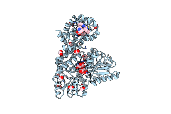
Structure Of Apo Mbp-Mcl1 At 1.9A
Organism: Escherichia coli, Homo sapiens
Method: X-RAY DIFFRACTION Resolution:1.90 Å Release Date: 2015-05-06 Classification: APOPTOSIS Ligands: MG, EDO, FMT |
Organism: Escherichia coli, Homo sapiens
Method: X-RAY DIFFRACTION
Release Date: 2015-05-06
Ligands: MG, EDO, FMT
 |
Structure Of Mbp-Mcl1 Bound To Ligand 1 At 2.35A
Organism: Escherichia coli, Homo sapiens
Method: X-RAY DIFFRACTION Resolution:2.35 Å Release Date: 2015-05-06 Classification: APOPTOSIS Ligands: 865, EDO, FMT |
Organism: Escherichia coli, Homo sapiens
Method: X-RAY DIFFRACTION
Release Date: 2015-05-06
Ligands: 865, EDO, FMT
 |
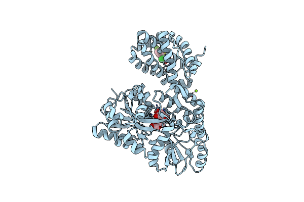
Structure Of Mbp-Mcl1 Bound To Ligand 2 At 1.55A
Organism: Escherichia coli, Homo sapiens
Method: X-RAY DIFFRACTION Resolution:1.55 Å Release Date: 2015-05-06 Classification: APOPTOSIS Ligands: 19H, MG, FMT, NA, EDO |
Organism: Escherichia coli, Homo sapiens
Method: X-RAY DIFFRACTION
Release Date: 2015-05-06
Ligands: 19H, MG, FMT, NA, EDO
 |
Structure Of Mbp-Mcl1 Bound To Ligand 4 At 2.4A
Organism: Escherichia coli, Homo sapiens
Method: X-RAY DIFFRACTION Resolution:2.40 Å Release Date: 2015-05-06 Classification: APOPTOSIS Ligands: CL, MG, 3R4 |
Organism: Escherichia coli, Homo sapiens
Method: X-RAY DIFFRACTION
Release Date: 2015-05-06
Ligands: CL, MG, 3R4
 |
The Structure Of Mbp-Mcl1 Bound To Ligand 5 At 1.9A
Organism: Escherichia coli, Homo sapiens
Method: X-RAY DIFFRACTION Resolution:1.90 Å Release Date: 2015-05-06 Classification: APOPTOSIS Ligands: MG, EDO, FMT, 3R6 |
Organism: Escherichia coli, Homo sapiens
Method: X-RAY DIFFRACTION
Release Date: 2015-05-06
Ligands: MG, EDO, FMT, 3R6
 |
The Structure Of Mbp-Mcl1 Bound To Ligand 6 At 2.0A
Organism: Escherichia coli, Homo sapiens
Method: X-RAY DIFFRACTION Resolution:2.00 Å Release Date: 2015-05-06 Classification: APOPTOSIS Ligands: 3R7, FMT, MG |
Organism: Escherichia coli, Homo sapiens
Method: X-RAY DIFFRACTION
Release Date: 2015-05-06
Ligands: 3R7, FMT, MG
 |
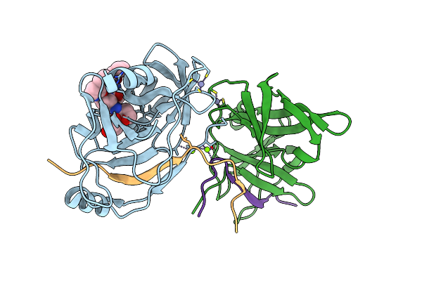
Protein-Ligand Complex Of A Novel Macrocyclic Hcv Ns3 Protease Inhibitor Derived From Amino Cyclic Boronates
Organism: Hepatitis c virus
Method: X-RAY DIFFRACTION Resolution:3.30 Å Release Date: 2011-08-17 Classification: HYDROLASE/HYDROLASE REGULATOR Ligands: TR8, ZN, MG |
Organism: Hepatitis c virus
Method: X-RAY DIFFRACTION
Release Date: 2011-08-17
Ligands: TR8, ZN, MG
 |
Crystal Structure Of Hcv Ns3 Protease With A Boronate Inhibitor
Organism: Hepatitis c virus
Method: X-RAY DIFFRACTION Resolution:2.48 Å Release Date: 2010-06-02 Classification: HYDROLASE Ligands: ZN, MG, BBQ |
Organism: Hepatitis c virus
Method: X-RAY DIFFRACTION
Release Date: 2010-06-02
Ligands: ZN, MG, BBQ