

Search Count: 2,709
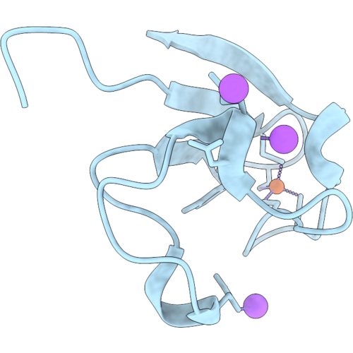 |
Pyrococcus Abyssi Rubredoxin @ 0.43 Angstrom Resolution, Transferable Aspherical Atom Model (Taam) Refinement
Organism: Pyrococcus abyssi ge5
Method: X-RAY DIFFRACTION Resolution:0.43 Å Release Date: 2026-09-16 Classification: ELECTRON TRANSPORT Ligands: FE, NA |
Organism: Pyrococcus abyssi ge5
Method: X-RAY DIFFRACTION
Release Date: 2026-09-16
Ligands: FE, NA
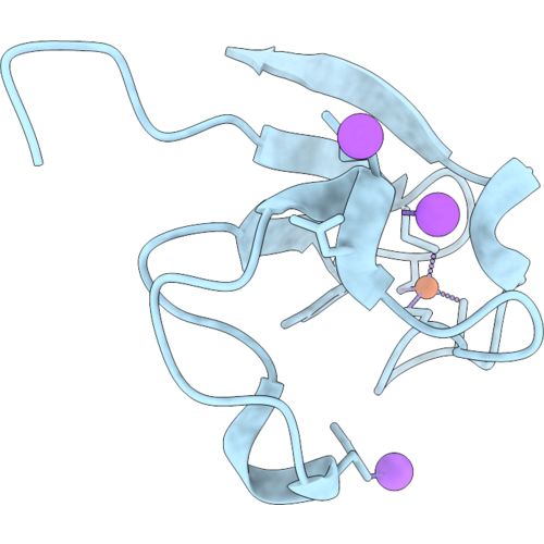 |
Pyrococcus Abyssi Rubredoxin @ 0.43 Angstrom Resolution, Independent Atom Model (Iam) Refinement
Organism: Pyrococcus abyssi ge5
Method: X-RAY DIFFRACTION Resolution:0.43 Å Release Date: 2026-09-16 Classification: ELECTRON TRANSPORT Ligands: FE, NA |
Organism: Pyrococcus abyssi ge5
Method: X-RAY DIFFRACTION
Release Date: 2026-09-16
Ligands: FE, NA
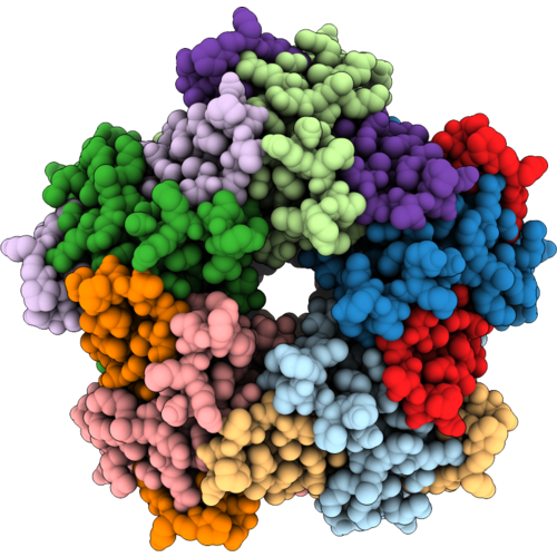 |
Single Particle Reconstruction Of Rhodospirillum Rubrum Encapsulated Ferritin In Encapsulin Nano Compartment
Organism: Rhodospirillum rubrum
Method: ELECTRON MICROSCOPY Release Date: 2026-09-02 Classification: OXIDOREDUCTASE Ligands: FE |
Organism: Rhodospirillum rubrum
Method: ELECTRON MICROSCOPY
Release Date: 2026-09-02
Ligands: FE
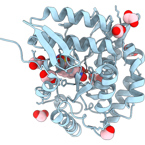 |
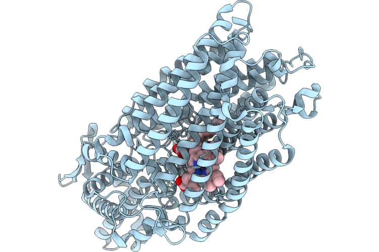
Crystal Structure Of Phenylalanine Hydroxylase (Pah) With Belinostat
Organism: Homo sapiens
Method: X-RAY DIFFRACTION Resolution:1.94 Å Release Date: 2026-09-02 Classification: OXIDOREDUCTASE Ligands: FE, 5OG, EDO |
Organism: Homo sapiens
Method: X-RAY DIFFRACTION
Release Date: 2026-09-02
Ligands: FE, 5OG, EDO
 |
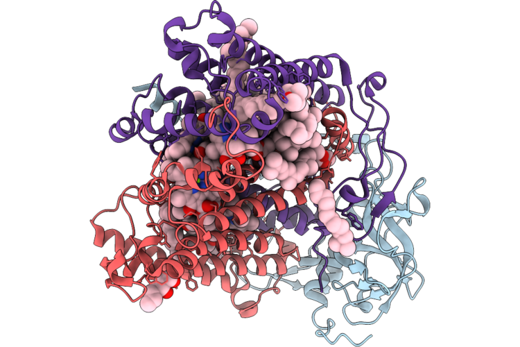
Cryo-Em Structure Of Native Monomeric Quinol-Dependent Nitric Oxide Reductase From Achromobacter Xylosoxidans.
Organism: Achromobacter xylosoxidans
Method: ELECTRON MICROSCOPY Release Date: 2026-08-26 Classification: MEMBRANE PROTEIN Ligands: CA, HEM, CU, FE |
Organism: Achromobacter xylosoxidans
Method: ELECTRON MICROSCOPY
Release Date: 2026-08-26
Ligands: CA, HEM, CU, FE
 |
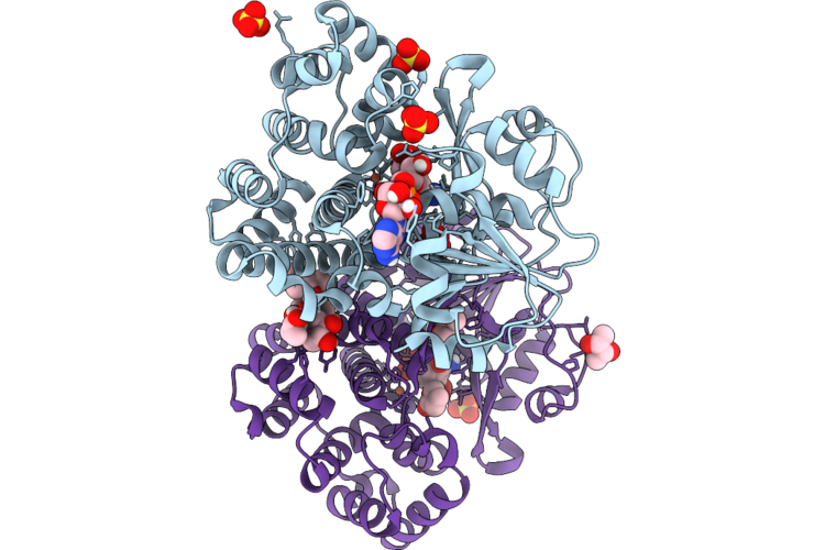
Structural And Electrostatic Analysis Of A Reaction Center Variant With High-Yield Charge Separation Along The Alternative Electron Transfer Path
Organism: Cereibacter sphaeroides
Method: X-RAY DIFFRACTION Resolution:2.70 Å Release Date: 2026-08-26 Classification: PHOTOSYNTHESIS Ligands: BCL, BPH, HTO, UQ, LDA, FE, SPO, CDL, U10, MG |
Organism: Cereibacter sphaeroides
Method: X-RAY DIFFRACTION
Release Date: 2026-08-26
Ligands: BCL, BPH, HTO, UQ, LDA, FE, SPO, CDL, U10, MG
 |
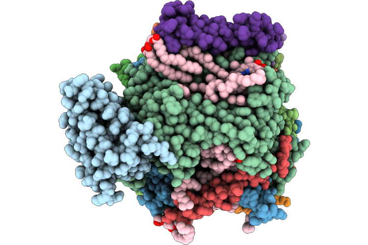
Crystal Structure Of A Omega-Hydroxyacid Iron-Containing Dehydrogenase
Organism: [clostridium] viride
Method: X-RAY DIFFRACTION Resolution:1.73 Å Release Date: 2026-08-19 Classification: OXIDOREDUCTASE Ligands: NAD, FE, GBL, BU3, SO4 |
Organism: [clostridium] viride
Method: X-RAY DIFFRACTION
Release Date: 2026-08-19
Ligands: NAD, FE, GBL, BU3, SO4
 |
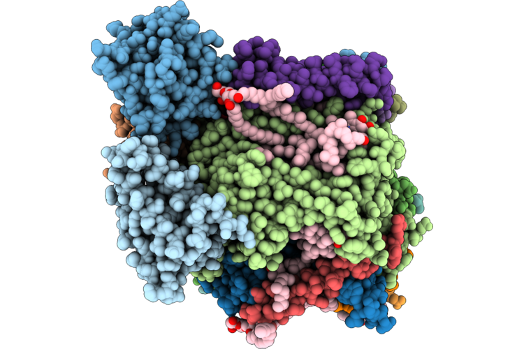
Cryo-Em Structure Of Psii Intermediate Psb27-Psii
Organism: Thermosynechococcus vestitus bp-1
Method: ELECTRON MICROSCOPY Release Date: 2026-08-19 Classification: PHOTOSYNTHESIS Ligands: FE, MN, CL, BCR, CLA, LHG, PHO, PL9, BCT, LMG, HEM |
Organism: Thermosynechococcus vestitus bp-1
Method: ELECTRON MICROSCOPY
Release Date: 2026-08-19
Ligands: FE, MN, CL, BCR, CLA, LHG, PHO, PL9, BCT, LMG, HEM
 |
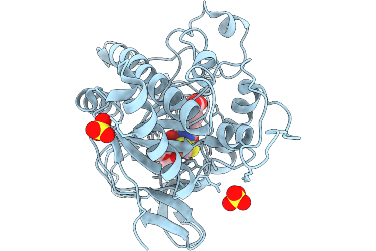
Cryo-Em Structure Of Psii Intermediate Psb32-Psii
Organism: Thermosynechococcus vestitus bp-1
Method: ELECTRON MICROSCOPY Release Date: 2026-08-19 Classification: PHOTOSYNTHESIS Ligands: FE, CL, BCR, CLA, PHO, PL9, LHG, LMG, BCT, HEM |
Organism: Thermosynechococcus vestitus bp-1
Method: ELECTRON MICROSCOPY
Release Date: 2026-08-19
Ligands: FE, CL, BCR, CLA, PHO, PL9, LHG, LMG, BCT, HEM
 |
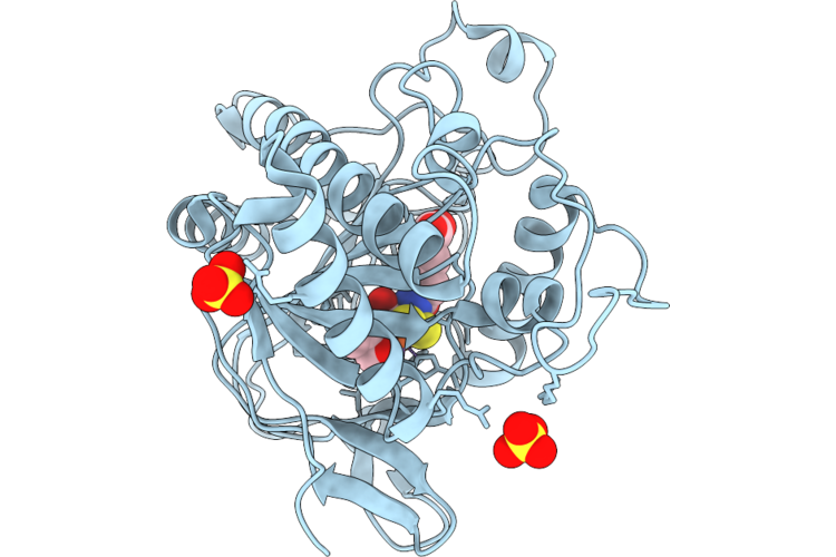
Isopenicillin N Synthase Co-Crystallised With Fe And Acdv After 6S O2 Exposure
Organism: Aspergillus nidulans fgsc a4
Method: X-RAY DIFFRACTION Resolution:1.50 Å Release Date: 2026-08-12 Classification: OXIDOREDUCTASE Ligands: A1IZG, A1I0E, SO4, OXY, FE, IO0, ACV |
Organism: Aspergillus nidulans fgsc a4
Method: X-RAY DIFFRACTION
Release Date: 2026-08-12
Ligands: A1IZG, A1I0E, SO4, OXY, FE, IO0, ACV
 |
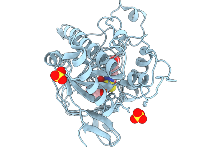
Isopenicillin N Synthase Co-Crystallised With Fe And Acdv After 8S O2 Exposure
Organism: Aspergillus nidulans fgsc a4
Method: X-RAY DIFFRACTION Resolution:1.60 Å Release Date: 2026-08-12 Classification: OXIDOREDUCTASE Ligands: A1IZG, IO0, OXY, A1I0E, SO4, FE |
Organism: Aspergillus nidulans fgsc a4
Method: X-RAY DIFFRACTION
Release Date: 2026-08-12
Ligands: A1IZG, IO0, OXY, A1I0E, SO4, FE
 |
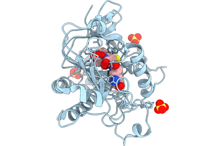
Isopenicillin N Synthase Co-Crystallised With Fe And Acdv After 10S O2 Exposure
Organism: Aspergillus nidulans fgsc a4
Method: X-RAY DIFFRACTION Resolution:1.70 Å Release Date: 2026-08-12 Classification: OXIDOREDUCTASE Ligands: IO0, OXY, A1I0E, A1IZG, SO4, FE |
Organism: Aspergillus nidulans fgsc a4
Method: X-RAY DIFFRACTION
Release Date: 2026-08-12
Ligands: IO0, OXY, A1I0E, A1IZG, SO4, FE
 |
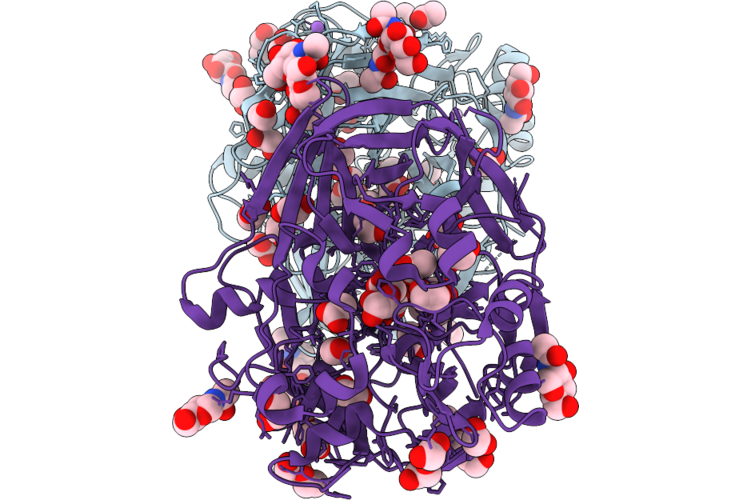
Isopenicillin N Synthase Co-Crystallised With Fe And Acdv After 10S O2 Exposure
Organism: Aspergillus nidulans fgsc a4
Method: X-RAY DIFFRACTION Resolution:1.68 Å Release Date: 2026-08-12 Classification: OXIDOREDUCTASE Ligands: SO4, FE, IP1, A1I0E, A1IZG, IO0, OXY |
Organism: Aspergillus nidulans fgsc a4
Method: X-RAY DIFFRACTION
Release Date: 2026-08-12
Ligands: SO4, FE, IP1, A1I0E, A1IZG, IO0, OXY
 |
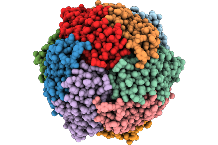
X-Ray Crystal Structure Of The Beta-Carotene Oxygenase Like G (Bcolg) From Lancelet Floridae
Organism: Branchiostoma floridae
Method: X-RAY DIFFRACTION Resolution:2.40 Å Release Date: 2026-08-12 Classification: OXIDOREDUCTASE Ligands: NAG, FE, NA, EDO, ACT, PEG, ETE, GOL |
Organism: Branchiostoma floridae
Method: X-RAY DIFFRACTION
Release Date: 2026-08-12
Ligands: NAG, FE, NA, EDO, ACT, PEG, ETE, GOL
 |
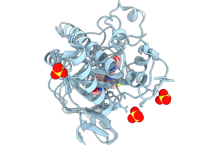
Sub-Tomogram Averaged Structure Of E. Coli Dna Protection During Starvation Protein (Dps)
Organism: Escherichia coli
Method: ELECTRON MICROSCOPY Resolution:3.00 Å Release Date: 2026-08-05 Classification: DNA BINDING PROTEIN Ligands: FE |
Organism: Escherichia coli
Method: ELECTRON MICROSCOPY
Release Date: 2026-08-05
Ligands: FE
 |
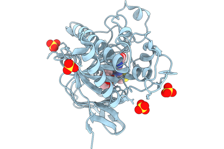
Isopenicillin N Synthase Q280A And L282A Variant In Complex With Fe And Acv Under Anaerobic Conditions.
Organism: Aspergillus nidulans fgsc a4
Method: X-RAY DIFFRACTION Resolution:1.68 Å Release Date: 2026-07-29 Classification: OXIDOREDUCTASE Ligands: SO4, ACV, TRS, FE |
Organism: Aspergillus nidulans fgsc a4
Method: X-RAY DIFFRACTION
Release Date: 2026-07-29
Ligands: SO4, ACV, TRS, FE
 |
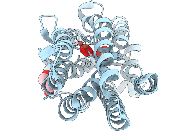
Isopenicillin N Synthase S281A Variant In Complex With Fe And Acv Under Anaerobic Conditions.
Organism: Aspergillus nidulans fgsc a4
Method: X-RAY DIFFRACTION Resolution:1.38 Å Release Date: 2026-07-29 Classification: OXIDOREDUCTASE Ligands: GOL, SO4, ACV, FE |
Organism: Aspergillus nidulans fgsc a4
Method: X-RAY DIFFRACTION
Release Date: 2026-07-29
Ligands: GOL, SO4, ACV, FE
 |
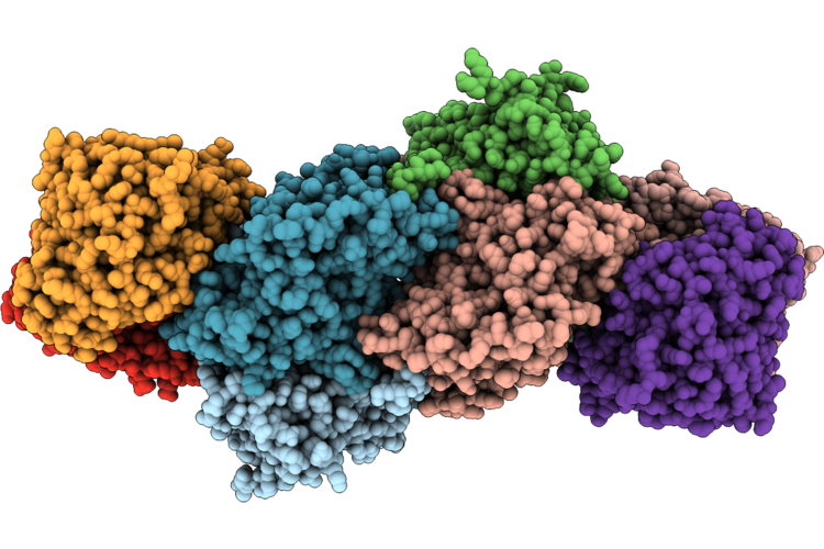
Crystal Structure Of U-1,2 Peroxo Intermediate From Paba Synthase Nitrosomonas Urea Cadd (Nucadd)
Organism: Nitrosomonas ureae
Method: X-RAY DIFFRACTION Resolution:2.07 Å Release Date: 2026-07-22 Classification: OXIDOREDUCTASE Ligands: PER, GOL, FE, SO4 |
Organism: Nitrosomonas ureae
Method: X-RAY DIFFRACTION
Release Date: 2026-07-22
Ligands: PER, GOL, FE, SO4
 |
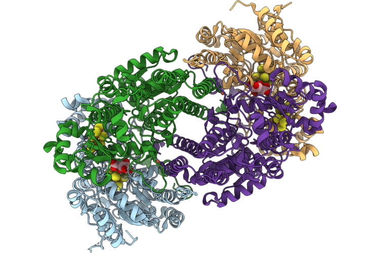
Cryoem Structure Of Gluconacetobacter Diazotrophicus Nitrogenase Complex (2:1 Fep:Mofep) Inhibited By Befx (C2 Symmetry)
Organism: Gluconacetobacter diazotrophicus pa1 5
Method: ELECTRON MICROSCOPY Resolution:2.73 Å Release Date: 2026-07-15 Classification: OXIDOREDUCTASE Ligands: HCA, ICS, CLF, FE, SF4, MG, ADP, 0BE |
Organism: Gluconacetobacter diazotrophicus pa1 5
Method: ELECTRON MICROSCOPY
Release Date: 2026-07-15
Ligands: HCA, ICS, CLF, FE, SF4, MG, ADP, 0BE
 |
Cryoem Structure Of Gluconacetobacter Diazotrophicus Mofep (C2 Symmetry)
Organism: Gluconacetobacter diazotrophicus pa1 5
Method: ELECTRON MICROSCOPY Resolution:2.15 Å Release Date: 2026-07-15 Classification: OXIDOREDUCTASE Ligands: FE, CLF, HCA, ICS |
Organism: Gluconacetobacter diazotrophicus pa1 5
Method: ELECTRON MICROSCOPY
Release Date: 2026-07-15
Ligands: FE, CLF, HCA, ICS