

Search Count: 19
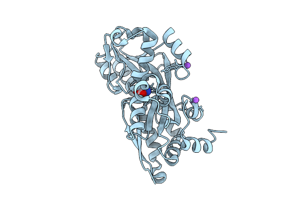 |
Vc0430/Vcglup Bound To Glutamate
Organism: Vibrio cholerae
Method: X-RAY DIFFRACTION Resolution:2.08 Å Release Date: 2025-12-24 Classification: TRANSPORT PROTEIN |
Organism: Vibrio cholerae
Method: X-RAY DIFFRACTION
Release Date: 2025-12-24
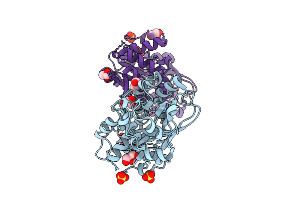 |
Vca0144 From Vibrio Cholerae
Organism: Vibrio cholerae
Method: X-RAY DIFFRACTION Resolution:1.80 Å Release Date: 2025-12-24 Classification: TRANSPORT PROTEIN Ligands: GOL, SO4 |
Organism: Vibrio cholerae
Method: X-RAY DIFFRACTION
Release Date: 2025-12-24
Ligands: GOL, SO4
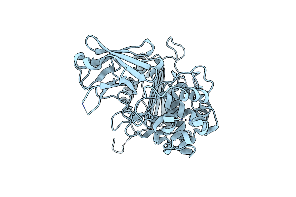 |
Crystal Structure Of Pbp Cyclase Ulm16
Organism: Streptomyces sp. kcb13f003
Method: X-RAY DIFFRACTION Resolution:2.06 Å Release Date: 2023-11-08 Classification: HYDROLASE |
Organism: Streptomyces sp. kcb13f003
Method: X-RAY DIFFRACTION
Release Date: 2023-11-08
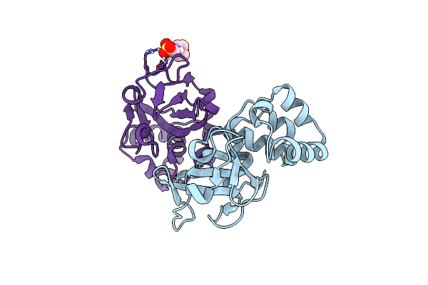 |
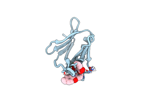
Structure Of The Bacterial Toxin, Teca, An Asparagine Deamidase From Alcaligenes Faecalis.
Organism: Alcaligenes faecalis
Method: X-RAY DIFFRACTION Resolution:1.84 Å Release Date: 2022-11-02 Classification: TOXIN Ligands: MES |
Organism: Alcaligenes faecalis
Method: X-RAY DIFFRACTION
Release Date: 2022-11-02
Ligands: MES
 |
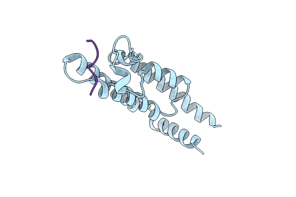
Structure Of Xl5-Ligated Hrpn13 Pru Domain
Organism: Homo sapiens
Method: SOLUTION NMR Release Date: 2021-12-15 Classification: PROTEIN BINDING Ligands: XAY |
Organism: Homo sapiens
Method: SOLUTION NMR
Release Date: 2021-12-15
Ligands: XAY
 |
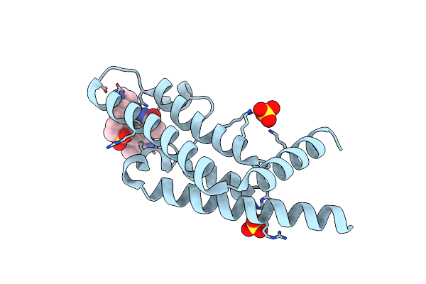
Atad2 Bromodomain Complexed With Histone H4K5Ac (Res 1-10) Ligand
Organism: Homo sapiens
Method: X-RAY DIFFRACTION Resolution:1.60 Å Release Date: 2021-09-22 Classification: NUCLEAR PROTEIN |
Organism: Homo sapiens
Method: X-RAY DIFFRACTION
Release Date: 2021-09-22
 |
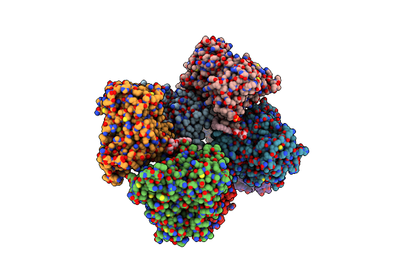
Atad2B Bromodomain In Complex With 4-({[(3R,4R)-4-{[3-Methyl-5-(5-Methylpyridin-3-Yl)-2-Oxo-1,2-Dihydro-1,7-Naphthyridin-8-Yl]Amino}Piperidin-3-Yl]Oxy}Methyl)-1Lambda~6~-Thiane-1,1-Dione (Compound 38)
Organism: Homo sapiens
Method: X-RAY DIFFRACTION Resolution:2.40 Å Release Date: 2020-10-28 Classification: NUCLEAR PROTEIN Ligands: SO4, YD4 |
Organism: Homo sapiens
Method: X-RAY DIFFRACTION
Release Date: 2020-10-28
Ligands: SO4, YD4
 |
Crystal Structure Of Human P450 2C9*2 Genetic Variant In Complex With Losartan
Organism: Homo sapiens
Method: X-RAY DIFFRACTION Resolution:3.12 Å Release Date: 2020-09-30 Classification: OXIDOREDUCTASE Ligands: HEM, LSN, CM5, SO4 |
Organism: Homo sapiens
Method: X-RAY DIFFRACTION
Release Date: 2020-09-30
Ligands: HEM, LSN, CM5, SO4
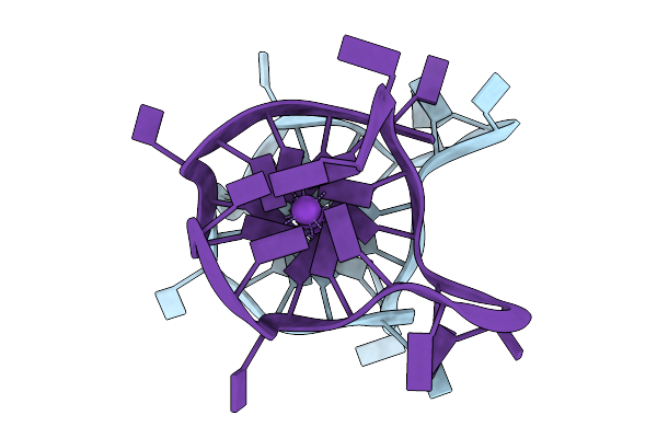 |
Kras G-Quadruplex G16T Mutant.
Organism: Homo sapiens
Method: X-RAY DIFFRACTION Resolution:1.60 Å Release Date: 2020-04-15 Classification: DNA Ligands: K |
Organism: Homo sapiens
Method: X-RAY DIFFRACTION
Release Date: 2020-04-15
Ligands: K
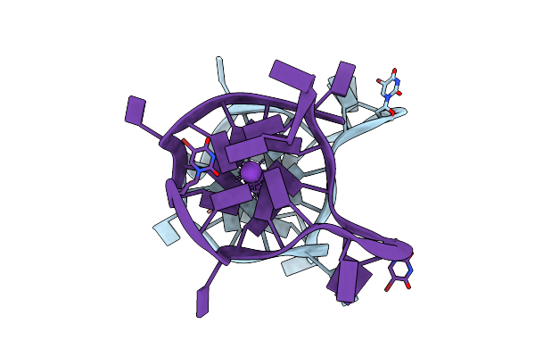 |
Kras G-Quadruplex G16T Mutant With Bromo Uracil Replacing T8 And T16.
Organism: Homo sapiens
Method: X-RAY DIFFRACTION Resolution:1.80 Å Release Date: 2020-04-15 Classification: DNA Ligands: K |
Organism: Homo sapiens
Method: X-RAY DIFFRACTION
Release Date: 2020-04-15
Ligands: K
 |
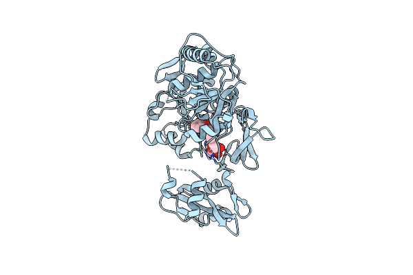
Synaptotagmin 1 C2A Beta4 Chimera With Enhanced Pip2 Binding Function
Organism: Rattus norvegicus
Method: X-RAY DIFFRACTION Resolution:1.83 Å Release Date: 2019-04-17 Classification: EXOCYTOSIS |
Organism: Rattus norvegicus
Method: X-RAY DIFFRACTION
Release Date: 2019-04-17
 |
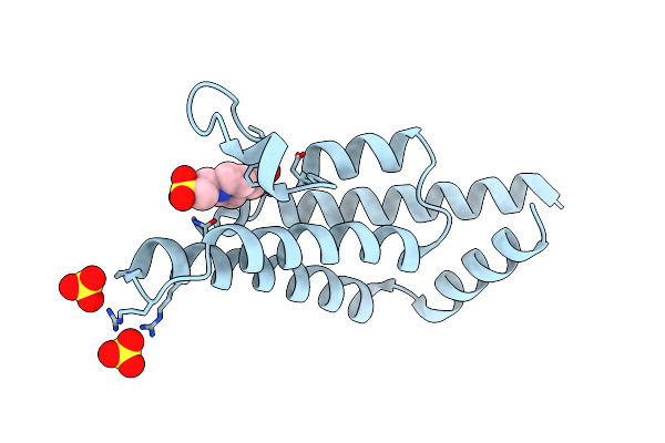
Wild-Type E. Coli Mene With Bound M Phenylether-Linked Analogue Of Osb-Ams
Organism: Escherichia coli (strain k12)
Method: X-RAY DIFFRACTION Resolution:1.83 Å Release Date: 2019-04-10 Classification: LIGASE Ligands: KOY |
Organism: Escherichia coli (strain k12)
Method: X-RAY DIFFRACTION
Release Date: 2019-04-10
Ligands: KOY
 |
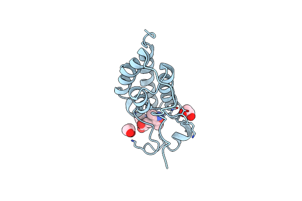
Crystal Structure Of The Bromodomain Of Human Atad2 With A Disulfide Bridge
Organism: Homo sapiens
Method: X-RAY DIFFRACTION Resolution:1.93 Å Release Date: 2018-12-19 Classification: PROTEIN BINDING Ligands: SO4, EPE |
Organism: Homo sapiens
Method: X-RAY DIFFRACTION
Release Date: 2018-12-19
Ligands: SO4, EPE
 |
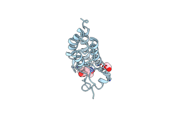
Crystal Structure Of The First Bromodomain Of Human Brd4 In Complex With A 3,5-Dimethylisoxazol Ligand
Organism: Homo sapiens
Method: X-RAY DIFFRACTION Resolution:1.34 Å Release Date: 2018-04-18 Classification: TRANSCRIPTION Ligands: EDO, E5Q |
Organism: Homo sapiens
Method: X-RAY DIFFRACTION
Release Date: 2018-04-18
Ligands: EDO, E5Q
 |
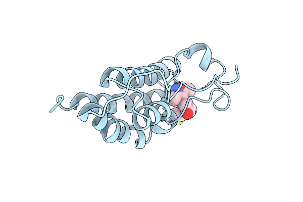
Crystal Structure Of The First Bromodomain Of Human Brd4 In Complex With A 3,5-Dimethylisoxazol Ligand
Organism: Homo sapiens
Method: X-RAY DIFFRACTION Resolution:1.28 Å Release Date: 2018-04-18 Classification: TRANSCRIPTION Ligands: EDO, E5T |
Organism: Homo sapiens
Method: X-RAY DIFFRACTION
Release Date: 2018-04-18
Ligands: EDO, E5T
 |
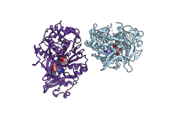
Crystal Structure Of The First Bromodomain Of Human Brd4 In Complex With A 3,5-Dimethylisoxazol Ligand
Organism: Homo sapiens
Method: X-RAY DIFFRACTION Resolution:1.34 Å Release Date: 2018-04-18 Classification: TRANSCRIPTION Ligands: E5W |
Organism: Homo sapiens
Method: X-RAY DIFFRACTION
Release Date: 2018-04-18
Ligands: E5W
 |
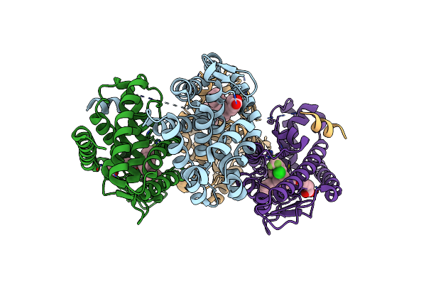
R195K E. Coli Mene With Bound Osb-Ams
Organism: Escherichia coli (strain k12)
Method: X-RAY DIFFRACTION Resolution:2.40 Å Release Date: 2015-10-07 Classification: LIGASE Ligands: MG, 4YB |
Organism: Escherichia coli (strain k12)
Method: X-RAY DIFFRACTION
Release Date: 2015-10-07
Ligands: MG, 4YB
 |
Hrxralpha & Mlxralpha With An Indole Pharmacophore, Sb786875
Organism: Homo sapiens, Mus musculus
Method: X-RAY DIFFRACTION Resolution:2.06 Å Release Date: 2009-02-10 Classification: TRANSCRIPTION Ligands: REA, LX2 |
Organism: Homo sapiens, Mus musculus
Method: X-RAY DIFFRACTION
Release Date: 2009-02-10
Ligands: REA, LX2
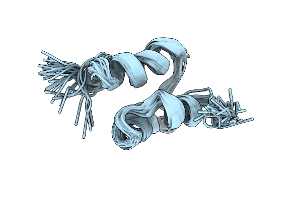 |
Nmr Structure Of The Uba Domain Of P62 (Sqstm1)
Organism: Homo sapiens
Method: SOLUTION NMR Release Date: 2008-02-19 Classification: SIGNALING PROTEIN |
Organism: Homo sapiens
Method: SOLUTION NMR
Release Date: 2008-02-19