

Search Count: 73
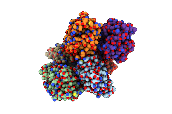 |
The X-Ray Structure Of Juvenile Hormone Diol Kinase From The Silk Worm Bombyx Mori.
Organism: Bombyx mori
Method: X-RAY DIFFRACTION Resolution:1.99 Å Release Date: 2021-12-22 Classification: METAL BINDING PROTEIN Ligands: CA |
Organism: Bombyx mori
Method: X-RAY DIFFRACTION
Release Date: 2021-12-22
Ligands: CA
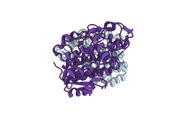 |
The X-Ray Structure Of L-Threonine Dehydrogenase From The Common Hospital Pathogen Clostridium Difficile.
Organism: Clostridioides difficile
Method: X-RAY DIFFRACTION Resolution:2.60 Å Release Date: 2021-06-16 Classification: OXIDOREDUCTASE |
Organism: Clostridioides difficile
Method: X-RAY DIFFRACTION
Release Date: 2021-06-16
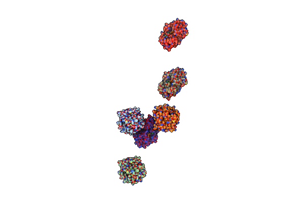 |
Three-Dimensional Structure Of L-Threonine 3-Dehydrogenase From Trypanosoma Brucei Refined To 1.77 Angstroms
Organism: Trypanosoma brucei
Method: X-RAY DIFFRACTION Resolution:1.77 Å Release Date: 2018-01-17 Classification: OXIDOREDUCTASE Ligands: NAD, ACT, CL, NA, GOL |
Organism: Trypanosoma brucei
Method: X-RAY DIFFRACTION
Release Date: 2018-01-17
Ligands: NAD, ACT, CL, NA, GOL
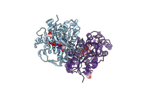 |
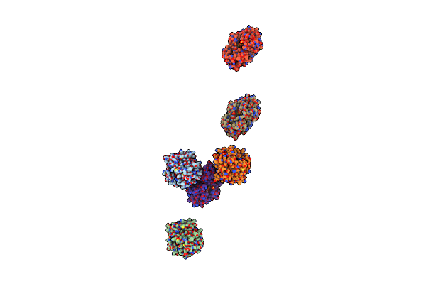
Three-Dimensional Structure Of L-Threonine 3-Dehydrogenase From Trypanosoma Brucei Bound To Nadh And L-Threonine Refined To 1.72 Angstroms
Organism: Trypanosoma brucei
Method: X-RAY DIFFRACTION Resolution:1.72 Å Release Date: 2018-01-10 Classification: OXIDOREDUCTASE Ligands: NAI, THR, GOL, NA |
Organism: Trypanosoma brucei
Method: X-RAY DIFFRACTION
Release Date: 2018-01-10
Ligands: NAI, THR, GOL, NA
 |
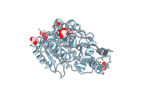
Three-Dimensional Structure Of L-Threonine 3-Dehydrogenase From Trypanosoma Brucei Bound To Nad+ Refined To 2.3 Angstroms
Organism: Trypanosoma brucei
Method: X-RAY DIFFRACTION Resolution:2.30 Å Release Date: 2017-11-15 Classification: OXIDOREDUCTASE Ligands: NAD, GOL, SO4 |
Organism: Trypanosoma brucei
Method: X-RAY DIFFRACTION
Release Date: 2017-11-15
Ligands: NAD, GOL, SO4
 |
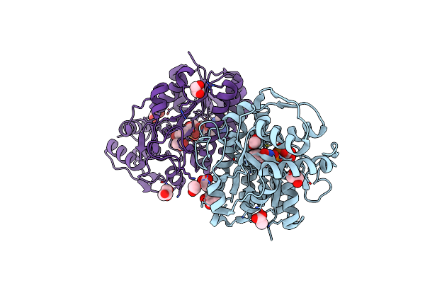
Three-Dimensional Structure Of L-Threonine 3-Dehydrogenase From Trypanosoma Brucei Refined To 2.1 Angstroms
Organism: Trypanosoma brucei
Method: X-RAY DIFFRACTION Resolution:2.10 Å Release Date: 2017-11-15 Classification: OXIDOREDUCTASE Ligands: GOL |
Organism: Trypanosoma brucei
Method: X-RAY DIFFRACTION
Release Date: 2017-11-15
Ligands: GOL
 |
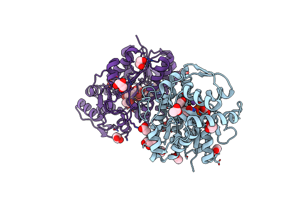
Three-Dimensional Structure Of L-Threonine 3-Dehydrogenase From Trypanosoma Brucei Showing Different Active Site Loop Conformations Between Dimer Subunits, Refined To 1.9 Angstroms
Organism: Trypanosoma brucei
Method: X-RAY DIFFRACTION Resolution:1.90 Å Release Date: 2017-11-15 Classification: OXIDOREDUCTASE Ligands: NAD, GOL, ACT |
Organism: Trypanosoma brucei
Method: X-RAY DIFFRACTION
Release Date: 2017-11-15
Ligands: NAD, GOL, ACT
 |
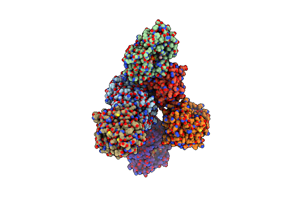
Three-Dimensional Structure Of L-Threonine 3-Dehydrogenase From Trypanosoma Brucei Bound To Nad+ Refined To 2.2 Angstroms
Organism: Trypanosoma brucei
Method: X-RAY DIFFRACTION Resolution:2.20 Å Release Date: 2017-11-15 Classification: OXIDOREDUCTASE Ligands: NAD, ACT, GOL, NA |
Organism: Trypanosoma brucei
Method: X-RAY DIFFRACTION
Release Date: 2017-11-15
Ligands: NAD, ACT, GOL, NA
 |
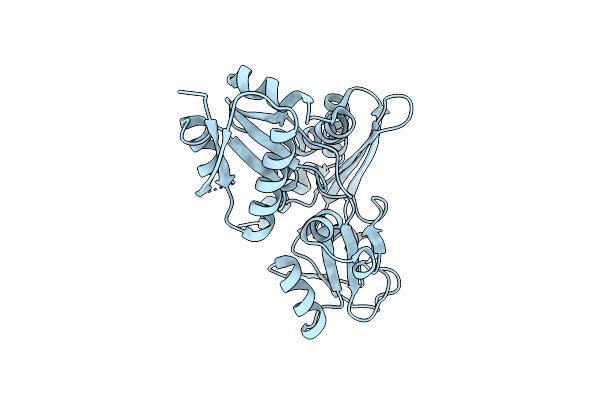
Three-Dimensional Structure Of L-Threonine 3-Dehydrogenase From Trypanosoma Brucei Bound To Nad+ And L-Allo-Threonine Refined To 2.23 Angstroms
Organism: Trypanosoma brucei
Method: X-RAY DIFFRACTION Resolution:2.26 Å Release Date: 2017-11-15 Classification: OXIDOREDUCTASE Ligands: NAD, GOL, ACT, ALO |
Organism: Trypanosoma brucei
Method: X-RAY DIFFRACTION
Release Date: 2017-11-15
Ligands: NAD, GOL, ACT, ALO
 |
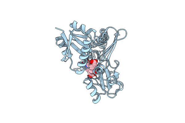
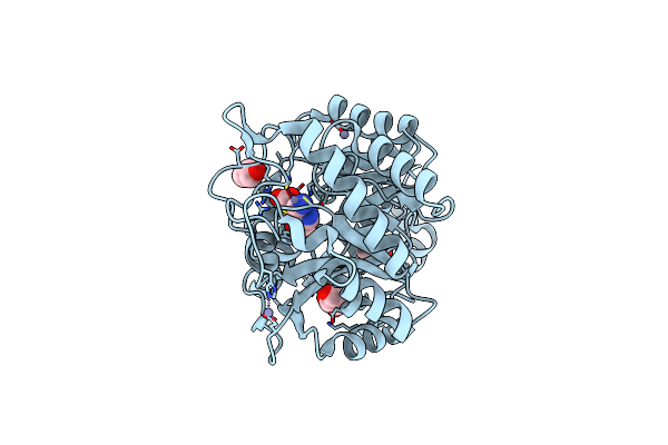
Bacillus Megaterium Porphobilinogen Deaminase D82A Mutant
Organism: Bacillus megaterium
Method: X-RAY DIFFRACTION Resolution:2.69 Å Release Date: 2017-10-11 Classification: TRANSFERASE |
Organism: Bacillus megaterium
Method: X-RAY DIFFRACTION
Release Date: 2017-10-11
 |
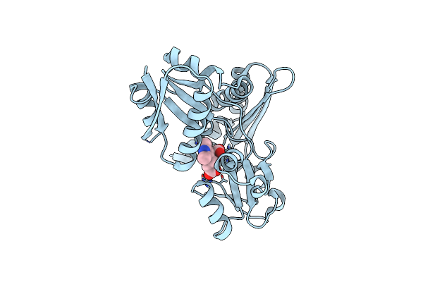
Bacillus Megaterium Porphobilinogen Deaminase D82E Mutant
Organism: Bacillus megaterium
Method: X-RAY DIFFRACTION Resolution:1.81 Å Release Date: 2017-09-06 Classification: TRANSFERASE Ligands: DPM |
Organism: Bacillus megaterium
Method: X-RAY DIFFRACTION
Release Date: 2017-09-06
Ligands: DPM
 |
Bacillus Megaterium Porphobilinogen Deaminase D82N Mutant
Organism: Bacillus megaterium
Method: X-RAY DIFFRACTION Resolution:1.87 Å Release Date: 2017-09-06 Classification: TRANSFERASE Ligands: AWQ |
Organism: Bacillus megaterium
Method: X-RAY DIFFRACTION
Release Date: 2017-09-06
Ligands: AWQ
 |
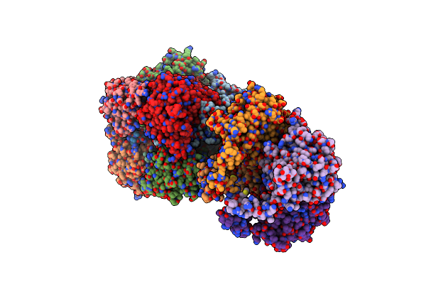
Product-Complex Of E.Coli 5-Amino Laevulinic Acid Dehydratase
Organism: Escherichia coli
Method: X-RAY DIFFRACTION Resolution:2.10 Å Release Date: 2016-12-07 Classification: LYASE Ligands: PBG, ZN, GOL |
Organism: Escherichia coli
Method: X-RAY DIFFRACTION
Release Date: 2016-12-07
Ligands: PBG, ZN, GOL
 |
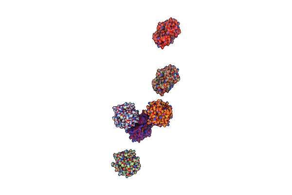
Pyrobaculum Calidifontis 5-Aminolaevulinic Acid Dehydratase
Organism: Pyrobaculum calidifontis (strain jcm 11548 / va1)
Method: X-RAY DIFFRACTION Resolution:3.47 Å Release Date: 2016-10-12 Classification: LYASE Ligands: ZN |
Organism: Pyrobaculum calidifontis (strain jcm 11548 / va1)
Method: X-RAY DIFFRACTION
Release Date: 2016-10-12
Ligands: ZN
 |
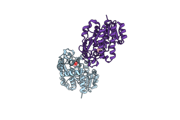
L-Threonine Dehydrogenase From Trypanosoma Brucei With Nad And The Inhibitor Pyruvate Bound.
Organism: Trypanosoma brucei
Method: X-RAY DIFFRACTION Resolution:2.10 Å Release Date: 2016-07-27 Classification: OXIDOREDUCTASE Ligands: NAD, PYR, BME, ACT, NA |
Organism: Trypanosoma brucei
Method: X-RAY DIFFRACTION
Release Date: 2016-07-27
Ligands: NAD, PYR, BME, ACT, NA
 |
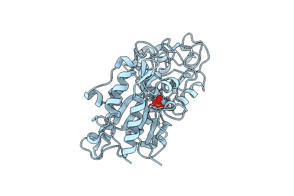
L-Threonine Dehydrogenase From Trypanosoma Brucei.
Organism: Trypanosoma brucei
Method: X-RAY DIFFRACTION Resolution:1.45 Å Release Date: 2016-06-22 Classification: OXIDOREDUCTASE Ligands: ACT |
Organism: Trypanosoma brucei
Method: X-RAY DIFFRACTION
Release Date: 2016-06-22
Ligands: ACT
 |
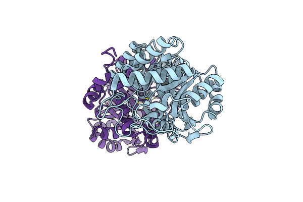
Phosphate-Binding Protein From Stenotrophomonas Maltophilia.
Organism: Stenotrophomonas maltophilia r551-3
Method: X-RAY DIFFRACTION Resolution:1.10 Å Release Date: 2016-05-04 Classification: TRANSPORT PROTEIN Ligands: PO4 |
Organism: Stenotrophomonas maltophilia r551-3
Method: X-RAY DIFFRACTION
Release Date: 2016-05-04
Ligands: PO4
 |
X-Ray Structure Of Human Recombinant 5-Aminolaevulinic Acid Dehydratase (Hralad).
Organism: Homo sapiens
Method: X-RAY DIFFRACTION Resolution:2.80 Å Release Date: 2016-01-27 Classification: LYASE Ligands: ZN |
Organism: Homo sapiens
Method: X-RAY DIFFRACTION
Release Date: 2016-01-27
Ligands: ZN
 |
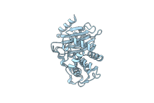
The X-Ray Structure Of Octameric Human Native 5-Aminolaevulinic Acid Dehydratase.
Organism: Homo sapiens
Method: X-RAY DIFFRACTION Resolution:2.83 Å Release Date: 2016-01-27 Classification: LYASE Ligands: ZN, SO4, DAV |
Organism: Homo sapiens
Method: X-RAY DIFFRACTION
Release Date: 2016-01-27
Ligands: ZN, SO4, DAV
 |
100K Neutron Ligand Free: Exploring The Mechanism Of Beta-Lactam Ring Protonation In The Class A Beta-Lactamase Acylation Mechanism Using Neutron And X-Ray Crystallography
Organism: Escherichia coli
Method: NEUTRON DIFFRACTION Resolution:1.70 Å Release Date: 2015-12-16 Classification: HYDROLASE |
Organism: Escherichia coli
Method: NEUTRON DIFFRACTION
Release Date: 2015-12-16