

Search Count: 40
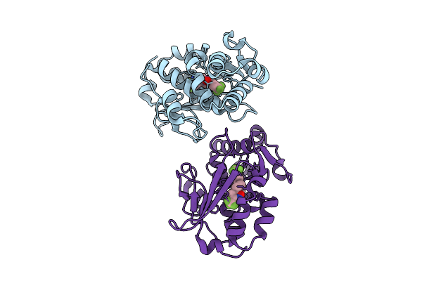 |
Crystal Structure Of The Mycobacterium Tuberculosis Regulator Virs (N-Terminal Fragment 4-208) In Complex With The Drug Candidate Alpibectir
Organism: Mycobacterium tuberculosis h37rv
Method: X-RAY DIFFRACTION Resolution:1.49 Å Release Date: 2025-10-29 Classification: TRANSCRIPTION Ligands: YPQ |
Organism: Mycobacterium tuberculosis h37rv
Method: X-RAY DIFFRACTION
Release Date: 2025-10-29
Ligands: YPQ
 |
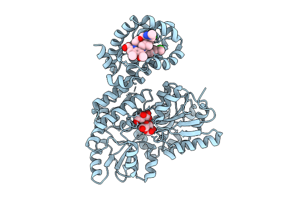
Myeloid Cell Leukemia-1 (Mcl-1) Complexed With Compound 8
Organism: Homo sapiens
Method: X-RAY DIFFRACTION Resolution:2.07 Å Release Date: 2025-09-17 Classification: APOPTOSIS Ligands: A1CL3 |
Organism: Homo sapiens
Method: X-RAY DIFFRACTION
Release Date: 2025-09-17
Ligands: A1CL3
 |
Myeloid Cell Leukemia-1 (Mcl-1) Complexed With Compound 13
Organism: Homo sapiens
Method: X-RAY DIFFRACTION Resolution:1.95 Å Release Date: 2025-09-17 Classification: APOPTOSIS Ligands: A1CL6 |
Organism: Homo sapiens
Method: X-RAY DIFFRACTION
Release Date: 2025-09-17
Ligands: A1CL6
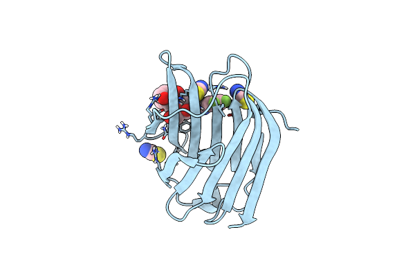 |
Co-Crystal Structure Of Galectin-3 With An Inhibitor
Organism: Homo sapiens
Method: X-RAY DIFFRACTION Resolution:1.45 Å Release Date: 2025-03-26 Classification: SUGAR BINDING PROTEIN Ligands: SCN, A1IB3 |
Organism: Homo sapiens
Method: X-RAY DIFFRACTION
Release Date: 2025-03-26
Ligands: SCN, A1IB3
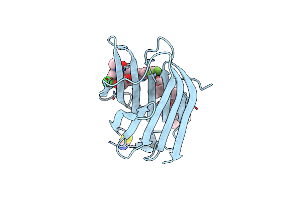 |
Co-Crystal Structure Of Galectin-3 With An Inhibitor
Organism: Homo sapiens
Method: X-RAY DIFFRACTION Resolution:1.78 Å Release Date: 2025-03-26 Classification: SUGAR BINDING PROTEIN Ligands: 2PE, A1IB4, SCN |
Organism: Homo sapiens
Method: X-RAY DIFFRACTION
Release Date: 2025-03-26
Ligands: 2PE, A1IB4, SCN
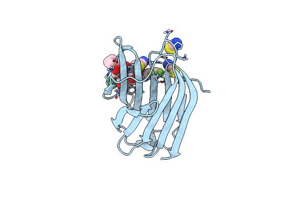 |
Galectin-3 With A Bound Inhibitor
Organism: Homo sapiens
Method: X-RAY DIFFRACTION Resolution:1.25 Å Release Date: 2025-01-29 Classification: SUGAR BINDING PROTEIN Ligands: A1H1W, SCN |
Organism: Homo sapiens
Method: X-RAY DIFFRACTION
Release Date: 2025-01-29
Ligands: A1H1W, SCN
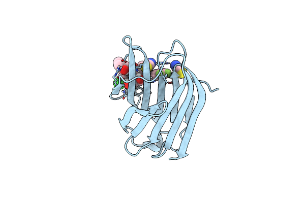 |
Galectin-3 With A Bound Inhibitor
Organism: Homo sapiens
Method: X-RAY DIFFRACTION Resolution:1.20 Å Release Date: 2025-01-29 Classification: SUGAR BINDING PROTEIN Ligands: A1H1Y, SCN |
Organism: Homo sapiens
Method: X-RAY DIFFRACTION
Release Date: 2025-01-29
Ligands: A1H1Y, SCN
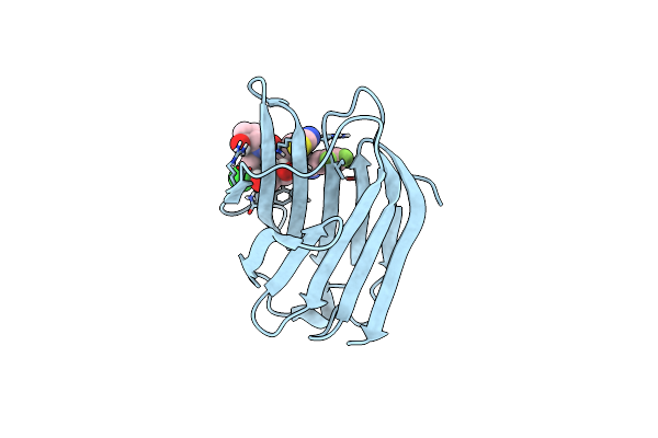 |
Galectin-3 With A Bound Inhibitor
Organism: Homo sapiens
Method: X-RAY DIFFRACTION Resolution:1.35 Å Release Date: 2025-01-29 Classification: SUGAR BINDING PROTEIN Ligands: A1H1X, SCN |
Organism: Homo sapiens
Method: X-RAY DIFFRACTION
Release Date: 2025-01-29
Ligands: A1H1X, SCN
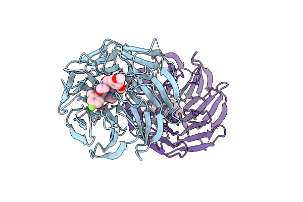 |
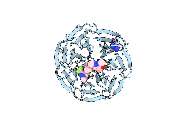
Crystal Structure Of Human Wd Repeat-Containing Protein 5 In Complex With 4-(3,5-Dimethoxybenzyl)-9-(4-Fluoro-2-Methylphenyl)-7-((2-Imino-3-Methyl-2,3-Dihydro-1H-Imidazol-1-Yl)Methyl)-3,4-Dihydrobenzo[F][1,4]Oxazepin-5(2H)-One (Compound 8)
Organism: Homo sapiens
Method: X-RAY DIFFRACTION Resolution:2.01 Å Release Date: 2023-12-20 Classification: TRANSCRIPTION Ligands: VV3 |
Organism: Homo sapiens
Method: X-RAY DIFFRACTION
Release Date: 2023-12-20
Ligands: VV3
 |
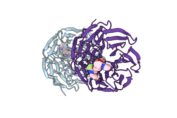
Wd Repeat-Containing Protein 5 Complexed With 4-(7-((1H-Imidazol-1-Yl)Methyl)-5-(1-Methyl-3-(Trifluoromethyl)-1H-Pyrazol-4-Yl)-1-Oxo-3,4-Dihydroisoquinolin-2(1H)-Yl)-6-Ethyl-N-Methylquinoline-8-Carboxamide (Compound 10)
Organism: Homo sapiens
Method: X-RAY DIFFRACTION Resolution:1.55 Å Release Date: 2023-01-11 Classification: TRANSCRIPTION Ligands: BEN, UY9 |
Organism: Homo sapiens
Method: X-RAY DIFFRACTION
Release Date: 2023-01-11
Ligands: BEN, UY9
 |
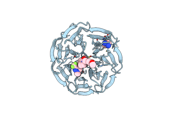
Discovery Of Potent Orally Bioavailable Wd Repeat Domain 5 (Wdr5) Inhibitors Using A Pharmacophore-Based Optimization
Organism: Homo sapiens
Method: X-RAY DIFFRACTION Resolution:1.81 Å Release Date: 2022-05-04 Classification: Transcription/Transcription Inhibitor Ligands: MBU |
Organism: Homo sapiens
Method: X-RAY DIFFRACTION
Release Date: 2022-05-04
Ligands: MBU
 |
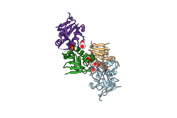
Wdr5 Bound To 2-(3,5-Dimethoxybenzyl)-7-((2-Methyl-1H-Imidazol-1-Yl)Methyl)-5-(1-Methyl-3-(Trifluoromethyl)-1H-Pyrazol-4-Yl)-3,4-Dihydroisoquinolin-1(2H)-One
Organism: Homo sapiens
Method: X-RAY DIFFRACTION Resolution:1.90 Å Release Date: 2022-04-27 Classification: Transcription/Transcription Inhibitor Ligands: BEN, M4U |
Organism: Homo sapiens
Method: X-RAY DIFFRACTION
Release Date: 2022-04-27
Ligands: BEN, M4U
 |
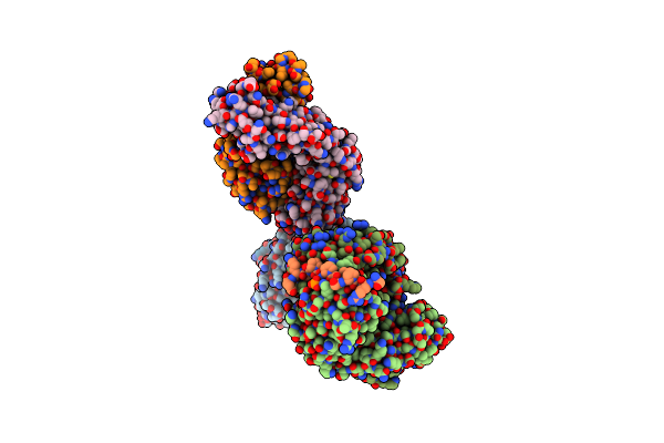
Crystal Structure Of Dna-Binding Domain Of Ompr Of Two-Component System Of Acinetobacter Baumannii
Organism: Acinetobacter baumannii (strain sdf)
Method: X-RAY DIFFRACTION Resolution:2.20 Å Release Date: 2021-08-11 Classification: DNA BINDING PROTEIN Ligands: SO4, PEG, GOL |
Organism: Acinetobacter baumannii (strain sdf)
Method: X-RAY DIFFRACTION
Release Date: 2021-08-11
Ligands: SO4, PEG, GOL
 |
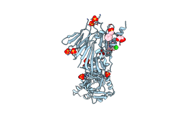
Crystal Structure Of Anti-Tau Antibody Pt3 Fab+Pt212/Pt217-Tau Peptide
Organism: Mus musculus, Homo sapiens
Method: X-RAY DIFFRACTION Resolution:2.00 Å Release Date: 2020-09-23 Classification: IMMUNE SYSTEM Ligands: GOL |
Organism: Mus musculus, Homo sapiens
Method: X-RAY DIFFRACTION
Release Date: 2020-09-23
Ligands: GOL
 |
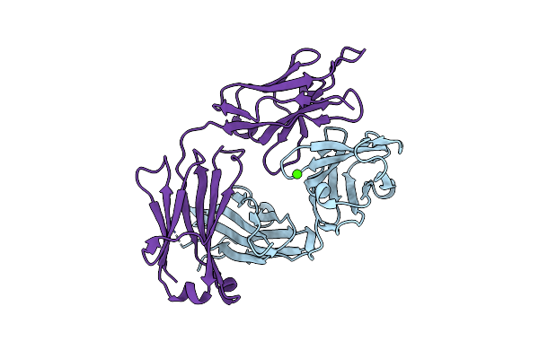
Crystal Structure Of Inhibitor Jnj-53718678 In Complex With Prefusion Rsv F Glycoprotein
Organism: Human respiratory syncytial virus, Human immunodeficiency virus 1
Method: X-RAY DIFFRACTION Resolution:2.50 Å Release Date: 2017-08-02 Classification: VIRAL PROTEIN/INHIBITOR Ligands: NAG, NHE, SO4, 6YA |
Organism: Human respiratory syncytial virus, Human immunodeficiency virus 1
Method: X-RAY DIFFRACTION
Release Date: 2017-08-02
Ligands: NAG, NHE, SO4, 6YA
 |
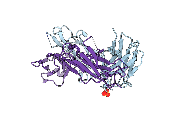
Crystal Structure Of Anti-Tau Antibody At8 Fab
Organism: Mus musculus
Method: X-RAY DIFFRACTION Resolution:2.10 Å Release Date: 2016-02-24 Classification: IMMUNE SYSTEM Ligands: CA |
Organism: Mus musculus
Method: X-RAY DIFFRACTION
Release Date: 2016-02-24
Ligands: CA
 |
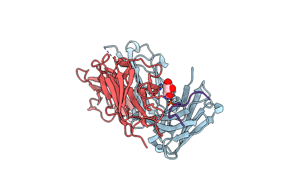
Structure Of Anti-Tau At8 Fab In The Presence Of Phosphopeptide
Organism: Mus musculus
Method: X-RAY DIFFRACTION Resolution:2.40 Å Release Date: 2016-02-24 Classification: IMMUNE SYSTEM Ligands: SO4 |
Organism: Mus musculus
Method: X-RAY DIFFRACTION
Release Date: 2016-02-24
Ligands: SO4
 |
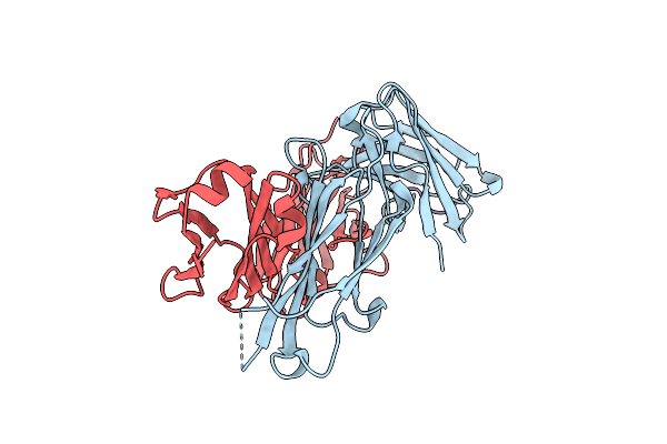
Anti-Tau At8 Fab With Doubly Phosphorylated Tau Peptide
Organism: Mus musculus, Homo sapiens
Method: X-RAY DIFFRACTION Resolution:1.64 Å Release Date: 2016-02-24 Classification: IMMUNE SYSTEM Ligands: GOL |
Organism: Mus musculus, Homo sapiens
Method: X-RAY DIFFRACTION
Release Date: 2016-02-24
Ligands: GOL
 |
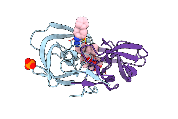
Anti-Tau At8 Fab With Triply Phosphorylated Tau Peptide
Organism: Mus musculus, Homo sapiens
Method: X-RAY DIFFRACTION Resolution:1.50 Å Release Date: 2016-02-24 Classification: IMMUNE SYSTEM |
Organism: Mus musculus, Homo sapiens
Method: X-RAY DIFFRACTION
Release Date: 2016-02-24
 |
Crystal Structure Of Wild-Type Hiv-1 Protease In Complex With Tmc310911
Organism: Human immunodeficiency virus 1
Method: X-RAY DIFFRACTION Resolution:1.90 Å Release Date: 2011-09-21 Classification: HYDROLASE/HYDROLASE INHIBITOR Ligands: 74T, PO4 |
Organism: Human immunodeficiency virus 1
Method: X-RAY DIFFRACTION
Release Date: 2011-09-21
Ligands: 74T, PO4