

Search Count: 19
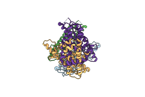 |
Cryoem Structure Of Shedu From Bacillus Cereus
Organism: Bacillus cereus b4264
Method: ELECTRON MICROSCOPY Release Date: 2024-07-31 Classification: DNA BINDING PROTEIN |
Organism: Bacillus cereus b4264
Method: ELECTRON MICROSCOPY
Release Date: 2024-07-31
 |
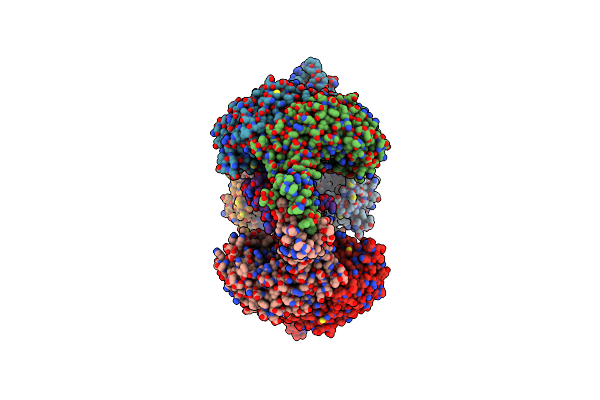
Cryoem Structure Of Octamer Assembly Of Shedu Nuclease Domain From Bacillus Cereus
Organism: Bacillus cereus b4264
Method: ELECTRON MICROSCOPY Release Date: 2024-07-31 Classification: DNA BINDING PROTEIN |
Organism: Bacillus cereus b4264
Method: ELECTRON MICROSCOPY
Release Date: 2024-07-31
 |
Cryoem Structure Of Locally-Refined Tetramer Of Shedu Nuclease Domain From Bacillus Cereus
Organism: Bacillus cereus b4264
Method: ELECTRON MICROSCOPY Release Date: 2024-07-31 Classification: DNA BINDING PROTEIN |
Organism: Bacillus cereus b4264
Method: ELECTRON MICROSCOPY
Release Date: 2024-07-31
 |
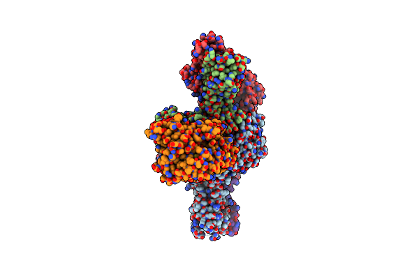
Cryoem Structure Of Aria-Ocr Complex
Organism: Escherichia coli b185, Escherichia phage t7
Method: ELECTRON MICROSCOPY Release Date: 2024-06-26 Classification: IMMUNE SYSTEM Ligands: ATP |
Organism: Escherichia coli b185, Escherichia phage t7
Method: ELECTRON MICROSCOPY
Release Date: 2024-06-26
Ligands: ATP
 |
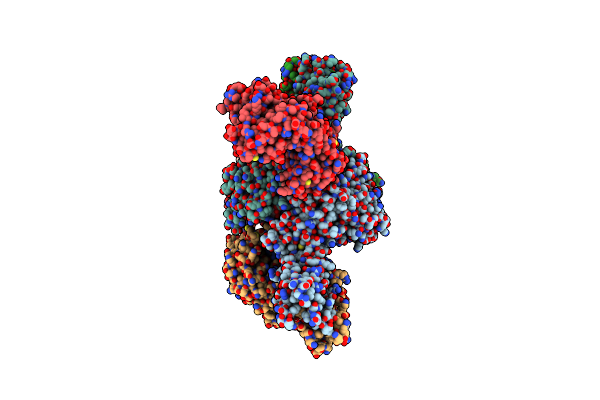
Cryoem Structure Of Aria-Arib Complex (Form I)
Organism: Escherichia coli b185
Method: ELECTRON MICROSCOPY Release Date: 2024-06-26 Classification: IMMUNE SYSTEM Ligands: ATP, MG |
Organism: Escherichia coli b185
Method: ELECTRON MICROSCOPY
Release Date: 2024-06-26
Ligands: ATP, MG
 |
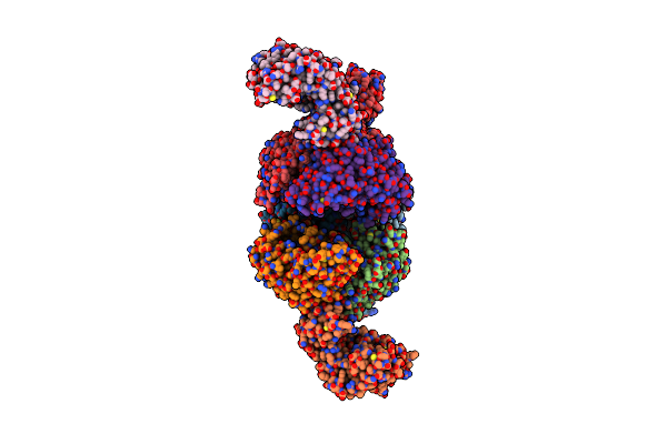
Cryoem Structure Of Aria-Arib Complex (Form Ii)
Organism: Escherichia coli b185
Method: ELECTRON MICROSCOPY Release Date: 2024-06-26 Classification: IMMUNE SYSTEM Ligands: ATP |
Organism: Escherichia coli b185
Method: ELECTRON MICROSCOPY
Release Date: 2024-06-26
Ligands: ATP
 |
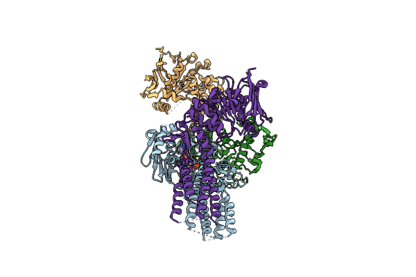
Cryoem Structure Of Aria-Arib Complex (Form Iii)
Organism: Escherichia coli b185
Method: ELECTRON MICROSCOPY Release Date: 2024-06-26 Classification: IMMUNE SYSTEM Ligands: ATP |
Organism: Escherichia coli b185
Method: ELECTRON MICROSCOPY
Release Date: 2024-06-26
Ligands: ATP
 |
Cryoem Structure Of Aria (E393Q) Sensory Subunit
Organism: Escherichia coli b185
Method: ELECTRON MICROSCOPY Release Date: 2024-06-26 Classification: IMMUNE SYSTEM Ligands: ATP |
Organism: Escherichia coli b185
Method: ELECTRON MICROSCOPY
Release Date: 2024-06-26
Ligands: ATP
 |
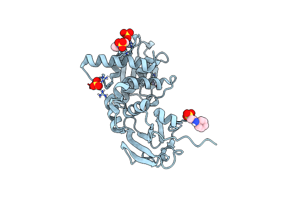
Structure Of Bacteriophage Pa1C Gp2
Organism: Pseudomonas phage pa1c
Method: X-RAY DIFFRACTION Resolution:2.63 Å Release Date: 2023-05-10 Classification: VIRAL PROTEIN |
Organism: Pseudomonas phage pa1c
Method: X-RAY DIFFRACTION
Release Date: 2023-05-10
 |
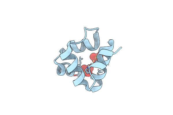
Structure Of Thauera Sp. K11 Capp
Organism: Thauera sp. k11
Method: X-RAY DIFFRACTION Resolution:1.35 Å Release Date: 2022-09-14 Classification: HYDROLASE Ligands: NHE, ZN, SO4 |
Organism: Thauera sp. k11
Method: X-RAY DIFFRACTION
Release Date: 2022-09-14
Ligands: NHE, ZN, SO4
 |
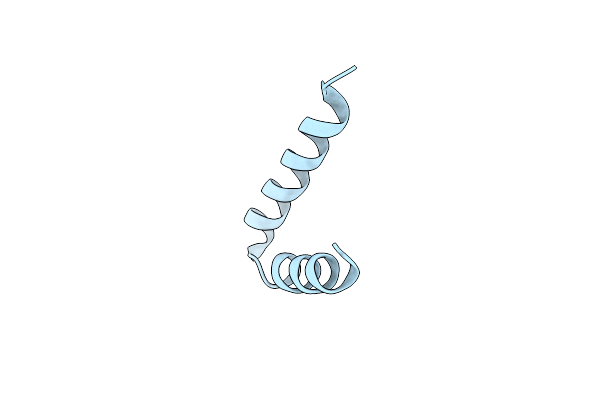
Structure Of E. Coli Ms115-1 Caph N-Terminal Domain
Organism: Escherichia coli
Method: X-RAY DIFFRACTION Resolution:1.02 Å Release Date: 2022-09-14 Classification: DNA BINDING PROTEIN Ligands: SO4 |
Organism: Escherichia coli
Method: X-RAY DIFFRACTION
Release Date: 2022-09-14
Ligands: SO4
 |
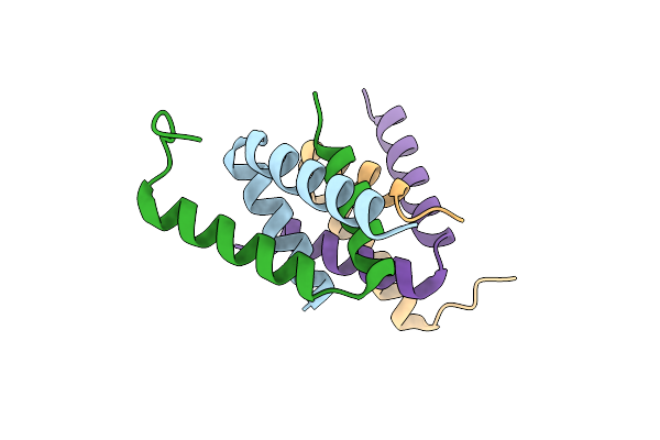
Structure Of E. Coli Caph C-Terminal Domain I99M Mutant
Organism: Escherichia coli
Method: X-RAY DIFFRACTION Resolution:1.26 Å Release Date: 2022-09-14 Classification: DNA BINDING PROTEIN |
Organism: Escherichia coli
Method: X-RAY DIFFRACTION
Release Date: 2022-09-14
 |
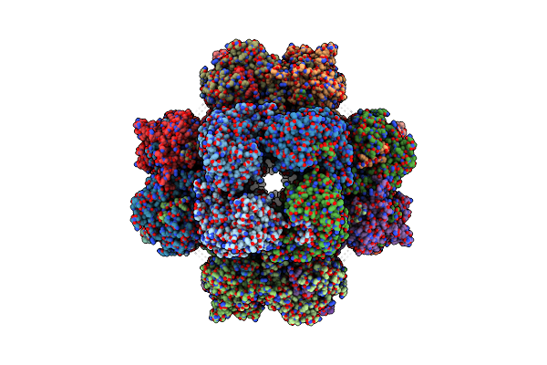
Structure Of E. Coli Caph C-Terminal Domain
Organism: Escherichia coli
Method: X-RAY DIFFRACTION Resolution:1.75 Å Release Date: 2022-09-14 Classification: DNA BINDING PROTEIN |
Organism: Escherichia coli
Method: X-RAY DIFFRACTION
Release Date: 2022-09-14
 |
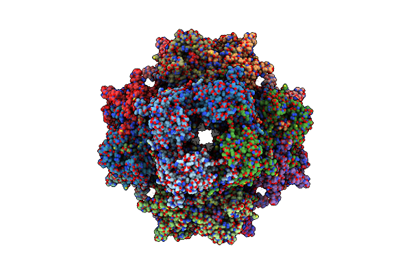
201Phi2-1 Chimallin Cubic (O, 24Mer) Assembly
Organism: Pseudomonas phage 201phi2-1
Method: ELECTRON MICROSCOPY Release Date: 2022-07-27 Classification: STRUCTURAL PROTEIN |
Organism: Pseudomonas phage 201phi2-1
Method: ELECTRON MICROSCOPY
Release Date: 2022-07-27
 |
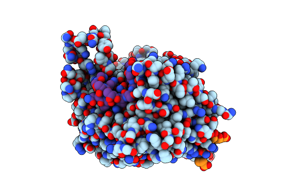
201Phi2-1 Chimallin Localized Tetramer Reconstruction
Organism: Pseudomonas phage 201phi2-1
Method: ELECTRON MICROSCOPY Release Date: 2022-07-27 Classification: STRUCTURAL PROTEIN |
Organism: Pseudomonas phage 201phi2-1
Method: ELECTRON MICROSCOPY
Release Date: 2022-07-27
 |
201Phi2-1 Chimallin C1 Localized Reconstruction
Organism: Pseudomonas phage 201phi2-1
Method: ELECTRON MICROSCOPY Release Date: 2022-07-27 Classification: STRUCTURAL PROTEIN |
Organism: Pseudomonas phage 201phi2-1
Method: ELECTRON MICROSCOPY
Release Date: 2022-07-27
 |
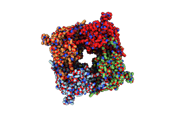
Goslar Chimallin Cubic (O, 24Mer) Assembly
Organism: Escherichia phage vb_ecom_goslar
Method: ELECTRON MICROSCOPY Release Date: 2022-07-27 Classification: STRUCTURAL PROTEIN |
Organism: Escherichia phage vb_ecom_goslar
Method: ELECTRON MICROSCOPY
Release Date: 2022-07-27
 |
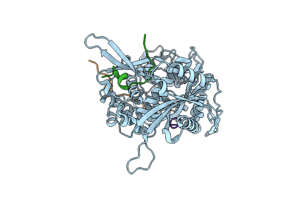
Goslar Chimallin C4 Tetramer Localized Reconstruction
Organism: Escherichia phage vb_ecom_goslar
Method: ELECTRON MICROSCOPY Release Date: 2022-07-27 Classification: STRUCTURAL PROTEIN |
Organism: Escherichia phage vb_ecom_goslar
Method: ELECTRON MICROSCOPY
Release Date: 2022-07-27
 |
Goslar Chimallin C1 Localized Reconstruction
Organism: Escherichia phage vb_ecom_goslar
Method: ELECTRON MICROSCOPY Release Date: 2022-07-27 Classification: STRUCTURAL PROTEIN |
Organism: Escherichia phage vb_ecom_goslar
Method: ELECTRON MICROSCOPY
Release Date: 2022-07-27