

Search Count: 25
 |
Inactive Mu-Opioid Receptor With Nb6M, Nabfab, And Isoquinuclidine Compound #33
Organism: Homo sapiens
Method: ELECTRON MICROSCOPY Release Date: 2025-02-12 Classification: MEMBRANE PROTEIN |
Organism: Homo sapiens
Method: ELECTRON MICROSCOPY
Release Date: 2025-02-12
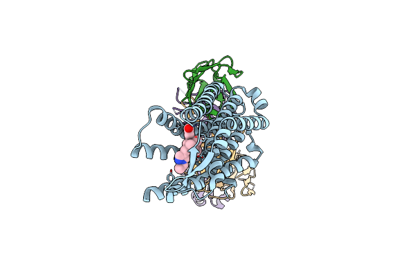 |
Inactive Mu-Opioid Receptor With Nb6M, Nabfab, And Isoquinuclidine Compound #020_E1
Organism: Homo sapiens, Synthetic construct
Method: ELECTRON MICROSCOPY Release Date: 2025-02-12 Classification: MEMBRANE PROTEIN Ligands: A1BNM |
Organism: Homo sapiens, Synthetic construct
Method: ELECTRON MICROSCOPY
Release Date: 2025-02-12
Ligands: A1BNM
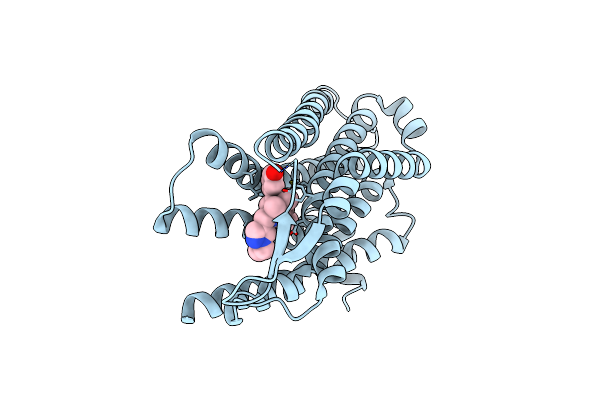 |
Locally-Refined Inactive Mu-Opioid Receptor With Nb6M, Nabfab, And Isoquinuclidine Compound #020_E1
Organism: Homo sapiens
Method: ELECTRON MICROSCOPY Release Date: 2025-02-12 Classification: MEMBRANE PROTEIN Ligands: A1BNM |
Organism: Homo sapiens
Method: ELECTRON MICROSCOPY
Release Date: 2025-02-12
Ligands: A1BNM
 |
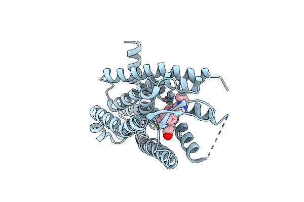
Inactive Kappa-Opioid Receptor With Nb6M, Nabfab, And Isoquinuclidine Compound #020_E1
Organism: Mus musculus, Synthetic construct
Method: ELECTRON MICROSCOPY Release Date: 2025-02-12 Classification: MEMBRANE PROTEIN Ligands: A1BNM |
Organism: Mus musculus, Synthetic construct
Method: ELECTRON MICROSCOPY
Release Date: 2025-02-12
Ligands: A1BNM
 |
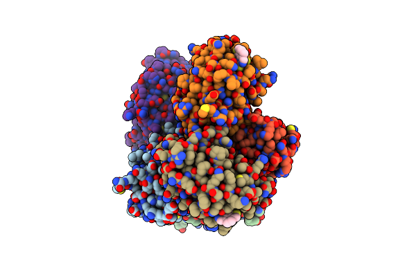
Locally-Refined Inactive Kappa-Opioid Receptor With Nb6M, Nabfab, And Isoquinuclidine Compound #020_E1
Organism: Mus musculus
Method: ELECTRON MICROSCOPY Release Date: 2025-02-12 Classification: MEMBRANE PROTEIN Ligands: A1BNM |
Organism: Mus musculus
Method: ELECTRON MICROSCOPY
Release Date: 2025-02-12
Ligands: A1BNM
 |
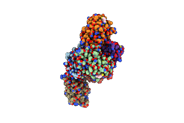
Structure Of Map Kinase Phosphatase 5 In Complex With 3,3-Dimethyl-1-((9-Propyl-5,6-Dihydrothieno[3,4-H]Quinazolin-2-Yl)Thio)Butan-2-One, An Allosteric Inhibitor
Organism: Homo sapiens
Method: X-RAY DIFFRACTION Resolution:2.30 Å Release Date: 2022-10-05 Classification: HYDROLASE/INHIBITOR Ligands: L8K |
Organism: Homo sapiens
Method: X-RAY DIFFRACTION
Release Date: 2022-10-05
Ligands: L8K
 |
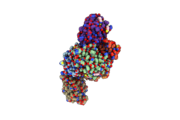
Structure Of Map Kinase Phosphatase 5 In Complex With 3,3-Dimethyl-1-((9-Propyl-5,6-Dihydrothieno[2,3-H]Quinazolin-2-Yl)Thio)Butan-2-One, An Allosteric Inhibitor
Organism: Homo sapiens
Method: X-RAY DIFFRACTION Resolution:3.14 Å Release Date: 2022-10-05 Classification: HYDROLASE/INHIBITOR Ligands: L8R |
Organism: Homo sapiens
Method: X-RAY DIFFRACTION
Release Date: 2022-10-05
Ligands: L8R
 |
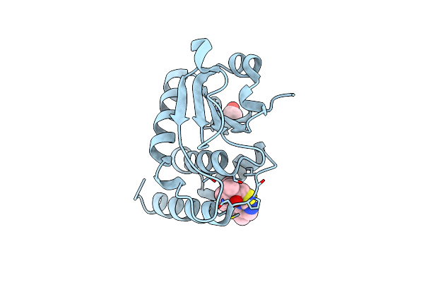
Structure Of Map Kinase Phosphatase 5 In Complex With 3,3-Dimethyl-1-((5,6-Dihydrobenzo[H]Quinazolin-2-Yl)Thio)Butan-2-One, An Allosteric Inhibitor
Organism: Homo sapiens
Method: X-RAY DIFFRACTION Resolution:2.51 Å Release Date: 2022-10-05 Classification: HYDROLASE/INHIBITOR Ligands: NUN, DTT |
Organism: Homo sapiens
Method: X-RAY DIFFRACTION
Release Date: 2022-10-05
Ligands: NUN, DTT
 |
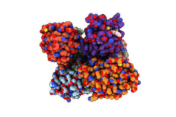
Structure Of Map Kinase Phosphatase 5 In Complex With 3,3-Dimethyl-1-((5,6-Dihydropyrido[2,3-H]Quinazolin-2-Yl)Thio)Butan-2-One, An Allosteric Inhibitor
Organism: Homo sapiens
Method: X-RAY DIFFRACTION Resolution:1.80 Å Release Date: 2022-10-05 Classification: HYDROLASE/INHIBITOR Ligands: NUU, ACT |
Organism: Homo sapiens
Method: X-RAY DIFFRACTION
Release Date: 2022-10-05
Ligands: NUU, ACT
 |
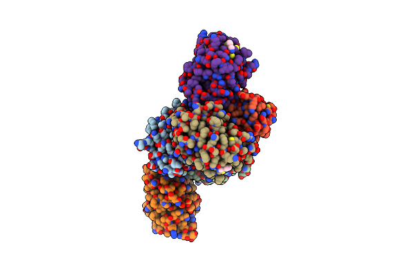
Structure Of Map Kinase Phosphatase 5 In Complex With 3,3-Dimethyl-1-((9-Chloro-5,6-Dihydrobenzo[H]Quinazolin-2-Yl)Thio)Butan-2-One, An Allosteric Inhibitor
Organism: Homo sapiens
Method: X-RAY DIFFRACTION Resolution:3.00 Å Release Date: 2022-10-05 Classification: HYDROLASE/INHIBITOR Ligands: NV3 |
Organism: Homo sapiens
Method: X-RAY DIFFRACTION
Release Date: 2022-10-05
Ligands: NV3
 |
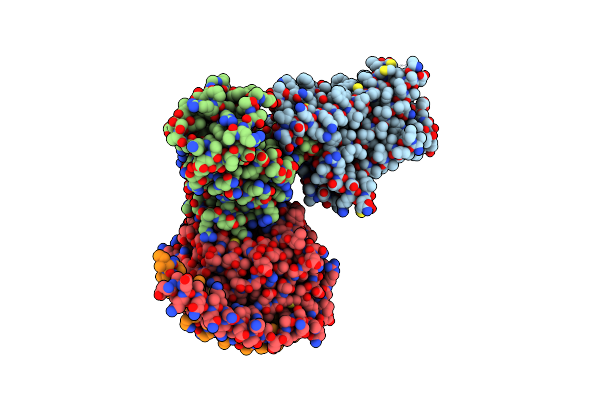
Structure Of Map Kinase Phosphatase 5 In Complex With 3,3-Dimethyl-1-((9-Propyl-5,6-Dihydrothieno[3,4-H]Quinazolin-2-Yl)Thio)Butan-2-One, An Allosteric Inhibitor
Organism: Homo sapiens
Method: X-RAY DIFFRACTION Resolution:2.70 Å Release Date: 2022-10-05 Classification: HYDROLASE/INHIBITOR Ligands: NVF |
Organism: Homo sapiens
Method: X-RAY DIFFRACTION
Release Date: 2022-10-05
Ligands: NVF
 |
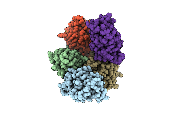
5-Ht2Ar Bound To A Novel Agonist In Complex With A Mini-Gq Protein And An Active-State Stabilizing Single-Chain Variable Fragment (Scfv16) Obtained By Cryo-Electron Microscopy (Cryoem)
Organism: Homo sapiens
Method: ELECTRON MICROSCOPY Release Date: 2022-07-06 Classification: MEMBRANE PROTEIN Ligands: 3IQ |
Organism: Homo sapiens
Method: ELECTRON MICROSCOPY
Release Date: 2022-07-06
Ligands: 3IQ
 |
Structure Of Map Kinase Phosphatase 5 In Complex With 3,3-Dimethyl-1-((9-(Methylthio)-5,6-Dihydrothieno[3,4-H]Quinazolin-2-Yl)Thio)Butan-2-One, An Allosteric Inhibitor
Organism: Homo sapiens
Method: X-RAY DIFFRACTION Resolution:2.70 Å Release Date: 2020-08-19 Classification: Hydrolase/Hydrolase Inhibitor Ligands: CJA, DTT, ACT |
Organism: Homo sapiens
Method: X-RAY DIFFRACTION
Release Date: 2020-08-19
Ligands: CJA, DTT, ACT
 |
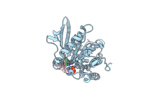
X-Ray Characterization Of Striatal-Enriched Protein Tyrosine Phosphatase Inhibitors
Organism: Homo sapiens
Method: X-RAY DIFFRACTION Resolution:2.15 Å Release Date: 2017-11-22 Classification: HYDROLASE Ligands: AXK |
Organism: Homo sapiens
Method: X-RAY DIFFRACTION
Release Date: 2017-11-22
Ligands: AXK
 |
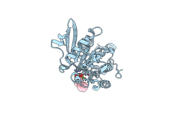
X-Ray Characterization Of Striatal-Enriched Protein Tyrosine Phosphatase Inhibitors
Organism: Homo sapiens
Method: X-RAY DIFFRACTION Resolution:2.10 Å Release Date: 2017-11-22 Classification: HYDROLASE Ligands: AY5 |
Organism: Homo sapiens
Method: X-RAY DIFFRACTION
Release Date: 2017-11-22
Ligands: AY5
 |
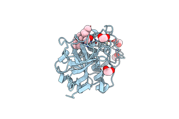
X-Ray Characterization Of Striatal-Enriched Protein Tyrosine Phosphatase Inhibitors
Organism: Homo sapiens
Method: X-RAY DIFFRACTION Resolution:2.05 Å Release Date: 2017-11-22 Classification: HYDROLASE Ligands: AY8 |
Organism: Homo sapiens
Method: X-RAY DIFFRACTION
Release Date: 2017-11-22
Ligands: AY8
 |
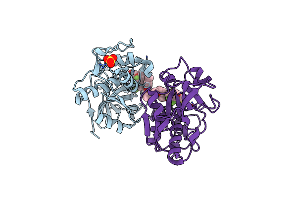
The Crystal Structure Of Cruzain In Complex With A Tetrafluorophenoxymethyl Ketone Inhibitor
Organism: Trypanosoma cruzi
Method: X-RAY DIFFRACTION Resolution:1.20 Å Release Date: 2010-03-02 Classification: HYDROLASE Ligands: KB2, EDO |
Organism: Trypanosoma cruzi
Method: X-RAY DIFFRACTION
Release Date: 2010-03-02
Ligands: KB2, EDO
 |
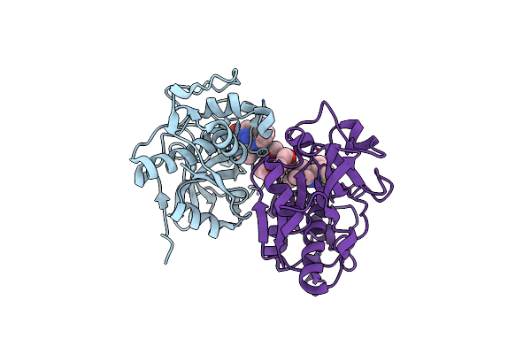
The Structure Of Cathepsin S With A Novel 2-Arylphenoxyacetaldehyde Inhibitor Derived By The Substrate Activity Screening (Sas) Method
Organism: Homo sapiens
Method: X-RAY DIFFRACTION Resolution:1.60 Å Release Date: 2007-05-22 Classification: HYDROLASE Ligands: SO4, TF5, PEU |
Organism: Homo sapiens
Method: X-RAY DIFFRACTION
Release Date: 2007-05-22
Ligands: SO4, TF5, PEU
 |
Crystal Structure Of Cathepsin S In Complex With A Nonpeptidic Inhibitor.
Organism: Homo sapiens
Method: X-RAY DIFFRACTION Resolution:1.50 Å Release Date: 2006-10-24 Classification: HYDROLASE Ligands: H7J, P15 |
Organism: Homo sapiens
Method: X-RAY DIFFRACTION
Release Date: 2006-10-24
Ligands: H7J, P15
 |
Crystal Structure Of Cathepsin S In Complex With A Nonpeptidic Inhibitor (Hexagonal Spacegroup)
Organism: Homo sapiens
Method: X-RAY DIFFRACTION Resolution:1.90 Å Release Date: 2006-10-24 Classification: HYDROLASE Ligands: SO4, H7J |
Organism: Homo sapiens
Method: X-RAY DIFFRACTION
Release Date: 2006-10-24
Ligands: SO4, H7J