

Search Count: 30
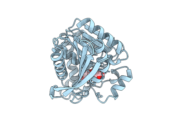 |
M. Tuberculosis Meets European Lead Factory: Identification And Structural Characterization Of Novel Rv0183 Inhibitors Using X-Ray Crystallography: Elf5
Organism: Mycobacterium tuberculosis h37rv
Method: X-RAY DIFFRACTION Resolution:1.60 Å Release Date: 2025-08-13 Classification: HYDROLASE Ligands: 8KE, I3F, CL |
Organism: Mycobacterium tuberculosis h37rv
Method: X-RAY DIFFRACTION
Release Date: 2025-08-13
Ligands: 8KE, I3F, CL
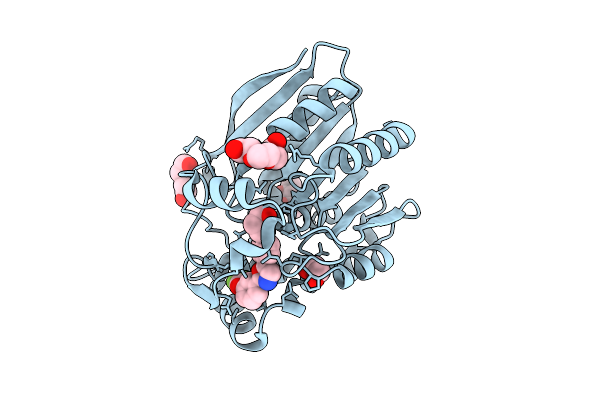 |
M. Tuberculosis Meets European Lead Factory: Identification And Structural Characterization Of Novel Rv0183 Inhibitors Using X-Ray Crystallography: Elf8
Organism: Mycobacterium tuberculosis h37rv
Method: X-RAY DIFFRACTION Resolution:1.80 Å Release Date: 2025-08-13 Classification: HYDROLASE Ligands: 8D5, MPD, EDO, PGE, PEG |
Organism: Mycobacterium tuberculosis h37rv
Method: X-RAY DIFFRACTION
Release Date: 2025-08-13
Ligands: 8D5, MPD, EDO, PGE, PEG
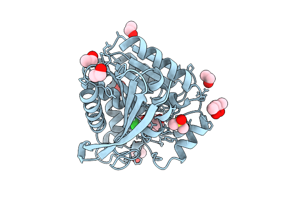 |
M. Tuberculosis Meets European Lead Factory: Identification And Structural Characterization Of Novel Rv0183 Inhibitors Using X-Ray Crystallography: Elf1
Organism: Mycobacterium tuberculosis h37rv
Method: X-RAY DIFFRACTION Resolution:1.25 Å Release Date: 2025-08-13 Classification: HYDROLASE Ligands: 7WW, PO4, MPD, DMS, EOH, EDO |
Organism: Mycobacterium tuberculosis h37rv
Method: X-RAY DIFFRACTION
Release Date: 2025-08-13
Ligands: 7WW, PO4, MPD, DMS, EOH, EDO
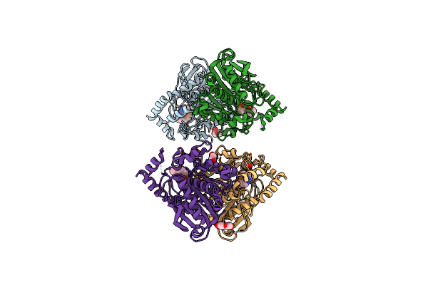 |
Structure Of Ester-Hydrolase Eh3 From The Metagenome Of Marine Sediments At Milazzo Harbor (Sicily, Italy) Complexed With A Derivative Of Bipyridine Phosphonate
Organism: Metagenome
Method: X-RAY DIFFRACTION Resolution:2.40 Å Release Date: 2023-07-19 Classification: HYDROLASE Ligands: PEG, ZK8, GOL |
Organism: Metagenome
Method: X-RAY DIFFRACTION
Release Date: 2023-07-19
Ligands: PEG, ZK8, GOL
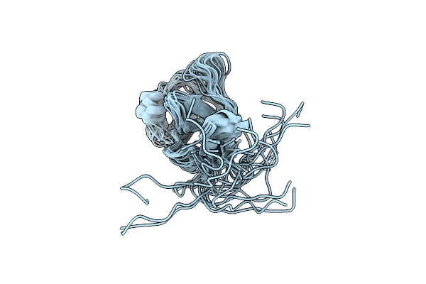 |
Nmr Solution Structure Of The Carbohydrate-Binding Module Family 5 (Cbm5) From Cellvibrio Japonicus Cjlpmo10A
Organism: Cellvibrio japonicus (strain ueda107)
Method: SOLUTION NMR Release Date: 2021-05-05 Classification: SUGAR BINDING PROTEIN |
Organism: Cellvibrio japonicus (strain ueda107)
Method: SOLUTION NMR
Release Date: 2021-05-05
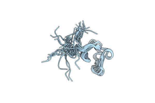 |
Nmr Solution Structure Of The Carbohydrate-Binding Module Family 73 (Cbm73) From Cellvibrio Japonicus Cjlpmo10A
Organism: Cellvibrio japonicus ueda107
Method: SOLUTION NMR Release Date: 2021-05-05 Classification: SUGAR BINDING PROTEIN |
Organism: Cellvibrio japonicus ueda107
Method: SOLUTION NMR
Release Date: 2021-05-05
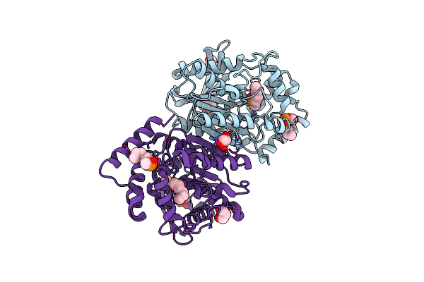 |
Structure Of Ester-Hydrolase Eh1Ab1 From The Metagenome Of Lake Arreo Complexed With A Derivative Of Methyl 4-Nitrophenyl Hexylphosphonate
Organism: Metagenome
Method: X-RAY DIFFRACTION Resolution:2.35 Å Release Date: 2019-12-25 Classification: HYDROLASE Ligands: GOL, MHH |
Organism: Metagenome
Method: X-RAY DIFFRACTION
Release Date: 2019-12-25
Ligands: GOL, MHH
 |
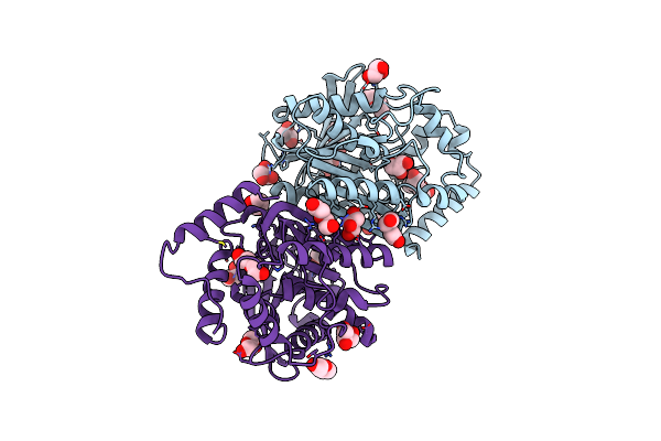
Structure Of Ester-Hydrolase Eh1Ab1 From The Metagenome Of Lake Arreo Complexed With A Derivative Of Bipyridine Phosphonate
Organism: Metagenome
Method: X-RAY DIFFRACTION Resolution:2.79 Å Release Date: 2019-12-25 Classification: HYDROLASE Ligands: GOL, ZK8, PEG |
Organism: Metagenome
Method: X-RAY DIFFRACTION
Release Date: 2019-12-25
Ligands: GOL, ZK8, PEG
 |
Structure Of Ester-Hydrolase Eh1Ab1 From The Metagenome Of Lake Arreo
Organism: Metagenome
Method: X-RAY DIFFRACTION Resolution:2.11 Å Release Date: 2019-11-27 Classification: HYDROLASE Ligands: PEG, GOL, MG |
Organism: Metagenome
Method: X-RAY DIFFRACTION
Release Date: 2019-11-27
Ligands: PEG, GOL, MG
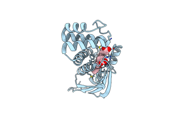 |
Crystal Structure Of Fe(Ii) Unliganded H-Nox Protein From K. Algicida
Organism: Kordia algicida ot-1
Method: X-RAY DIFFRACTION Resolution:1.90 Å Release Date: 2018-05-30 Classification: SIGNALING PROTEIN Ligands: HEM |
Organism: Kordia algicida ot-1
Method: X-RAY DIFFRACTION
Release Date: 2018-05-30
Ligands: HEM
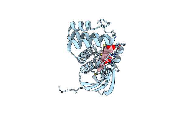 |
Crystal Structure Of Fe(Ii) Unliganded H-Nox Protein Mutant A71G From K. Algicida
Organism: Kordia algicida ot-1
Method: X-RAY DIFFRACTION Resolution:1.64 Å Release Date: 2018-05-30 Classification: SIGNALING PROTEIN Ligands: HEM |
Organism: Kordia algicida ot-1
Method: X-RAY DIFFRACTION
Release Date: 2018-05-30
Ligands: HEM
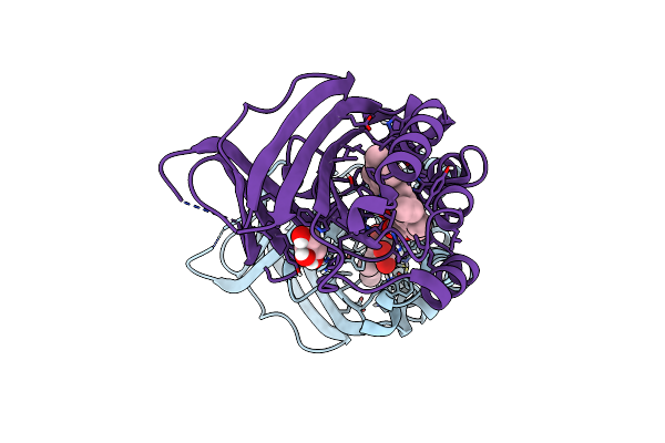 |
Joint Neutron/X-Ray Structure Of Dimeric Chlorite Dismutase From Cyanothece Sp. Pcc7425
Organism: Cyanothece sp. (strain pcc 7425 / atcc 29141)
Method: X-RAY DIFFRACTION, NEUTRON DIFFRACTION Resolution:2.000 Å, 2.350 Å Release Date: 2018-02-28 Classification: OXIDOREDUCTASE Ligands: HEM, OH, CL, GOL |
Organism: Cyanothece sp. (strain pcc 7425 / atcc 29141)
Method: X-RAY DIFFRACTION, NEUTRON DIFFRACTION
Release Date: 2018-02-28
Ligands: HEM, OH, CL, GOL
 |
Crystal Structure Of Dimeric Chlorite Dismutase From Cyanothece Sp. Pcc7425 At Ph 9.0 And 293 K.
Organism: Cyanothece sp. pcc 7425
Method: X-RAY DIFFRACTION Resolution:2.00 Å Release Date: 2018-01-31 Classification: OXIDOREDUCTASE Ligands: GOL, HEM, CL |
Organism: Cyanothece sp. pcc 7425
Method: X-RAY DIFFRACTION
Release Date: 2018-01-31
Ligands: GOL, HEM, CL
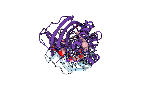 |
Crystal Structure Of Dimeric Chlorite Dismutase From Cyanothece Sp. Pcc7425 (Ph 6.5)
Organism: Cyanothece sp. pcc 7425
Method: X-RAY DIFFRACTION Resolution:1.30 Å Release Date: 2017-12-20 Classification: OXIDOREDUCTASE Ligands: HEM, SO4, GOL |
Organism: Cyanothece sp. pcc 7425
Method: X-RAY DIFFRACTION
Release Date: 2017-12-20
Ligands: HEM, SO4, GOL
 |
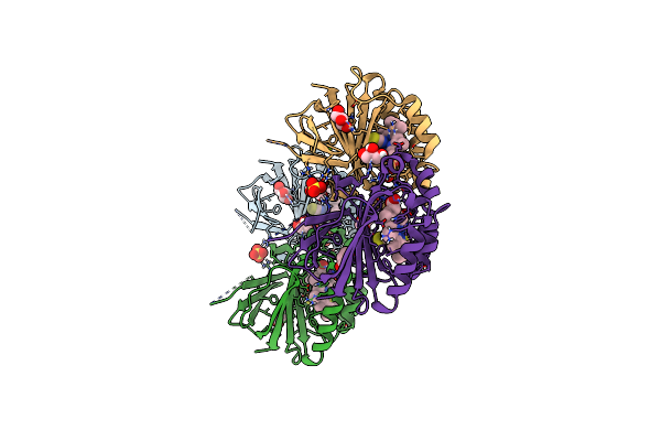
Crystal Structure Of Dimeric Chlorite Dismutase From Cyanothece Sp. Pcc7425 (Ph 8.5)
Organism: Cyanothece sp. (strain pcc 7425 / atcc 29141)
Method: X-RAY DIFFRACTION Resolution:1.55 Å Release Date: 2017-06-21 Classification: OXIDOREDUCTASE Ligands: MRD, HEM, OH, MPD |
Organism: Cyanothece sp. (strain pcc 7425 / atcc 29141)
Method: X-RAY DIFFRACTION
Release Date: 2017-06-21
Ligands: MRD, HEM, OH, MPD
 |
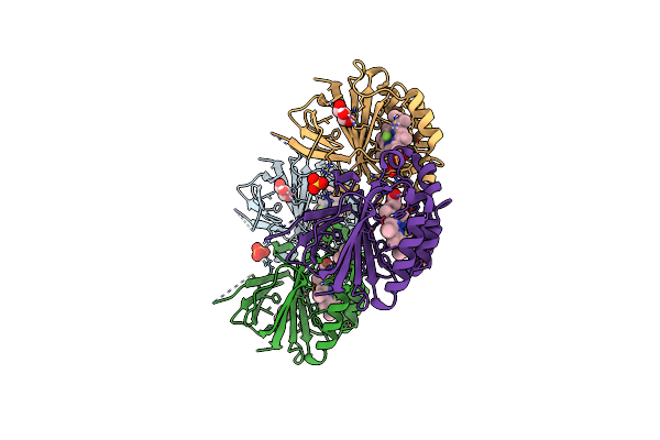
Crystal Structure Of Dimeric Chlorite Dismutase From Cyanothece Sp. Pcc7425 In Complex With Isothiocyanate
Organism: Cyanothece sp. (strain pcc 7425 / atcc 29141)
Method: X-RAY DIFFRACTION Resolution:1.28 Å Release Date: 2017-06-21 Classification: OXIDOREDUCTASE Ligands: IS8, HEM, GOL, SO4, MG |
Organism: Cyanothece sp. (strain pcc 7425 / atcc 29141)
Method: X-RAY DIFFRACTION
Release Date: 2017-06-21
Ligands: IS8, HEM, GOL, SO4, MG
 |
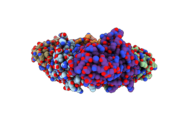
Crystal Structure Of Dimeric Chlorite Dismutase From Cyanothece Sp. Pcc7425 In Complex With Fluoride
Organism: Cyanothece sp. pcc 7425
Method: X-RAY DIFFRACTION Resolution:1.18 Å Release Date: 2017-06-21 Classification: OXIDOREDUCTASE Ligands: HEM, F, GOL, SO4 |
Organism: Cyanothece sp. pcc 7425
Method: X-RAY DIFFRACTION
Release Date: 2017-06-21
Ligands: HEM, F, GOL, SO4
 |
Crystal Structure Of Fe(Ii) Unliganded H-Nox Protein From C. Subterraneus
Organism: Caldanaerobacter subterraneus subsp. tengcongensis (strain dsm 15242 / jcm 11007 / nbrc 100824 / mb4)
Method: X-RAY DIFFRACTION Resolution:2.31 Å Release Date: 2016-07-06 Classification: SIGNALING PROTEIN Ligands: HEM |
 |
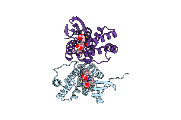
Crystal Structure Of Fe(Ii) No-Bound H-Nox Protein From C. Subterraneus
Organism: Caldanaerobacter subterraneus subsp. tengcongensis (strain dsm 15242 / jcm 11007 / nbrc 100824 / mb4)
Method: X-RAY DIFFRACTION Resolution:1.95 Å Release Date: 2016-07-06 Classification: SIGNALING PROTEIN Ligands: HEM, NO, IOD |
Organism: Caldanaerobacter subterraneus subsp. tengcongensis (strain dsm 15242 / jcm 11007 / nbrc 100824 / mb4)
Method: X-RAY DIFFRACTION
Release Date: 2016-07-06
Ligands: HEM, NO, IOD
 |
Crystal Structure Of Fe(Ii) Co-Bound H-Nox Protein From C. Subterraneus
Organism: Caldanaerobacter subterraneus subsp. tengcongensis (strain dsm 15242 / jcm 11007 / nbrc 100824 / mb4)
Method: X-RAY DIFFRACTION Resolution:1.95 Å Release Date: 2016-07-06 Classification: SIGNALING PROTEIN Ligands: HEM, CMO |
Organism: Caldanaerobacter subterraneus subsp. tengcongensis (strain dsm 15242 / jcm 11007 / nbrc 100824 / mb4)
Method: X-RAY DIFFRACTION
Release Date: 2016-07-06
Ligands: HEM, CMO