
Search Count: 76
All
Selected
 |
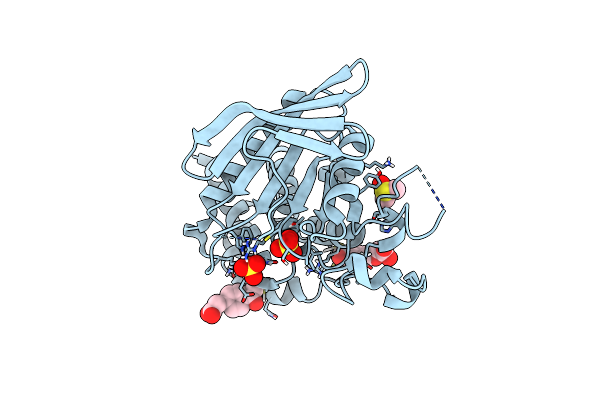
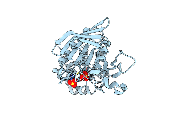
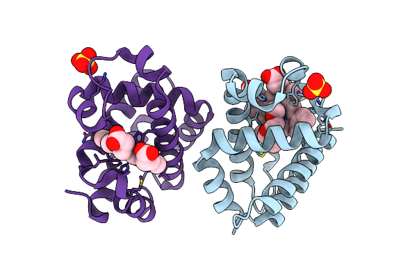
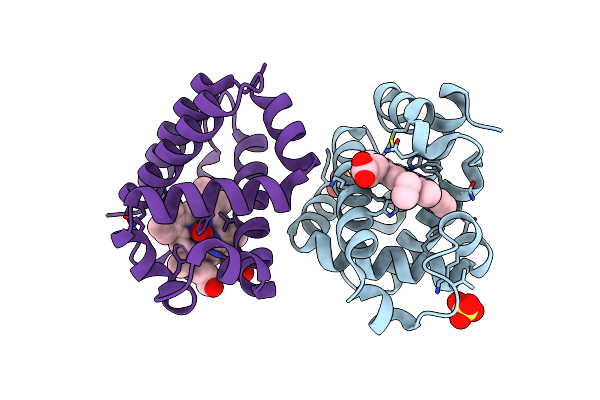
Crystal Structure Of Enterovirus D68 3C Protease Determined Via Sulfur Phasing
Organism: Enterovirus d68
Method: X-RAY DIFFRACTION Resolution:1.80 Å Release Date: 2026-04-01 Classification: VIRAL PROTEIN Ligands: CL |
 |
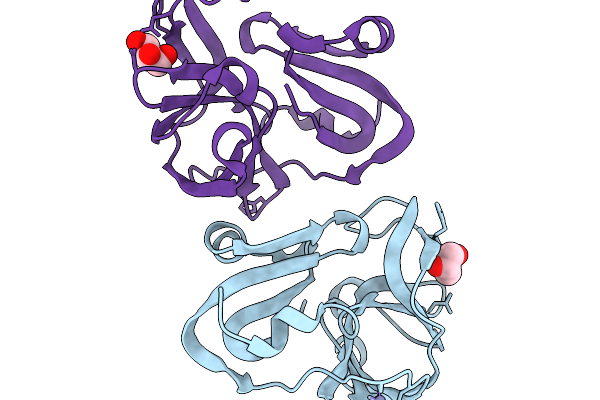
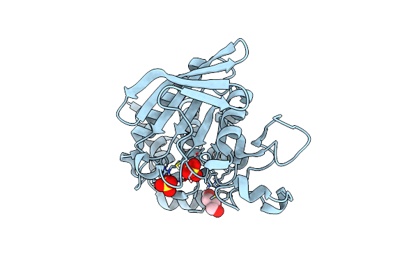
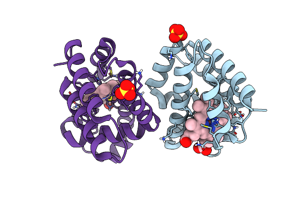
Crystal Structure Of Zika Virus Ns2B-Ns3 Protease Determined Via Sulfur Phasing
Organism: Zika virus
Method: X-RAY DIFFRACTION Resolution:1.77 Å Release Date: 2026-04-01 Classification: VIRAL PROTEIN |
 |
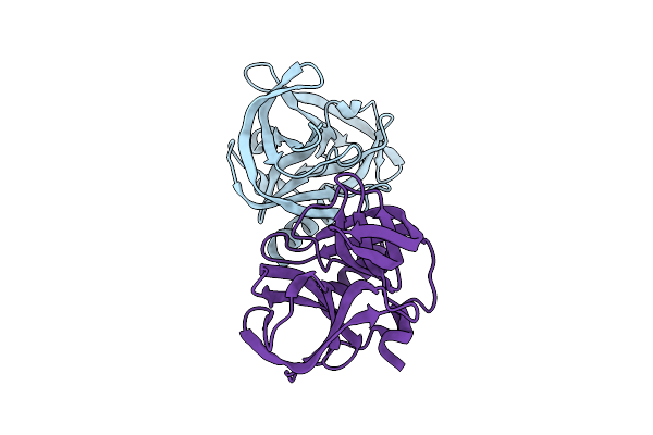
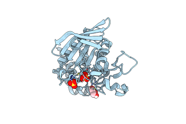
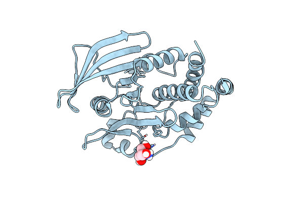
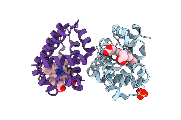
Crystal Structure Of Coxsackievirus A16 (G-10) 2A Protease Determined Via Sulfur Phasing
Organism: Coxsackievirus a16
Method: X-RAY DIFFRACTION Resolution:1.80 Å Release Date: 2026-03-25 Classification: VIRAL PROTEIN Ligands: GOL, ZN |
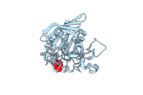 |
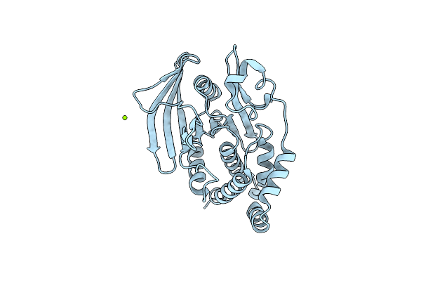
Organism: Homo sapiens
Method: X-RAY DIFFRACTION Resolution:1.27 Å Release Date: 2024-12-11 Classification: HYDROLASE Ligands: CIT |
 |
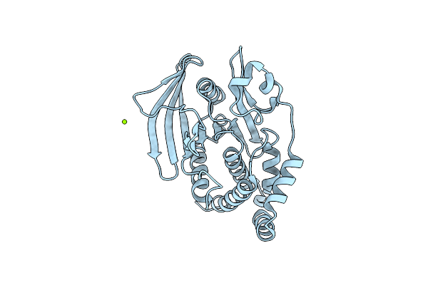
Step (Ptpn5) With Active-Site Disulfide Bond And Allosteric-Site Loop Shift
Organism: Homo sapiens
Method: X-RAY DIFFRACTION Resolution:1.75 Å Release Date: 2024-12-11 Classification: HYDROLASE Ligands: SO4 |
 |
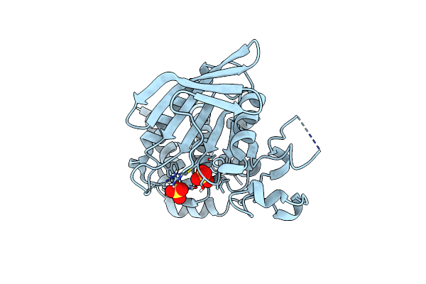
Step (Ptpn5) With Active-Site Disulfide Bond And Covalent Ligand Bound To Distal C505 And C518
Organism: Homo sapiens
Method: X-RAY DIFFRACTION Resolution:1.60 Å Release Date: 2024-12-11 Classification: HYDROLASE Ligands: A1BHT, DMS, SO4 |
 |
Crystal Structure Of Wild-Type Human Ptp1B (Ptpn1) At Room Temperature (298 K)
Organism: Homo sapiens
Method: X-RAY DIFFRACTION Resolution:1.94 Å Release Date: 2024-08-21 Classification: HYDROLASE Ligands: MG |
 |
Crystal Structure Of I19V Mutant Human Ptp1B (Ptpn1) At Room Temperature (298 K)
Organism: Homo sapiens
Method: X-RAY DIFFRACTION Resolution:1.99 Å Release Date: 2024-08-21 Classification: HYDROLASE Ligands: MG |
 |
Crystal Structure Of Q78R Mutant Human Ptp1B (Ptpn1) At Room Temperature (298 K)
Organism: Homo sapiens
Method: X-RAY DIFFRACTION Resolution:2.30 Å Release Date: 2024-08-21 Classification: HYDROLASE Ligands: MG |
 |
Crystal Structure Of D245G Mutant Human Ptp1B (Ptpn1) At Room Temperature (298 K)
Organism: Homo sapiens
Method: X-RAY DIFFRACTION Resolution:1.65 Å Release Date: 2024-08-21 Classification: HYDROLASE Ligands: MG |
 |
Crystal Structure Of Human Step (Ptpn5) At Cryogenic Temperature (100 K) And Ambient Pressure (0.1 Mpa)
Organism: Homo sapiens
Method: X-RAY DIFFRACTION Resolution:1.71 Å Release Date: 2023-06-21 Classification: HYDROLASE Ligands: SO4, GOL |
 |
Crystal Structure Of Human Step (Ptpn5) At Physiological Temperature (310 K) And Ambient Pressure (0.1 Mpa)
Organism: Homo sapiens
Method: X-RAY DIFFRACTION Resolution:1.96 Å Release Date: 2023-06-21 Classification: HYDROLASE Ligands: SO4 |
 |
Crystal Structure Of Human Step (Ptpn5) At Cryogenic Temperature (100 K) And High Pressure (205 Mpa)
Organism: Homo sapiens
Method: X-RAY DIFFRACTION Resolution:1.84 Å Release Date: 2023-06-21 Classification: HYDROLASE Ligands: SO4, GOL |
 |
Room-Temperature Serial Synchrotron Crystallography (Ssx) Structure Of Apo Ptp1B
Organism: Homo sapiens
Method: X-RAY DIFFRACTION Resolution:2.40 Å Release Date: 2022-08-17 Classification: HYDROLASE Ligands: TRS |
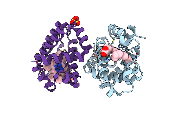 |
Serial Laue Crystallography Structure Of Dehaloperoxidase B From Amphitrite Ornata
Organism: Amphitrite ornata
Method: X-RAY DIFFRACTION Resolution:2.01 Å Release Date: 2021-11-03 Classification: OXIDOREDUCTASE Ligands: SO4, HEM |
 |
Organism: Amphitrite ornata
Method: X-RAY DIFFRACTION Resolution:1.75 Å Release Date: 2021-10-20 Classification: METAL BINDING PROTEIN Ligands: HEM, SO4, OXY, PG0 |
 |
Organism: Amphitrite ornata
Method: X-RAY DIFFRACTION, NEUTRON DIFFRACTION Resolution:2.2000 Å, 2.2150 Å Release Date: 2021-10-13 Classification: OXIDOREDUCTASE Ligands: HEM, OXY, SO4, DOD |
 |
Organism: Amphitrite ornata
Method: X-RAY DIFFRACTION Resolution:1.45 Å Release Date: 2021-10-06 Classification: OXIDOREDUCTASE Ligands: HEM, SO4 |
 |
Organism: Amphitrite ornata
Method: X-RAY DIFFRACTION Resolution:1.85 Å Release Date: 2021-10-06 Classification: OXIDOREDUCTASE Ligands: HEM, SO4 |
 |
Organism: Amphitrite ornata
Method: X-RAY DIFFRACTION Resolution:1.85 Å Release Date: 2021-10-06 Classification: OXIDOREDUCTASE Ligands: HEM, SO4, OXY |