

Search Count: 533
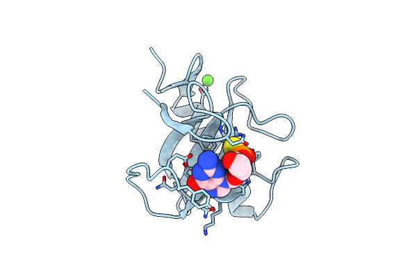 |
Ribonuclease T1 Complexed With 2',3'-Cgps, 1 Day
Organism: Aspergillus oryzae
Method: X-RAY DIFFRACTION Resolution:2.20 Å Release Date: 1998-02-25 Classification: ENDORIBONUCLEASE Ligands: CA, SGP |
Organism: Aspergillus oryzae
Method: X-RAY DIFFRACTION
Release Date: 1998-02-25
Ligands: CA, SGP
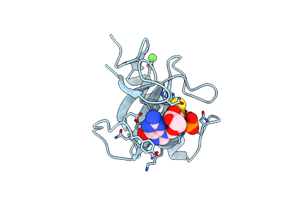 |
Ribonuclease T1 Complexed With 2',3'-Cgps + 3'-Gmp, 4 Days
Organism: Aspergillus oryzae
Method: X-RAY DIFFRACTION Resolution:1.90 Å Release Date: 1998-08-12 Classification: ENDORIBONUCLEASE Ligands: CA, SGP, 3GP |
Organism: Aspergillus oryzae
Method: X-RAY DIFFRACTION
Release Date: 1998-08-12
Ligands: CA, SGP, 3GP
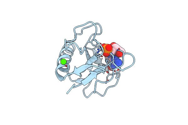 |
Ribonuclease 1 Complex With 2'Gmp
Organism: Aspergillus oryzae
Method: X-RAY DIFFRACTION Resolution:1.90 Å Release Date: 1999-02-16 Classification: ENDORIBONUCLEASE Ligands: CA, 2GP |
Organism: Aspergillus oryzae
Method: X-RAY DIFFRACTION
Release Date: 1999-02-16
Ligands: CA, 2GP
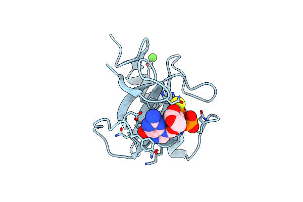 |
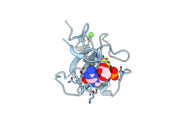
Ribonuclease T1/2',3'-Cgps And 3'-Gmp, 2 Days
Organism: Aspergillus oryzae
Method: X-RAY DIFFRACTION Resolution:1.80 Å Release Date: 1998-08-12 Classification: ENDORIBONUCLEASE Ligands: CA, SGP, 3GP |
Organism: Aspergillus oryzae
Method: X-RAY DIFFRACTION
Release Date: 1998-08-12
Ligands: CA, SGP, 3GP
 |
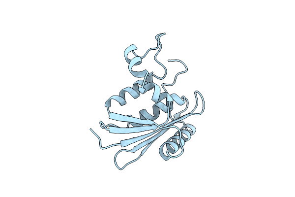
Ribonuclease T1 Complexed With 2',3'-Cgps + 3'-Gmp, 7 Days
Organism: Aspergillus oryzae
Method: X-RAY DIFFRACTION Resolution:1.65 Å Release Date: 1998-08-12 Classification: ENDORIBONUCLEASE Ligands: CA, 3GP, SGP |
Organism: Aspergillus oryzae
Method: X-RAY DIFFRACTION
Release Date: 1998-08-12
Ligands: CA, 3GP, SGP
 |
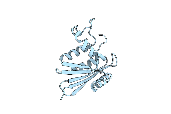
E. Coli Ribonuclease Hi D134H Mutant
Organism: Escherichia coli
Method: X-RAY DIFFRACTION Resolution:1.90 Å Release Date: 1997-03-12 Classification: ENDORIBONUCLEASE |
Organism: Escherichia coli
Method: X-RAY DIFFRACTION
Release Date: 1997-03-12
 |
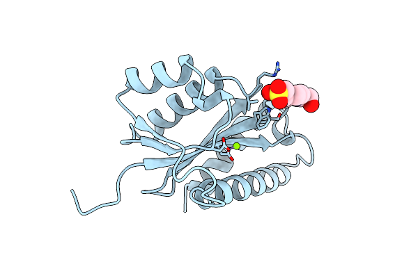
E. Coli Ribonuclease Hi D134A Mutant
Organism: Escherichia coli
Method: X-RAY DIFFRACTION Resolution:1.80 Å Release Date: 1997-03-12 Classification: ENDORIBONUCLEASE |
Organism: Escherichia coli
Method: X-RAY DIFFRACTION
Release Date: 1997-03-12
 |
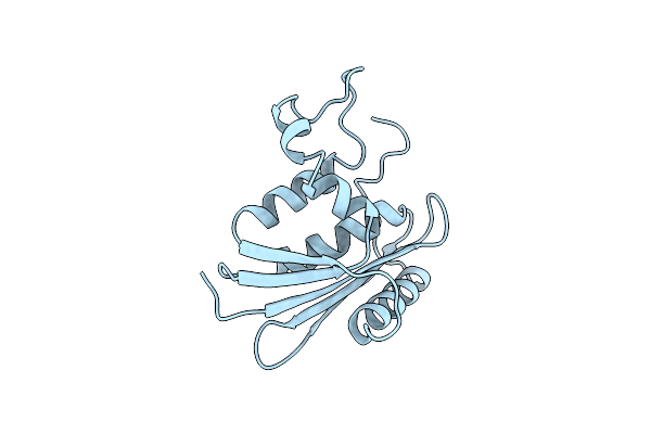
Asv Integrase Core Domain With Mg(Ii) Cofactor And Hepes Ligand, High Mg Concentration Form
Organism: Rous sarcoma virus (strain schmidt-ruppin)
Method: X-RAY DIFFRACTION Resolution:1.70 Å Release Date: 1996-04-03 Classification: ENDORIBONUCLEASE Ligands: MG, EPE |
Organism: Rous sarcoma virus (strain schmidt-ruppin)
Method: X-RAY DIFFRACTION
Release Date: 1996-04-03
Ligands: MG, EPE
 |
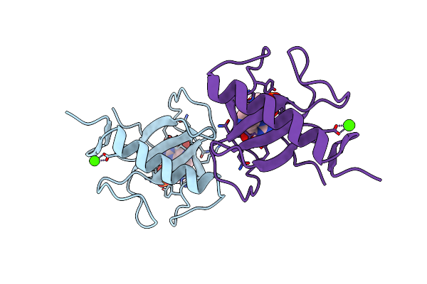
E. Coli Ribonuclease Hi D134N Mutant
Organism: Escherichia coli
Method: X-RAY DIFFRACTION Resolution:1.90 Å Release Date: 1997-03-12 Classification: ENDORIBONUCLEASE |
Organism: Escherichia coli
Method: X-RAY DIFFRACTION
Release Date: 1997-03-12
 |
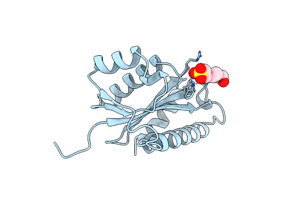
Ribonuclease T1, Phe 100 To Ala Mutant Complexed With 2' Gmp
Organism: Aspergillus oryzae
Method: X-RAY DIFFRACTION Resolution:1.80 Å Release Date: 1996-08-17 Classification: ENDORIBONUCLEASE Ligands: CA, 2GP |
Organism: Aspergillus oryzae
Method: X-RAY DIFFRACTION
Release Date: 1996-08-17
Ligands: CA, 2GP
 |
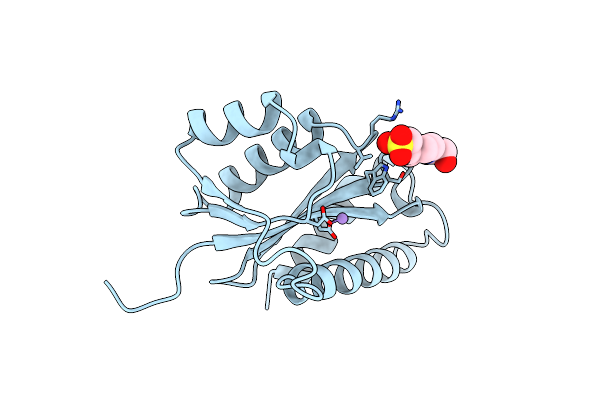
Asv Integrase Core Domain With Mg(Ii) Cofactor And Hepes Ligand, Low Mg Concentration Form
Organism: Rous sarcoma virus (strain schmidt-ruppin)
Method: X-RAY DIFFRACTION Resolution:2.20 Å Release Date: 1996-04-03 Classification: ENDORIBONUCLEASE Ligands: EPE |
Organism: Rous sarcoma virus (strain schmidt-ruppin)
Method: X-RAY DIFFRACTION
Release Date: 1996-04-03
Ligands: EPE
 |
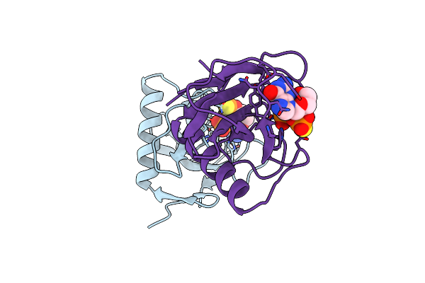
Asv Integrase Core Domain With Mn(Ii) Cofactor And Hepes Ligand, High Mg Concentration Form
Organism: Rous sarcoma virus (strain schmidt-ruppin)
Method: X-RAY DIFFRACTION Resolution:2.05 Å Release Date: 1996-04-03 Classification: ENDORIBONUCLEASE Ligands: MN, EPE |
Organism: Rous sarcoma virus (strain schmidt-ruppin)
Method: X-RAY DIFFRACTION
Release Date: 1996-04-03
Ligands: MN, EPE
 |
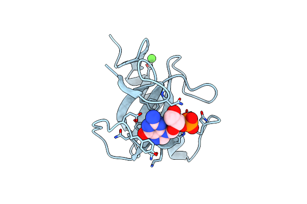
Ribonuclease T1/2',3'-Cgps, Non-Productive
Organism: Aspergillus oryzae
Method: X-RAY DIFFRACTION Resolution:2.00 Å Release Date: 1998-03-18 Classification: ENDORIBONUCLEASE Ligands: PO4, SGP |
Organism: Aspergillus oryzae
Method: X-RAY DIFFRACTION
Release Date: 1998-03-18
Ligands: PO4, SGP
 |
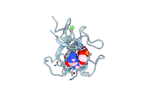
Ribonuclease T1/3'-Gmp, 9 Weeks
Organism: Aspergillus oryzae
Method: X-RAY DIFFRACTION Resolution:1.80 Å Release Date: 1998-03-18 Classification: ENDORIBONUCLEASE Ligands: CA, 3GP |
Organism: Aspergillus oryzae
Method: X-RAY DIFFRACTION
Release Date: 1998-03-18
Ligands: CA, 3GP
 |
Ribonuclease T1/3'-Gmp, 15 Weeks
Organism: Aspergillus oryzae
Method: X-RAY DIFFRACTION Resolution:2.20 Å Release Date: 1998-03-18 Classification: ENDORIBONUCLEASE Ligands: CA, 3GP |
Organism: Aspergillus oryzae
Method: X-RAY DIFFRACTION
Release Date: 1998-03-18
Ligands: CA, 3GP
 |
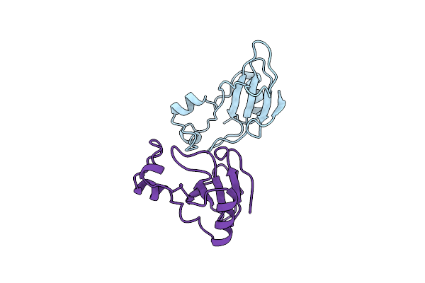
Ribonuclease T1: Free His92Gln Mutant
Organism: Aspergillus oryzae
Method: X-RAY DIFFRACTION Resolution:1.70 Å Release Date: 1998-07-15 Classification: ENDORIBONUCLEASE Ligands: CA |
Organism: Aspergillus oryzae
Method: X-RAY DIFFRACTION
Release Date: 1998-07-15
Ligands: CA
 |
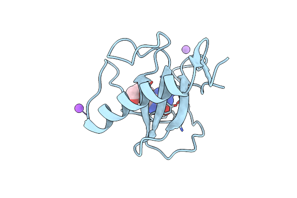
Structure Of Binase Mutant His 101 Asn
Organism: Bacillus intermedius
Method: X-RAY DIFFRACTION Resolution:2.20 Å Release Date: 1997-03-12 Classification: ENDORIBONUCLEASE |
Organism: Bacillus intermedius
Method: X-RAY DIFFRACTION
Release Date: 1997-03-12
 |
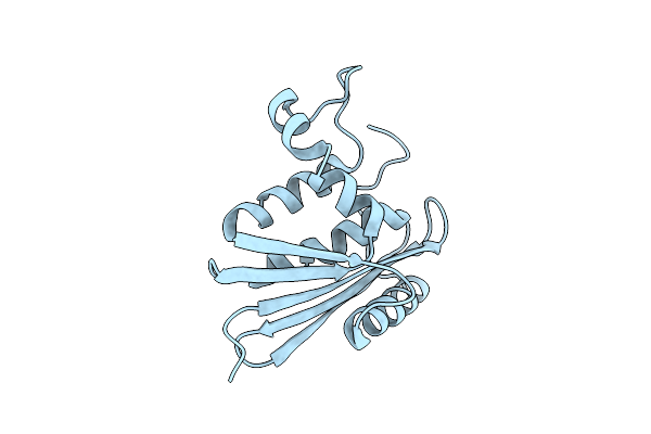
Ribonuclease T1 Carboxymethylated At Glu 58 In Complex With 2'Gmp
Organism: Aspergillus oryzae
Method: X-RAY DIFFRACTION Resolution:1.80 Å Release Date: 1996-07-11 Classification: HYDROLASE (ENDORIBONUCLEASE) |
Organism: Aspergillus oryzae
Method: X-RAY DIFFRACTION
Release Date: 1996-07-11
 |
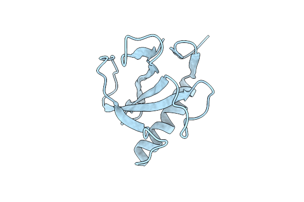
Structural Study Of Mutants Of Escherichia Coli Ribonuclease Hi With Enhanced Thermostability
Organism: Escherichia coli
Method: X-RAY DIFFRACTION Resolution:1.80 Å Release Date: 1994-01-31 Classification: HYDROLASE(ENDORIBONUCLEASE) |
Organism: Escherichia coli
Method: X-RAY DIFFRACTION
Release Date: 1994-01-31
 |
His 92 Ala Mutation In Ribonuclease T1 Induces Segmental Flexibility. An X-Ray Study
Organism: Aspergillus oryzae
Method: X-RAY DIFFRACTION Resolution:2.20 Å Release Date: 1992-01-15 Classification: HYDROLASE(ENDORIBONUCLEASE) |
Organism: Aspergillus oryzae
Method: X-RAY DIFFRACTION
Release Date: 1992-01-15