

Search Count: 29
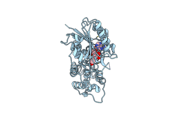 |
Crystal Structure Of Iridoid Synthase From Cantharanthus Roseus In Complex With Nadp+
Organism: Catharanthus roseus
Method: X-RAY DIFFRACTION Resolution:2.10 Å Release Date: 2015-11-25 Classification: OXIDOREDUCTASE Ligands: NAP |
Organism: Catharanthus roseus
Method: X-RAY DIFFRACTION
Release Date: 2015-11-25
Ligands: NAP
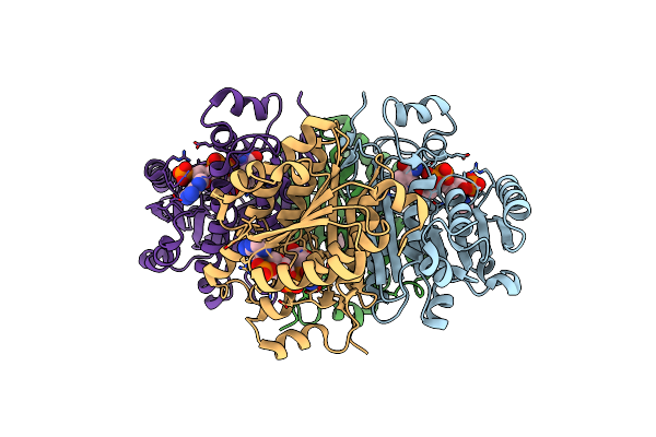 |
Substrate Fingerprint And The Structure Of Nadp+ Dependent Serine Dehydrogenase From Saccharomyces Cerevisiae Complexed With Nadp+
Organism: Saccharomyces cerevisiae
Method: X-RAY DIFFRACTION Resolution:2.60 Å Release Date: 2011-06-01 Classification: OXIDOREDUCTASE Ligands: NAP |
Organism: Saccharomyces cerevisiae
Method: X-RAY DIFFRACTION
Release Date: 2011-06-01
Ligands: NAP
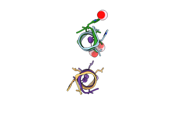 |
Gramicidin D Complex With Sodium Iodide
Organism: Bacillus brevis
Method: X-RAY DIFFRACTION Resolution:1.25 Å Release Date: 2010-07-28 Classification: ANTIBIOTIC |
Organism: Bacillus brevis
Method: X-RAY DIFFRACTION
Release Date: 2010-07-28
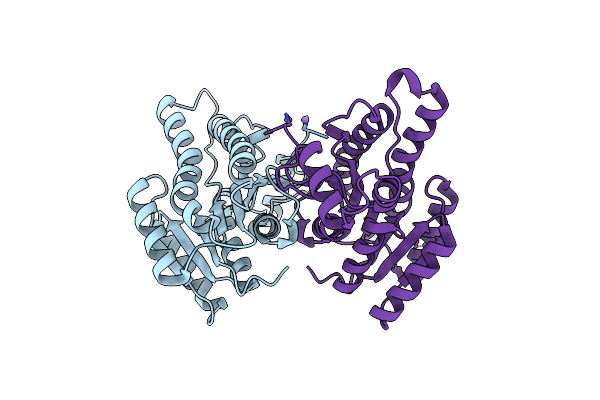 |
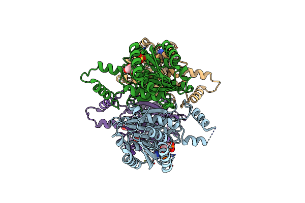
Apo Structure Of The Short Chain Oxidoreductase Q9Hya2 From Pseudomonas Aeruginosa Pao1 Containing An Atypical Catalytic Center
Organism: Pseudomonas aeruginosa
Method: X-RAY DIFFRACTION Resolution:2.32 Å Release Date: 2010-04-14 Classification: OXIDOREDUCTASE Ligands: CL, MN |
Organism: Pseudomonas aeruginosa
Method: X-RAY DIFFRACTION
Release Date: 2010-04-14
Ligands: CL, MN
 |
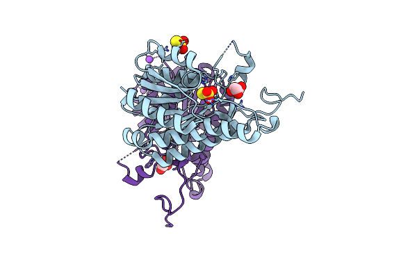
Nadph Bound Structure Of The Short Chain Oxidoreductase Q9Hya2 From Pseudomonas Aeruginosa Pao1 Containing An Atypical Catalytic Center
Organism: Pseudomonas aeruginosa
Method: X-RAY DIFFRACTION Resolution:2.30 Å Release Date: 2010-04-14 Classification: OXIDOREDUCTASE Ligands: NDP, CL, SO4, GOL |
Organism: Pseudomonas aeruginosa
Method: X-RAY DIFFRACTION
Release Date: 2010-04-14
Ligands: NDP, CL, SO4, GOL
 |
Fingerprint And Structural Analysis Of A Apo Scor Enzyme From Clostridium Thermocellum
Organism: Clostridium thermocellum atcc 27405
Method: X-RAY DIFFRACTION Resolution:1.70 Å Release Date: 2009-03-17 Classification: OXIDOREDUCTASE Ligands: GOL, THJ, NA |
Organism: Clostridium thermocellum atcc 27405
Method: X-RAY DIFFRACTION
Release Date: 2009-03-17
Ligands: GOL, THJ, NA
 |
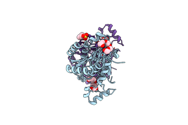
Fingerprint And Structural Analysis Of A Scor Enzyme With Its Bound Cofactor From Clostridium Thermocellum
Organism: Clostridium thermocellum atcc 27405
Method: X-RAY DIFFRACTION Resolution:2.10 Å Release Date: 2009-03-17 Classification: OXIDOREDUCTASE Ligands: NAD, GOL, THJ, NA |
Organism: Clostridium thermocellum atcc 27405
Method: X-RAY DIFFRACTION
Release Date: 2009-03-17
Ligands: NAD, GOL, THJ, NA
 |
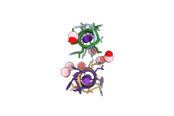
Structure Of Aquifex Aeolicus Fabg 3-Oxoacyl-(Acyl-Carrier Protein) Reductase
Organism: Aquifex aeolicus
Method: X-RAY DIFFRACTION Resolution:1.80 Å Release Date: 2007-05-01 Classification: OXIDOREDUCTASE Ligands: 1PE, MES |
Organism: Aquifex aeolicus
Method: X-RAY DIFFRACTION
Release Date: 2007-05-01
Ligands: 1PE, MES
 |
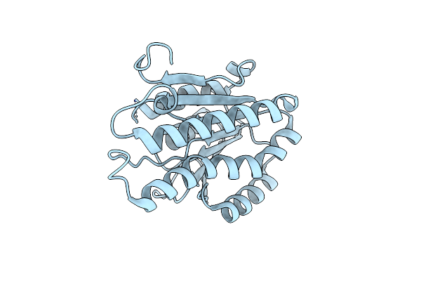
Gramicidin D Complex With Ki
Organism: Brevibacillus brevis
Method: X-RAY DIFFRACTION Resolution:0.80 Å Release Date: 2007-01-23 Classification: ANTIBIOTIC Ligands: K, IOD, MOH |
Organism: Brevibacillus brevis
Method: X-RAY DIFFRACTION
Release Date: 2007-01-23
Ligands: K, IOD, MOH
 |
Crystal Structure Of E. Coli Enta, A 2,3-Dihydrodihydroxy Benzoate Dehydrogenase
Organism: Escherichia coli
Method: X-RAY DIFFRACTION Resolution:2.00 Å Release Date: 2006-06-27 Classification: OXIDOREDUCTASE |
Organism: Escherichia coli
Method: X-RAY DIFFRACTION
Release Date: 2006-06-27
 |
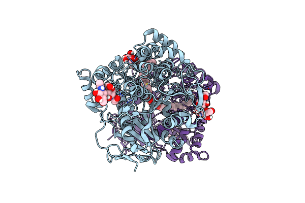
Gramicidin D From Bacillus Brevis (Ethanol Solvate)
Organism: Bacillus brevis
Method: X-RAY DIFFRACTION Resolution:1.14 Å Release Date: 2005-09-15 Classification: ANTIBIOTIC Ligands: RB, CL, EOH |
Organism: Bacillus brevis
Method: X-RAY DIFFRACTION
Release Date: 2005-09-15
Ligands: RB, CL, EOH
 |
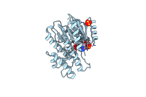
Cholesterol Esterase (Candida Cylindracea) Crystal Structure At 1.4A Resolution
Organism: Candida cylindracea
Method: X-RAY DIFFRACTION Resolution:1.40 Å Release Date: 2003-01-07 Classification: HYDROLASE Ligands: F23 |
Organism: Candida cylindracea
Method: X-RAY DIFFRACTION
Release Date: 2003-01-07
Ligands: F23
 |
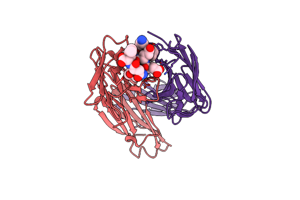
Crystal Structure Of Porcine Testicular Carbonyl Reductase/ 20Beta-Hydroxysteroid Dehydrogenase
Organism: Sus scrofa
Method: X-RAY DIFFRACTION Resolution:2.30 Å Release Date: 2002-11-13 Classification: OXIDOREDUCTASE Ligands: SO4, NDP |
Organism: Sus scrofa
Method: X-RAY DIFFRACTION
Release Date: 2002-11-13
Ligands: SO4, NDP
 |
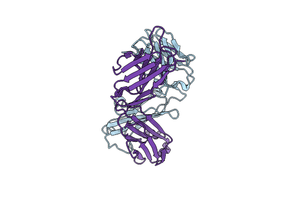
Fab Fragment Of Monoclonal Antibody (Lnkb-2) Against Human Interleukin-2 In Complex With Antigenic Peptide
Organism: Mus musculus
Method: X-RAY DIFFRACTION Resolution:2.60 Å Release Date: 2001-07-11 Classification: IMMUNE SYSTEM |
Organism: Mus musculus
Method: X-RAY DIFFRACTION
Release Date: 2001-07-11
 |
Fab (Lnkb-2) Of Monoclonal Antibody, Crystal Structure
Organism: Mus musculus
Method: X-RAY DIFFRACTION Resolution:2.20 Å Release Date: 2001-07-05 Classification: IMMUNE SYSTEM |
Organism: Mus musculus
Method: X-RAY DIFFRACTION
Release Date: 2001-07-05
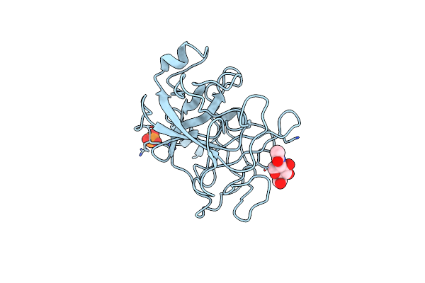 |
Bovine Duodenase(New Serine Protease), Crystal Structure
Organism: Bos taurus
Method: X-RAY DIFFRACTION Resolution:2.40 Å Release Date: 2001-04-14 Classification: HYDROLASE Ligands: NAG, PO4 |
Organism: Bos taurus
Method: X-RAY DIFFRACTION
Release Date: 2001-04-14
Ligands: NAG, PO4
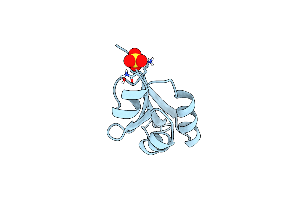 |
Ustilago Maydis Killer Toxin Kp6 Alpha-Subunit
Organism: Ustilago maydis
Method: X-RAY DIFFRACTION Resolution:1.80 Å Release Date: 1999-07-21 Classification: TOXIN Ligands: SO4 |
Organism: Ustilago maydis
Method: X-RAY DIFFRACTION
Release Date: 1999-07-21
Ligands: SO4
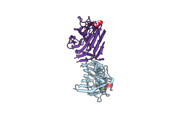 |
The Structure Of The Pea Lectin-D-Glucopyranose Complex
Organism: Pisum sativum
Method: X-RAY DIFFRACTION Resolution:1.90 Å Release Date: 1998-12-16 Classification: SUGAR BINDING PROTEIN Ligands: GLC, CA, MN |
Organism: Pisum sativum
Method: X-RAY DIFFRACTION
Release Date: 1998-12-16
Ligands: GLC, CA, MN
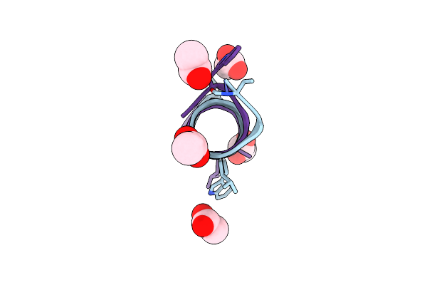 |
Gramicidin D From Bacillus Brevis (Active Form)
Organism: Brevibacillus brevis
Method: X-RAY DIFFRACTION Resolution:1.70 Å Release Date: 1998-10-14 Classification: ANTIBIOTIC Ligands: ACY |
Organism: Brevibacillus brevis
Method: X-RAY DIFFRACTION
Release Date: 1998-10-14
Ligands: ACY
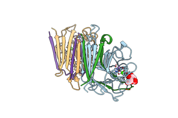 |
The Structure Of The Pea Lectin-D-Mannopyranose Complex
Organism: Pisum sativum
Method: X-RAY DIFFRACTION Resolution:2.10 Å Release Date: 1998-08-26 Classification: PLANT PROTEIN Ligands: CA, MN, MAN, BMA |
Organism: Pisum sativum
Method: X-RAY DIFFRACTION
Release Date: 1998-08-26
Ligands: CA, MN, MAN, BMA