

Search Count: 44
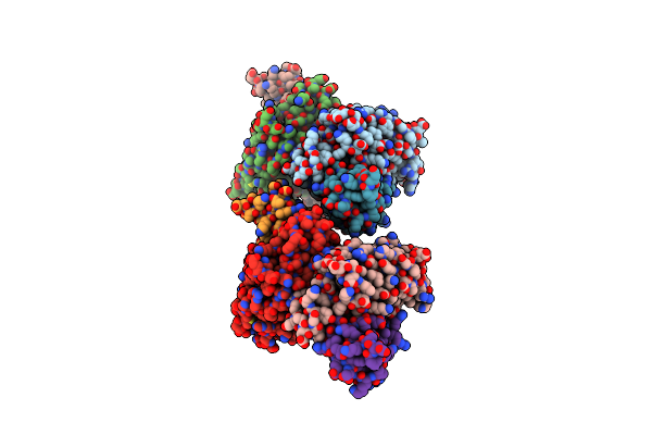 |
Mycobacterium Tuberculosis 3-Hydroxyl-Acp Dehydratase Hadab In Complex With 1,3-Diarylpyrazolyl-Acylsulfonamide Inhibitor
Organism: Mycobacterium tuberculosis
Method: X-RAY DIFFRACTION Resolution:2.40 Å Release Date: 2022-11-16 Classification: LYASE/INHIBITOR Ligands: CI7, EDO, PEG, GOL |
Organism: Mycobacterium tuberculosis
Method: X-RAY DIFFRACTION
Release Date: 2022-11-16
Ligands: CI7, EDO, PEG, GOL
 |
N-Terminal Bromodomain Of Human Brd4 With Ibet-Bd1 (Gsk778)
Organism: Homo sapiens
Method: X-RAY DIFFRACTION Resolution:1.28 Å Release Date: 2020-04-01 Classification: TRANSCRIPTION Ligands: GLU, LWB, EDO |
Organism: Homo sapiens
Method: X-RAY DIFFRACTION
Release Date: 2020-04-01
Ligands: GLU, LWB, EDO
 |
C-Terminal Bromodomain Of Human Brd2 With Ibet-Bd1 (Gsk778)
Organism: Homo sapiens
Method: X-RAY DIFFRACTION Resolution:1.60 Å Release Date: 2020-04-01 Classification: TRANSCRIPTION Ligands: LWB, EDO |
Organism: Homo sapiens
Method: X-RAY DIFFRACTION
Release Date: 2020-04-01
Ligands: LWB, EDO
 |
C-Terminal Bromodomain Of Human Brd2 With Ibet-Bd2 (Gsk046)
Organism: Homo sapiens
Method: X-RAY DIFFRACTION Resolution:1.60 Å Release Date: 2020-04-01 Classification: TRANSCRIPTION Ligands: EDO, LW5 |
Organism: Homo sapiens
Method: X-RAY DIFFRACTION
Release Date: 2020-04-01
Ligands: EDO, LW5
 |
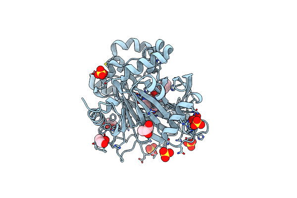
N-Terminal Bromodomain Of Human Brd4 With Ibet-Bd2 (Gsk046)
Organism: Homo sapiens
Method: X-RAY DIFFRACTION Resolution:1.60 Å Release Date: 2020-04-01 Classification: TRANSCRIPTION Ligands: EDO, LW5 |
Organism: Homo sapiens
Method: X-RAY DIFFRACTION
Release Date: 2020-04-01
Ligands: EDO, LW5
 |
Crystal Structure Of Human Kdm4D In Complex With 3-4-Methylthiophen-2- Yl Methylaminopyridine-4-Carboxylic Acid
Organism: Homo sapiens
Method: X-RAY DIFFRACTION Resolution:2.00 Å Release Date: 2016-11-09 Classification: INHIBITOR Ligands: ZN, FE2, GOL, YC8, SO4 |
Organism: Homo sapiens
Method: X-RAY DIFFRACTION
Release Date: 2016-11-09
Ligands: ZN, FE2, GOL, YC8, SO4
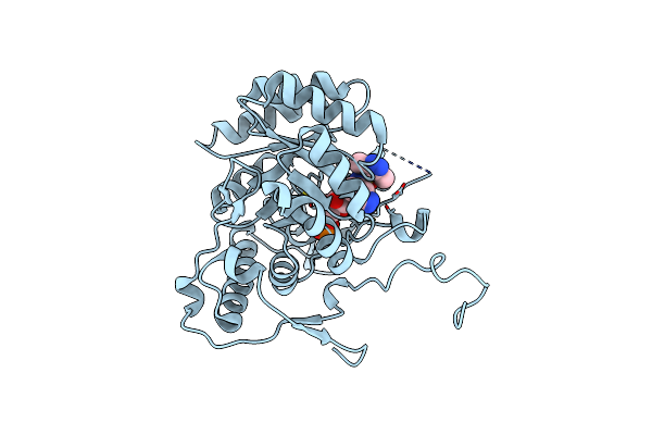 |
M. Thermoresistible Impdh In Complex With Imp And Compound 1
Organism: Mycobacterium thermoresistibile
Method: X-RAY DIFFRACTION Resolution:1.37 Å Release Date: 2016-10-19 Classification: OXIDOREDUCTASE Ligands: 6Q9, IMP |
Organism: Mycobacterium thermoresistibile
Method: X-RAY DIFFRACTION
Release Date: 2016-10-19
Ligands: 6Q9, IMP
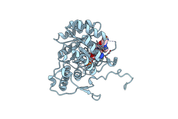 |
M. Thermoresistible Impdh In Complex With Imp And Compound 6
Organism: Mycobacterium thermoresistibile
Method: X-RAY DIFFRACTION Resolution:1.64 Å Release Date: 2016-10-19 Classification: OXIDOREDUCTASE Ligands: IMP, 6Q8 |
Organism: Mycobacterium thermoresistibile
Method: X-RAY DIFFRACTION
Release Date: 2016-10-19
Ligands: IMP, 6Q8
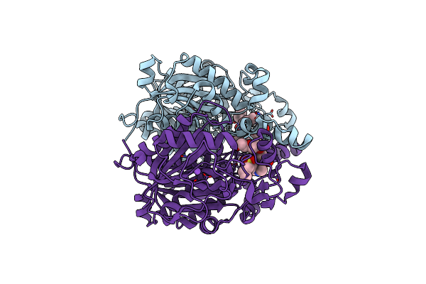 |
Gsk3011724A Cocrystallised With Mycobacterium Tuberculosis H37Rv Kasa
Organism: Mycobacterium tuberculosis (strain atcc 25618 / h37rv)
Method: X-RAY DIFFRACTION Resolution:2.13 Å Release Date: 2016-09-21 Classification: TRANSFERASE |
Organism: Mycobacterium tuberculosis (strain atcc 25618 / h37rv)
Method: X-RAY DIFFRACTION
Release Date: 2016-09-21
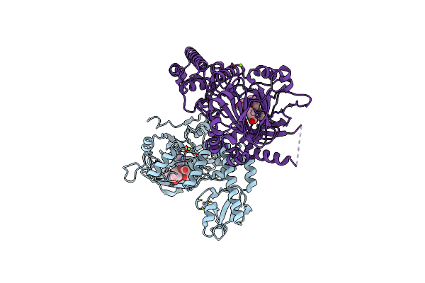 |
Cell Penetrant Inhibitors Of The Jmjd2 (Kdm4) And Jarid1 (Kdm5) Families Of Histone Lysine Demethylases
Organism: Homo sapiens
Method: X-RAY DIFFRACTION Resolution:2.05 Å Release Date: 2016-01-27 Classification: OXIDOREDUCTASE Ligands: YC8, ZN, MG, BCN, CO |
Organism: Homo sapiens
Method: X-RAY DIFFRACTION
Release Date: 2016-01-27
Ligands: YC8, ZN, MG, BCN, CO
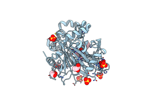 |
Crystal Structure Of Human Kdm4D In Complex With 3-(4- Phenylbutanamido)Pyridine-4-Carboxylic Acid
Organism: Homo sapiens
Method: X-RAY DIFFRACTION Resolution:2.00 Å Release Date: 2016-01-27 Classification: TRANSCRIPTION Ligands: ZN, FE2, GOL, YC8, SO4 |
Organism: Homo sapiens
Method: X-RAY DIFFRACTION
Release Date: 2016-01-27
Ligands: ZN, FE2, GOL, YC8, SO4
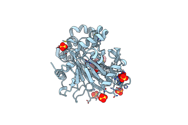 |
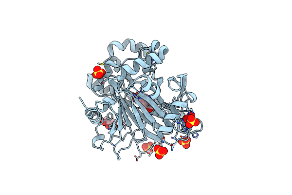
Crystal Structure Of Human Kdm4D In Complex With 3-4-Methylthiophen-2- Ylmethylaminopyridine-4-Carboxylic Acid
Organism: Homo sapiens
Method: X-RAY DIFFRACTION Resolution:1.98 Å Release Date: 2016-01-27 Classification: OXIDOREDUCTASE Ligands: ZN, CO, AUY, SO4 |
Organism: Homo sapiens
Method: X-RAY DIFFRACTION
Release Date: 2016-01-27
Ligands: ZN, CO, AUY, SO4
 |
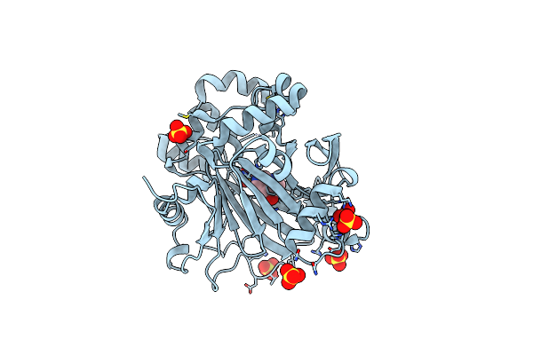
Crystal Structure Of Human Kdm4D In Complex With 3-Aminopyridine-4- Carboxylic Acid
Organism: Homo sapiens
Method: X-RAY DIFFRACTION Resolution:2.00 Å Release Date: 2016-01-27 Classification: OXIDOREDUCTASE Ligands: ZN, CO, GOL, 4SV, SO4 |
Organism: Homo sapiens
Method: X-RAY DIFFRACTION
Release Date: 2016-01-27
Ligands: ZN, CO, GOL, 4SV, SO4
 |
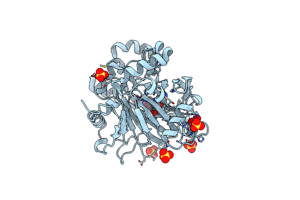
Crystal Structure Of Human Kdm4D In Complex With 3H,4H-Pyrido-3,4-D- Pyrimidin-4-One
Organism: Homo sapiens
Method: X-RAY DIFFRACTION Resolution:1.96 Å Release Date: 2016-01-27 Classification: OXIDOREDUCTASE Ligands: ZN, CO, N5J, SO4 |
Organism: Homo sapiens
Method: X-RAY DIFFRACTION
Release Date: 2016-01-27
Ligands: ZN, CO, N5J, SO4
 |
Crystal Structure Of Human Kdm4D In Complex With 2-1H-Pyrazol-4-Yloxy- 3H,4H-Pyrido-3,4-D-Pyrimidin-4-One
Organism: Homo sapiens
Method: X-RAY DIFFRACTION Resolution:1.91 Å Release Date: 2016-01-27 Classification: OXIDOREDUCTASE Ligands: ZN, CO, HA6, SO4 |
Organism: Homo sapiens
Method: X-RAY DIFFRACTION
Release Date: 2016-01-27
Ligands: ZN, CO, HA6, SO4
 |
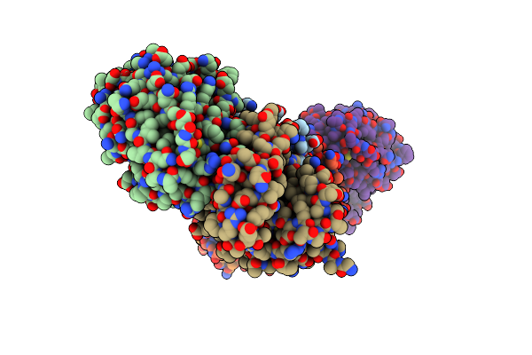
Crystal Structure Of M. Tuberculosis Echa6 (Apo, Trigonal Crystal Form)
Organism: Mycobacterium tuberculosis (strain atcc 25618 / h37rv)
Method: X-RAY DIFFRACTION Resolution:1.91 Å Release Date: 2016-01-20 Classification: LIPID BINDING PROTEIN |
Organism: Mycobacterium tuberculosis (strain atcc 25618 / h37rv)
Method: X-RAY DIFFRACTION
Release Date: 2016-01-20
 |
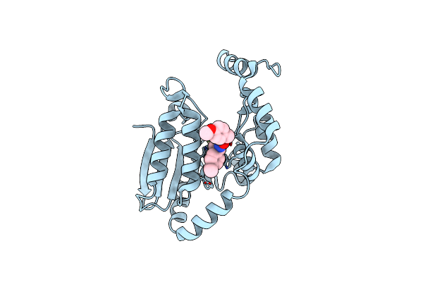
Crystal Structure Of M. Tuberculosis Echa6 Bound To C20-Coa
Organism: Mycobacterium tuberculosis (strain atcc 25618 / h37rv)
Method: X-RAY DIFFRACTION Resolution:2.44 Å Release Date: 2016-01-20 Classification: LIPID BINDING PROTEIN Ligands: 5F9 |
Organism: Mycobacterium tuberculosis (strain atcc 25618 / h37rv)
Method: X-RAY DIFFRACTION
Release Date: 2016-01-20
Ligands: 5F9
 |
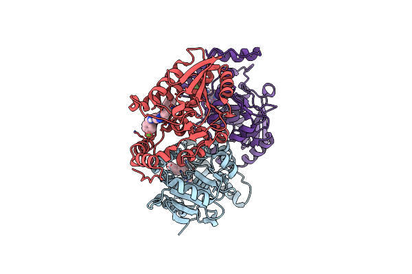
Crystal Structure Of M. Tuberculosis Echa6 Bound To Ligand Gsk366A
Organism: Mycobacterium tuberculosis
Method: X-RAY DIFFRACTION Resolution:1.70 Å Release Date: 2016-01-20 Classification: LIPID BINDING PROTEIN Ligands: 5FF |
Organism: Mycobacterium tuberculosis
Method: X-RAY DIFFRACTION
Release Date: 2016-01-20
Ligands: 5FF
 |
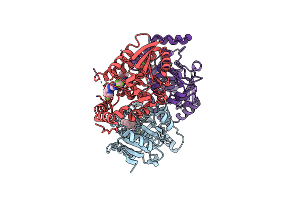
Crystal Structure Of M. Tuberculosis Echa6 Bound To Ligand Gsk059A.
Organism: Mycobacterium tuberculosis
Method: X-RAY DIFFRACTION Resolution:2.61 Å Release Date: 2016-01-20 Classification: LYASE Ligands: G59 |
Organism: Mycobacterium tuberculosis
Method: X-RAY DIFFRACTION
Release Date: 2016-01-20
Ligands: G59
 |
Crystal Structure Of M. Tuberculosis Echa6 Bound To Gsk572A
Organism: Mycobacterium tuberculosis
Method: X-RAY DIFFRACTION Resolution:2.23 Å Release Date: 2016-01-20 Classification: LIPID BINDING PROTEIN Ligands: 5FK |
Organism: Mycobacterium tuberculosis
Method: X-RAY DIFFRACTION
Release Date: 2016-01-20
Ligands: 5FK