
Search Count: 6
All
Selected
 |
Organism: Homo sapiens
Method: X-RAY DIFFRACTION Resolution:2.38 Å Release Date: 2020-09-09 Classification: LIPID BINDING PROTEIN Ligands: GOL, AOQ |
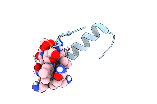 |
Organism: Homo sapiens
Method: SOLUTION NMR Release Date: 2016-09-21 Classification: HORMONE Ligands: CNC |
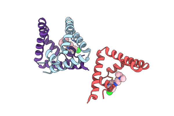 |
Organism: Homo sapiens
Method: X-RAY DIFFRACTION Resolution:2.13 Å Release Date: 2015-12-09 Classification: HYDROLASE ACTIVATOR Ligands: CLQ |
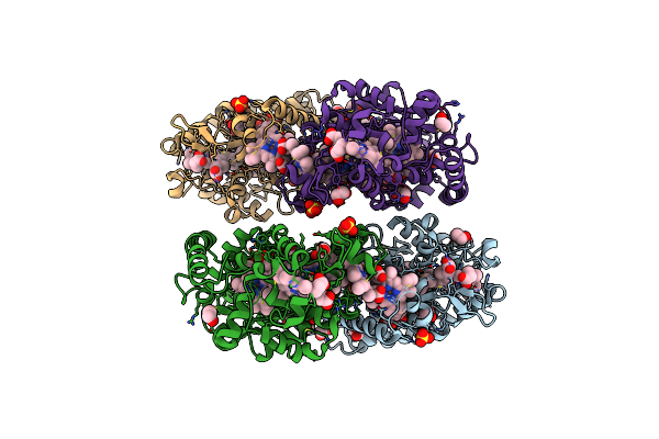 |
High Resolution Crystal Structure Of The Escherichia Coli Cytochrome C Nitrite Reductase.
Organism: Escherichia coli
Method: X-RAY DIFFRACTION Resolution:1.74 Å Release Date: 2008-03-25 Classification: OXIDOREDUCTASE Ligands: CA, HEC, SO4, EDO |
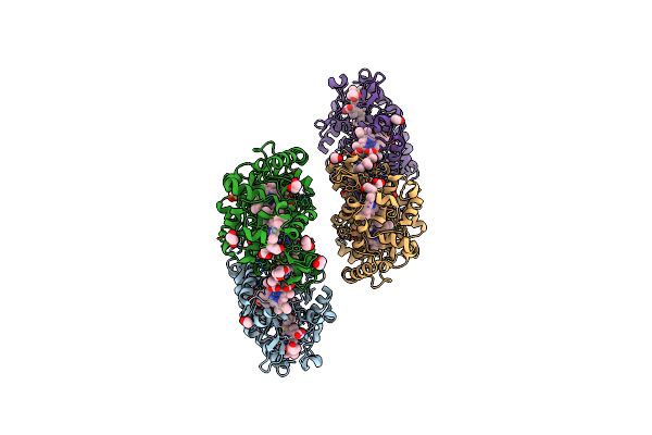 |
Organism: Escherichia coli k12
Method: X-RAY DIFFRACTION Resolution:2.04 Å Release Date: 2008-03-25 Classification: OXIDOREDUCTASE Ligands: CA, HEC, EDO |
 |
Crystal Structure Of Pseudomonas Aeruginosa Lectin 1 Determined By Single Wavelength Anomalous Scattering Phasing Method
Organism: Pseudomonas aeruginosa
Method: X-RAY DIFFRACTION Resolution:1.50 Å Release Date: 2002-12-11 Classification: SUGAR BINDING PROTEIN Ligands: CA |