

Search Count: 29
 |
Crystal Structure Of The Gh39 Enzyme From Xanthomonas Axonopodis Pv. Citri
Organism: Xanthomonas axonopodis pv. citri (strain 306)
Method: X-RAY DIFFRACTION Resolution:1.71 Å Release Date: 2020-07-22 Classification: HYDROLASE |
Organism: Xanthomonas axonopodis pv. citri (strain 306)
Method: X-RAY DIFFRACTION
Release Date: 2020-07-22
 |
Crystal Structure Of The Gh51 Arabinofuranosidase From Xanthomonas Axonopodis Pv. Citri
Organism: Xanthomonas axonopodis pv. citri (strain 306)
Method: X-RAY DIFFRACTION Resolution:1.91 Å Release Date: 2019-02-20 Classification: HYDROLASE Ligands: GOL |
Organism: Xanthomonas axonopodis pv. citri (strain 306)
Method: X-RAY DIFFRACTION
Release Date: 2019-02-20
Ligands: GOL
 |
Crystal Structure Of The Gh2 Exo-Beta-Mannanase From Xanthomonas Axonopodis Pv. Citri
Organism: Xanthomonas axonopodis pv. citri (strain 306)
Method: X-RAY DIFFRACTION Resolution:1.90 Å Release Date: 2018-07-18 Classification: HYDROLASE/CARBOHYDRATE Ligands: GOL, PEG, ACT |
Organism: Xanthomonas axonopodis pv. citri (strain 306)
Method: X-RAY DIFFRACTION
Release Date: 2018-07-18
Ligands: GOL, PEG, ACT
 |
Crystal Structure Of The Gh2 Exo-Beta-Mannanase From Xanthomonas Axonopodis Pv. Citri In Complex With Mannose
Organism: Xanthomonas axonopodis pv. citri (strain 306)
Method: X-RAY DIFFRACTION Resolution:2.13 Å Release Date: 2018-07-18 Classification: HYDROLASE/CARBOHYDRATE Ligands: BMA, ACT |
Organism: Xanthomonas axonopodis pv. citri (strain 306)
Method: X-RAY DIFFRACTION
Release Date: 2018-07-18
Ligands: BMA, ACT
 |
Crystal Structure Of The Nucleophile Mutant (E575A) Of The Gh2 Exo-Beta-Mannanase From Xanthomonas Axonopodis Pv. Citri
Organism: Xanthomonas axonopodis pv. citri (strain 306)
Method: X-RAY DIFFRACTION Resolution:2.00 Å Release Date: 2018-07-18 Classification: HYDROLASE/CARBOHYDRATE Ligands: BMA |
Organism: Xanthomonas axonopodis pv. citri (strain 306)
Method: X-RAY DIFFRACTION
Release Date: 2018-07-18
Ligands: BMA
 |
Crystal Structure Of The Acid-Base Mutant (E477A) Of The Gh2 Exo-Beta-Mannanase From Xanthomonas Axonopodis Pv. Citri
Organism: Xanthomonas axonopodis pv. citri (strain 306)
Method: X-RAY DIFFRACTION Resolution:2.20 Å Release Date: 2018-07-18 Classification: HYDROLASE/CARBOHYDRATE Ligands: BMA |
Organism: Xanthomonas axonopodis pv. citri (strain 306)
Method: X-RAY DIFFRACTION
Release Date: 2018-07-18
Ligands: BMA
 |
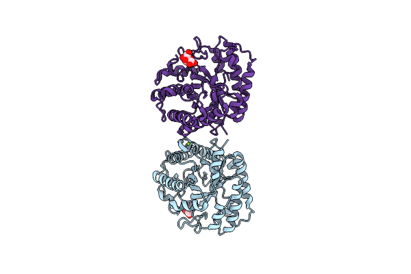
Crystal Structure Of Xeg5A, A Gh5 Xyloglucan-Specific Endo-Beta-1,4-Glucanase From Ruminal Metagenomic Library, In The Native Form
Organism: Uncultured bacterium
Method: X-RAY DIFFRACTION Resolution:1.79 Å Release Date: 2015-03-11 Classification: HYDROLASE Ligands: MG, TRS |
Organism: Uncultured bacterium
Method: X-RAY DIFFRACTION
Release Date: 2015-03-11
Ligands: MG, TRS
 |
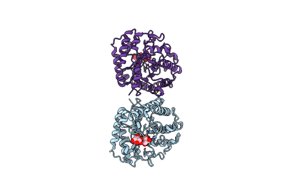
Crystal Structure Of Xeg5A, A Gh5 Xyloglucan-Specific Endo-Beta-1,4-Glucanase From Ruminal Metagenomic Library, In Complex With Glucose
Organism: Uncultured bacterium
Method: X-RAY DIFFRACTION Resolution:1.92 Å Release Date: 2015-03-11 Classification: HYDROLASE Ligands: BGC, MG |
Organism: Uncultured bacterium
Method: X-RAY DIFFRACTION
Release Date: 2015-03-11
Ligands: BGC, MG
 |
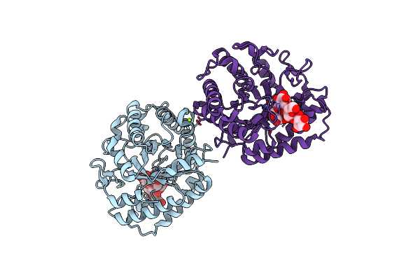
Crystal Structure Of Xeg5A, A Gh5 Xyloglucan-Specific Endo-Beta-1,4-Glucanase From Ruminal Metagenomic Library, In Complex With Glucose And Tris
Organism: Uncultured bacterium
Method: X-RAY DIFFRACTION Resolution:2.64 Å Release Date: 2015-03-11 Classification: HYDROLASE Ligands: TRS, BGC, MG |
Organism: Uncultured bacterium
Method: X-RAY DIFFRACTION
Release Date: 2015-03-11
Ligands: TRS, BGC, MG
 |
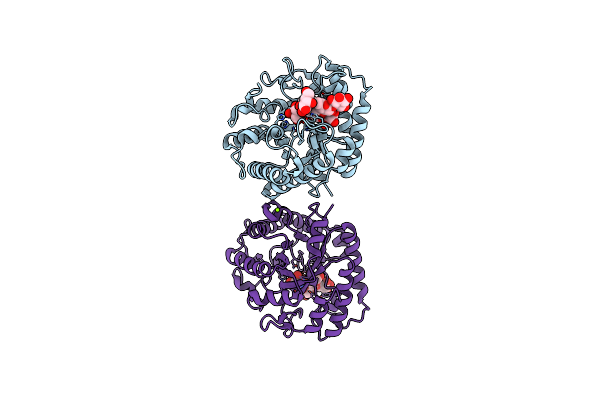
Crystal Structure Of Xeg5A, A Gh5 Xyloglucan-Specific Endo-Beta-1,4-Glucanase From Metagenomic Library, In Complex With A Xyloglucan Oligosaccharide
Organism: Uncultured bacterium
Method: X-RAY DIFFRACTION Resolution:2.15 Å Release Date: 2015-03-11 Classification: HYDROLASE Ligands: MG |
Organism: Uncultured bacterium
Method: X-RAY DIFFRACTION
Release Date: 2015-03-11
Ligands: MG
 |
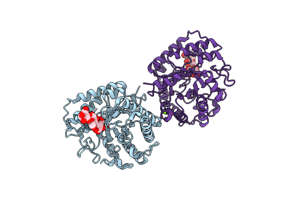
Crystal Structure Of Xeg5A, A Gh5 Xyloglucan-Specific Endo-Beta-1,4-Glucanase From Ruminal Metagenomic Library, In Complex With A Xyloglucan Oligosaccharide And Tris
Organism: Uncultured bacterium
Method: X-RAY DIFFRACTION Resolution:1.58 Å Release Date: 2015-03-11 Classification: HYDROLASE Ligands: TRS, MG |
Organism: Uncultured bacterium
Method: X-RAY DIFFRACTION
Release Date: 2015-03-11
Ligands: TRS, MG
 |
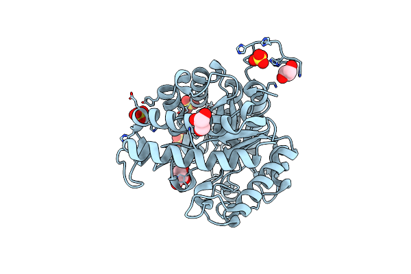
Crystal Structure Of Xeg5A, A Gh5 Xyloglucan-Specific Endo-Beta-1,4-Glucanase From Metagenomic Library, In Complex With Cellotriose
Organism: Uncultured bacterium
Method: X-RAY DIFFRACTION Resolution:2.40 Å Release Date: 2015-03-11 Classification: HYDROLASE Ligands: MG |
Organism: Uncultured bacterium
Method: X-RAY DIFFRACTION
Release Date: 2015-03-11
Ligands: MG
 |
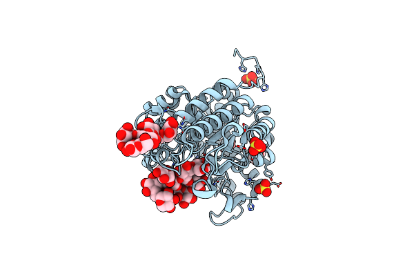
Crystal Structure Of Xeg5B, A Gh5 Xyloglucan-Specific Beta-1,4-Glucanase From Ruminal Metagenomic Library, In The Native Form
Organism: Uncultured bacterium
Method: X-RAY DIFFRACTION Resolution:1.72 Å Release Date: 2015-03-11 Classification: HYDROLASE Ligands: SO4, GOL |
Organism: Uncultured bacterium
Method: X-RAY DIFFRACTION
Release Date: 2015-03-11
Ligands: SO4, GOL
 |
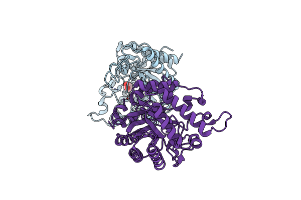
Crystal Structure Of Xeg5B, A Gh5 Xyloglucan-Specific Beta-1,4-Glucanase From Ruminal Metagenomic Library, In Complex With Xxlg
Organism: Uncultured bacterium
Method: X-RAY DIFFRACTION Resolution:1.15 Å Release Date: 2015-03-11 Classification: HYDROLASE Ligands: SO4, GOL |
Organism: Uncultured bacterium
Method: X-RAY DIFFRACTION
Release Date: 2015-03-11
Ligands: SO4, GOL
 |
Structure Of A Novel Gh10 Endoxylanase Retrieved From Sugarcane Soil Metagenome
Organism: Soil metagenome
Method: X-RAY DIFFRACTION Resolution:2.74 Å Release Date: 2013-10-23 Classification: HYDROLASE Ligands: GOL |
Organism: Soil metagenome
Method: X-RAY DIFFRACTION
Release Date: 2013-10-23
Ligands: GOL
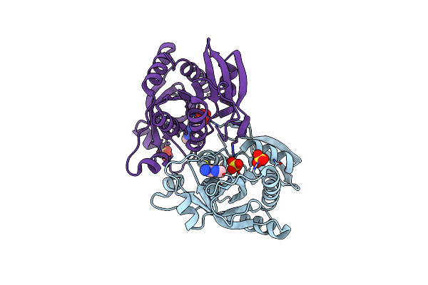 |
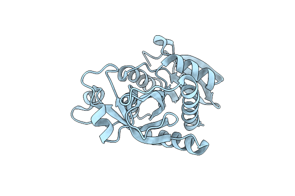
Crystal Structure Of The Hexameric Purine Nucleoside Phosphorylase From Bacillus Subtilis At Ph 4.2
Organism: Bacillus subtilis
Method: X-RAY DIFFRACTION Resolution:2.35 Å Release Date: 2012-09-26 Classification: TRANSFERASE Ligands: SO4, ADE |
Organism: Bacillus subtilis
Method: X-RAY DIFFRACTION
Release Date: 2012-09-26
Ligands: SO4, ADE
 |
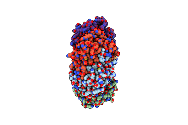
Crystal Structure Of The Hexameric Purine Nucleoside Phosphorylase From Bacillus Subtilis In Space Group P6322 At Ph 4.6
Organism: Bacillus subtilis
Method: X-RAY DIFFRACTION Resolution:2.65 Å Release Date: 2012-09-26 Classification: TRANSFERASE |
Organism: Bacillus subtilis
Method: X-RAY DIFFRACTION
Release Date: 2012-09-26
 |
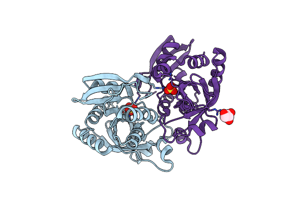
Crystal Structure Of The Hexameric Purine Nucleoside Phosphorylase From Bacillus Subtilis In Space Group P212121 At Ph 5.6
Organism: Bacillus subtilis
Method: X-RAY DIFFRACTION Resolution:1.61 Å Release Date: 2012-09-26 Classification: TRANSFERASE Ligands: SO4, GOL |
Organism: Bacillus subtilis
Method: X-RAY DIFFRACTION
Release Date: 2012-09-26
Ligands: SO4, GOL
 |
Crystal Structure Of The Hexameric Purine Nucleoside Phosphorylase From Bacillus Subtilis In Space Group H32 At Ph 7.5
Organism: Bacillus subtilis
Method: X-RAY DIFFRACTION Resolution:1.70 Å Release Date: 2012-09-26 Classification: TRANSFERASE Ligands: SO4, CL, GOL |
Organism: Bacillus subtilis
Method: X-RAY DIFFRACTION
Release Date: 2012-09-26
Ligands: SO4, CL, GOL
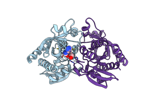 |
Crystal Structure Of The Hexameric Purine Nucleoside Phosphorylase From Bacillus Subtilis In Complex With Adenosine
Organism: Bacillus subtilis
Method: X-RAY DIFFRACTION Resolution:1.91 Å Release Date: 2012-09-26 Classification: TRANSFERASE Ligands: ADN |
Organism: Bacillus subtilis
Method: X-RAY DIFFRACTION
Release Date: 2012-09-26
Ligands: ADN