

Search Count: 11
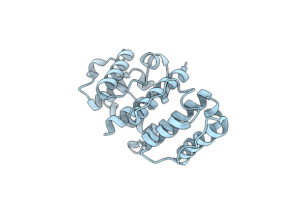 |
X-Ray Structure Of The Human Alpha-Actinin Isoform 3 At 2.2A Resolution
Organism: Homo sapiens
Method: X-RAY DIFFRACTION Resolution:2.19 Å Release Date: 2005-05-17 Classification: CONTRACTILE PROTEIN |
Organism: Homo sapiens
Method: X-RAY DIFFRACTION
Release Date: 2005-05-17
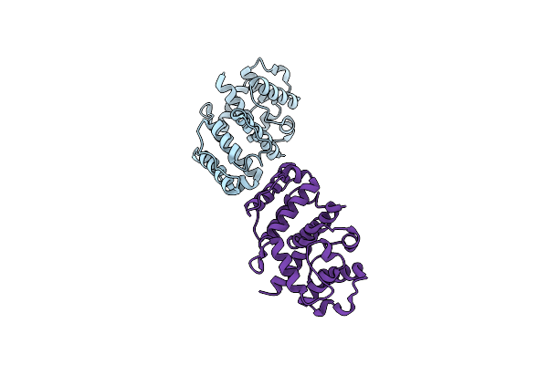 |
High Resolution Structure Of The Human Alpha-Actinin Isoform 3
Organism: Homo sapiens
Method: X-RAY DIFFRACTION Resolution:1.60 Å Release Date: 2005-05-17 Classification: CONTRACTILE PROTEIN |
Organism: Homo sapiens
Method: X-RAY DIFFRACTION
Release Date: 2005-05-17
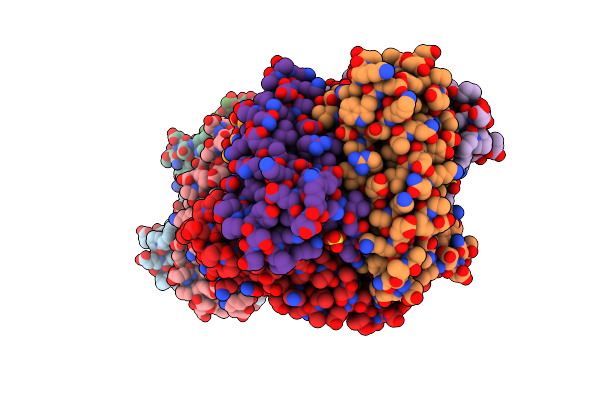 |
Crystal Structure Of Ni-Containing Superoxide Dismutase With Ni-Ligation Corresponding To The Oxidized State
Organism: Streptomyces seoulensis
Method: X-RAY DIFFRACTION Resolution:2.20 Å Release Date: 2004-05-18 Classification: OXIDOREDUCTASE Ligands: SO4, 3NI |
Organism: Streptomyces seoulensis
Method: X-RAY DIFFRACTION
Release Date: 2004-05-18
Ligands: SO4, 3NI
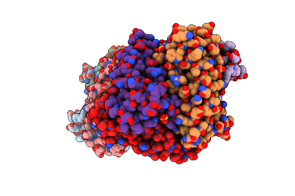 |
Crystal Structure Of Ni-Containing Superoxide Dismutase With Ni-Ligation Corresponding To The State After Partial X-Ray-Induced Reduction
Organism: Streptomyces seoulensis
Method: X-RAY DIFFRACTION Resolution:2.20 Å Release Date: 2004-05-18 Classification: OXIDOREDUCTASE Ligands: SO4, 3NI |
Organism: Streptomyces seoulensis
Method: X-RAY DIFFRACTION
Release Date: 2004-05-18
Ligands: SO4, 3NI
 |
Crystal Structure Of Ni-Containing Superoxide Dismutase With Ni-Ligation Corresponding To The State After Full X-Ray-Induced Reduction
Organism: Streptomyces seoulensis
Method: X-RAY DIFFRACTION Resolution:1.60 Å Release Date: 2004-05-18 Classification: OXIDOREDUCTASE Ligands: NI, SO4 |
Organism: Streptomyces seoulensis
Method: X-RAY DIFFRACTION
Release Date: 2004-05-18
Ligands: NI, SO4
 |
Crystal Structure Of Ni-Containing Superoxide Dismutase With Ni-Ligation Corresponding To The Thiosulfate-Reduced State
Organism: Streptomyces seoulensis
Method: X-RAY DIFFRACTION Resolution:2.10 Å Release Date: 2004-05-18 Classification: OXIDOREDUCTASE Ligands: NI, SO4, THJ |
Organism: Streptomyces seoulensis
Method: X-RAY DIFFRACTION
Release Date: 2004-05-18
Ligands: NI, SO4, THJ
 |
Crystal Structure Of Ni-Containing Superoxide Dismutase With Ni-Ligation Corresponding To The State After Full X-Ray-Induced Reduction
Organism: Streptomyces seoulensis
Method: X-RAY DIFFRACTION Resolution:1.68 Å Release Date: 2004-05-18 Classification: OXIDOREDUCTASE Ligands: NI, SO4, ACY |
Organism: Streptomyces seoulensis
Method: X-RAY DIFFRACTION
Release Date: 2004-05-18
Ligands: NI, SO4, ACY
 |
Crystal Structure Of Perchloric Acid Soluble Protein-A Translational Inhibitor
Organism: Rattus norvegicus
Method: X-RAY DIFFRACTION Resolution:1.80 Å Release Date: 2004-03-09 Classification: ALPHA-BETA STRUCTURE |
Organism: Rattus norvegicus
Method: X-RAY DIFFRACTION
Release Date: 2004-03-09
 |
Ph Domain From Bruton'S Tyrosine Kinase In Complex With Inositol 1,3,4,5-Tetrakisphosphate
Organism: Homo sapiens
Method: X-RAY DIFFRACTION Resolution:2.40 Å Release Date: 1999-06-15 Classification: TRANSFERASE Ligands: ZN, 4IP |
Organism: Homo sapiens
Method: X-RAY DIFFRACTION
Release Date: 1999-06-15
Ligands: ZN, 4IP
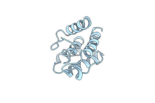 |
Calponin Homology (Ch) Domain From Human Beta-Spectrin
Organism: Homo sapiens
Method: X-RAY DIFFRACTION Resolution:2.00 Å Release Date: 1998-02-04 Classification: CYTOSKELETON |
Organism: Homo sapiens
Method: X-RAY DIFFRACTION
Release Date: 1998-02-04
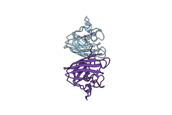 |
Three-Dimensional Structure Of Xenopus Laevis Cu,Zn Superoxide Dismutase B Determined By X-Ray Crystallography At 1.5 Angstroms Resolution
Organism: Xenopus laevis
Method: X-RAY DIFFRACTION Resolution:1.49 Å Release Date: 1995-07-10 Classification: OXIDOREDUCTASE (SUPEROXIDE ACCEPTOR) Ligands: CU, ZN |
Organism: Xenopus laevis
Method: X-RAY DIFFRACTION
Release Date: 1995-07-10
Ligands: CU, ZN