

Search Count: 36
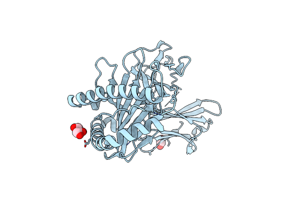 |
Crystal Structure Of A Single Chain Trimer Composed Of Hla-B*39:06 Y84C Variant, Beta-2Microglobulin, And Nrvmlpkaa Peptide From Nlrp2 (1 Molecule/Asymmetric Unit)
Organism: Homo sapiens
Method: X-RAY DIFFRACTION Resolution:1.70 Å Release Date: 2024-09-04 Classification: IMMUNE SYSTEM Ligands: EDO, GOL, CL |
Organism: Homo sapiens
Method: X-RAY DIFFRACTION
Release Date: 2024-09-04
Ligands: EDO, GOL, CL
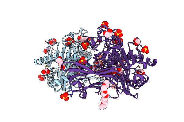 |
Crystal Structure Of A Single Chain Trimer Composed Of Hla-B*39:06 Y84C Variant, Beta-2Microglobulin, And Nrvmlpkaa Peptide From Nlrp2 (2 Molecules/Asymmetric Unit)
Organism: Homo sapiens
Method: X-RAY DIFFRACTION Resolution:1.70 Å Release Date: 2024-09-04 Classification: IMMUNE SYSTEM Ligands: SO4, EDO, PEG, CL, P3G, GOL |
Organism: Homo sapiens
Method: X-RAY DIFFRACTION
Release Date: 2024-09-04
Ligands: SO4, EDO, PEG, CL, P3G, GOL
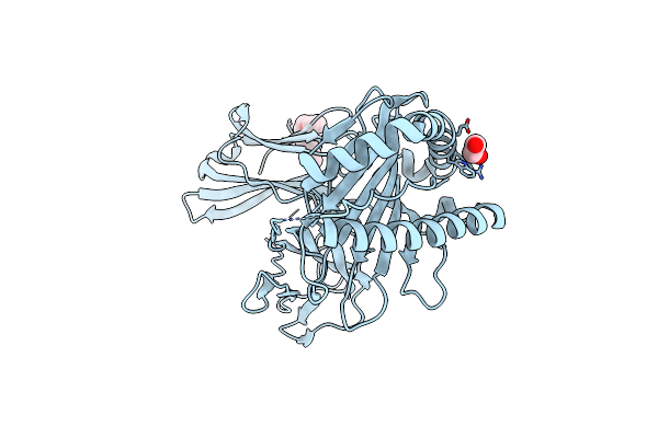 |
Crystal Structure Of A Single Chain Trimer Composed Of Hla-B*39:01 Y84C Variant, Beta-2Microglobulin, And Nrvmlpkaa Peptide From Nlrp2
Organism: Homo sapiens
Method: X-RAY DIFFRACTION Resolution:1.70 Å Release Date: 2024-09-04 Classification: IMMUNE SYSTEM Ligands: P4G, EDO |
Organism: Homo sapiens
Method: X-RAY DIFFRACTION
Release Date: 2024-09-04
Ligands: P4G, EDO
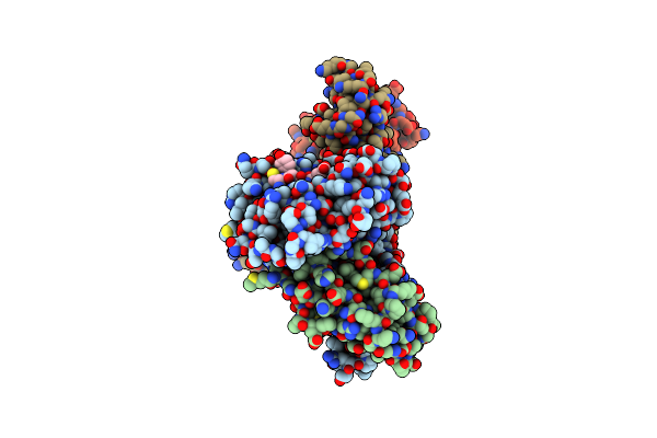 |
Crystal Structure Of H-2D In Complex With An Insulin Derived Peptide
Organism: Mus musculus
Method: X-RAY DIFFRACTION Resolution:2.34 Å Release Date: 2014-03-26 Classification: IMMUNE SYSTEM |
Organism: Mus musculus
Method: X-RAY DIFFRACTION
Release Date: 2014-03-26
 |
Crystal Structure Of H-2D In Complex With A Mimotopic Peptide
Organism: Mus musculus
Method: X-RAY DIFFRACTION Resolution:1.98 Å Release Date: 2014-03-26 Classification: IMMUNE SYSTEM Ligands: ZN, IMD, GOL, SO4 |
Organism: Mus musculus
Method: X-RAY DIFFRACTION
Release Date: 2014-03-26
Ligands: ZN, IMD, GOL, SO4
 |
Crystal Structure Of A Single Chain Construct Composed Of Mhc Class I H-2Kd, Beta-2Microglobulin And A Peptide Which Is An Autoantigen For Type 1 Diabetes
Organism: Mus musculus
Method: X-RAY DIFFRACTION Resolution:2.70 Å Release Date: 2011-07-06 Classification: IMMUNE SYSTEM |
Organism: Mus musculus
Method: X-RAY DIFFRACTION
Release Date: 2011-07-06
 |
Crystal Structure Of The Class I Mhc Molecule H-2Kwm7 With A Single Self Peptide Vndiferi
Organism: Mus musculus
Method: X-RAY DIFFRACTION Resolution:2.50 Å Release Date: 2010-01-12 Classification: IMMUNE SYSTEM |
Organism: Mus musculus
Method: X-RAY DIFFRACTION
Release Date: 2010-01-12
 |
Crystal Structure Of The Class I Mhc Molecule H-2Kwm7 With A Single Self Peptide Iqqsierl
Organism: Mus musculus
Method: X-RAY DIFFRACTION Resolution:2.10 Å Release Date: 2010-01-12 Classification: IMMUNE SYSTEM Ligands: CL |
Organism: Mus musculus
Method: X-RAY DIFFRACTION
Release Date: 2010-01-12
Ligands: CL
 |
Crystal Structure Of The Class I Mhc Molecule H-2Kwm7 With A Single Self Peptide Vndifeai
Organism: Mus musculus, Synthetic construct
Method: X-RAY DIFFRACTION Resolution:2.03 Å Release Date: 2010-01-12 Classification: IMMUNE SYSTEM |
Organism: Mus musculus, Synthetic construct
Method: X-RAY DIFFRACTION
Release Date: 2010-01-12
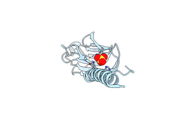 |
Crystal Structure Of Human Gitrl Variant
Organism: Saccharomyces cerevisiae, homo sapiens
Method: X-RAY DIFFRACTION Resolution:1.95 Å Release Date: 2007-11-20 Classification: IMMUNE SYSTEM Ligands: SO4 |
Organism: Saccharomyces cerevisiae, homo sapiens
Method: X-RAY DIFFRACTION
Release Date: 2007-11-20
Ligands: SO4
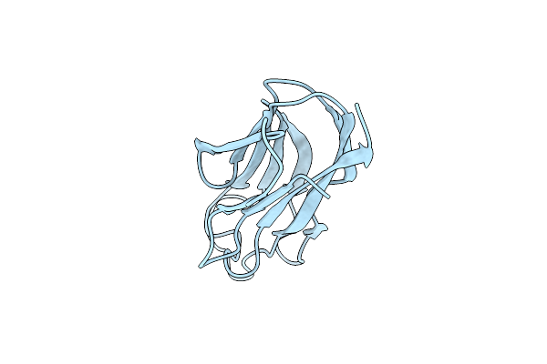 |
Crystal Structure Of Human Gitrl
Organism: Homo sapiens
Method: X-RAY DIFFRACTION Resolution:2.30 Å Release Date: 2007-11-13 Classification: SIGNALING PROTEIN |
Organism: Homo sapiens
Method: X-RAY DIFFRACTION
Release Date: 2007-11-13
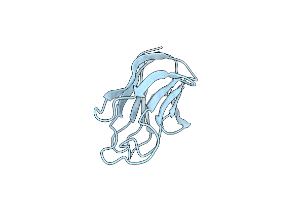 |
Crystal Structure Of Human Gitrl Mutant
Organism: Homo sapiens
Method: X-RAY DIFFRACTION Resolution:3.20 Å Release Date: 2007-11-13 Classification: CYTOKINE |
Organism: Homo sapiens
Method: X-RAY DIFFRACTION
Release Date: 2007-11-13
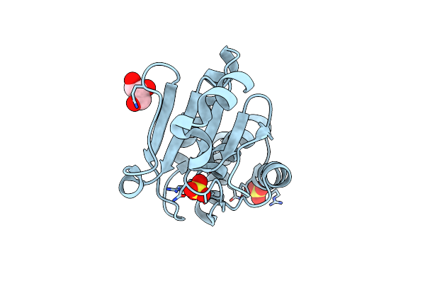 |
Crystal Structure Of Human Tyrosine Phosphatase-Like Serine/Threonine/Tyrosine-Interacting Protein
Organism: Homo sapiens
Method: X-RAY DIFFRACTION Resolution:1.60 Å Release Date: 2007-08-28 Classification: HYDROLASE Ligands: SO4, GOL |
Organism: Homo sapiens
Method: X-RAY DIFFRACTION
Release Date: 2007-08-28
Ligands: SO4, GOL
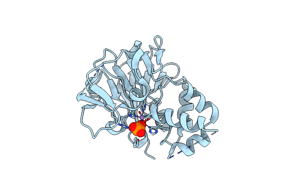 |
Crystal Structure Of A Putative Diadenosine Tetraphosphatase
Organism: Trypanosoma brucei
Method: X-RAY DIFFRACTION Resolution:2.05 Å Release Date: 2007-07-24 Classification: HYDROLASE Ligands: MN, PO4 |
Organism: Trypanosoma brucei
Method: X-RAY DIFFRACTION
Release Date: 2007-07-24
Ligands: MN, PO4
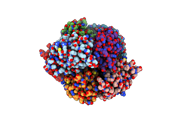 |
Crystal Structure Of Human Carboxy-Terminal Domain Rna Polymerase Ii Polypeptide A Small Phosphatase 2
Organism: Homo sapiens
Method: X-RAY DIFFRACTION Resolution:2.51 Å Release Date: 2007-06-19 Classification: HYDROLASE Ligands: MG |
Organism: Homo sapiens
Method: X-RAY DIFFRACTION
Release Date: 2007-06-19
Ligands: MG
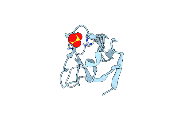 |
T Cell Immunoglobulin Mucin-3 Crystal Structure Revealed A Galectin-9-Independent Binding Surface
Organism: Mus musculus
Method: X-RAY DIFFRACTION Resolution:1.95 Å Release Date: 2007-04-10 Classification: SIGNALING PROTEIN Ligands: SO4 |
Organism: Mus musculus
Method: X-RAY DIFFRACTION
Release Date: 2007-04-10
Ligands: SO4
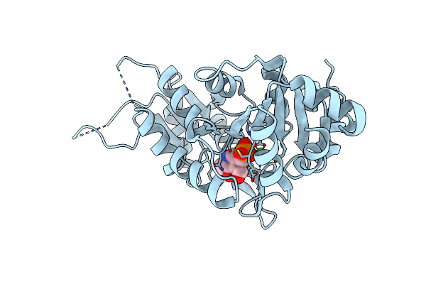 |
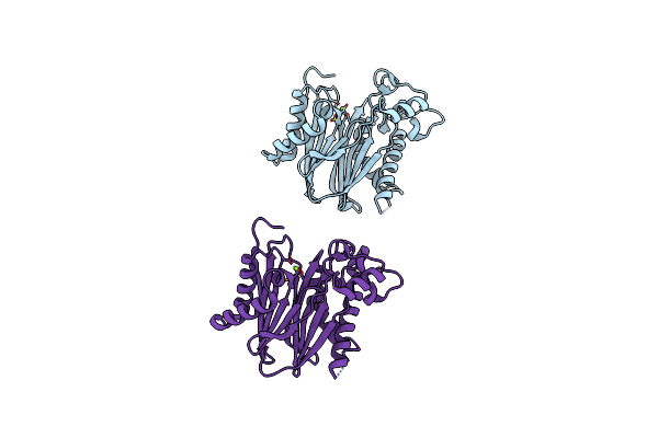
Crystal Structure Of Human Pyridoxal Phosphate Phosphatase With Plp
Organism: Homo sapiens
Method: X-RAY DIFFRACTION Resolution:2.25 Å Release Date: 2007-04-03 Classification: HYDROLASE Ligands: CA, PLP |
Organism: Homo sapiens
Method: X-RAY DIFFRACTION
Release Date: 2007-04-03
Ligands: CA, PLP
 |
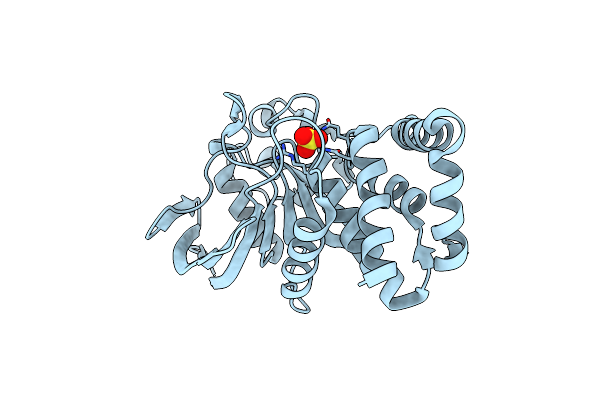
Crystal Structure Of The Serine/Threonine Phosphatase Domain Of Human Ppm1B
Organism: Homo sapiens
Method: X-RAY DIFFRACTION Resolution:1.82 Å Release Date: 2007-04-03 Classification: HYDROLASE Ligands: MG |
Organism: Homo sapiens
Method: X-RAY DIFFRACTION
Release Date: 2007-04-03
Ligands: MG
 |
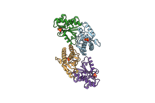
Crystal Structure Of The Human Tyrosine Receptor Phosphate Gamma
Organism: Homo sapiens
Method: X-RAY DIFFRACTION Resolution:1.70 Å Release Date: 2007-04-03 Classification: HYDROLASE Ligands: SO4 |
Organism: Homo sapiens
Method: X-RAY DIFFRACTION
Release Date: 2007-04-03
Ligands: SO4
 |
Crystal Structure Of Acid Phosphatase 1 (Acp1) From Mus Musculus
Organism: Mus musculus
Method: X-RAY DIFFRACTION Resolution:1.90 Å Release Date: 2007-03-20 Classification: HYDROLASE Ligands: PO4 |
Organism: Mus musculus
Method: X-RAY DIFFRACTION
Release Date: 2007-03-20
Ligands: PO4