

Search Count: 26
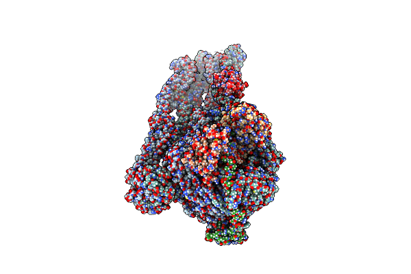 |
Mukbef In A Nucleotide-Bound State With Open Neck Gate (Heads Core)
Organism: Photorhabdus thracensis, Escherichia coli
Method: ELECTRON MICROSCOPY Release Date: 2025-03-26 Classification: DNA BINDING PROTEIN Ligands: MG, ATP |
Organism: Photorhabdus thracensis, Escherichia coli
Method: ELECTRON MICROSCOPY
Release Date: 2025-03-26
Ligands: MG, ATP
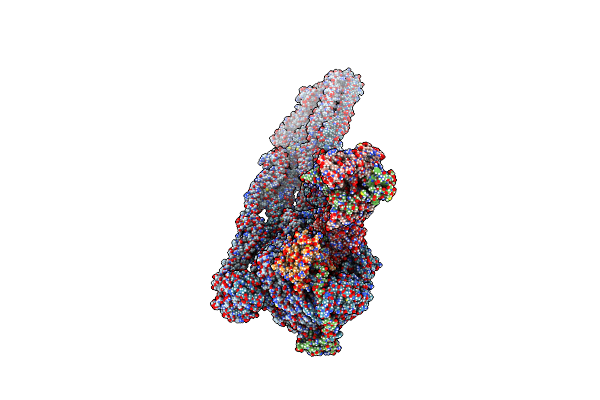 |
Mukbef In A Nucleotide-Bound State With Open Neck Gate (Monomer)
Organism: Photorhabdus thracensis, Escherichia coli
Method: ELECTRON MICROSCOPY Release Date: 2025-03-26 Classification: DNA BINDING PROTEIN Ligands: MG, ATP |
Organism: Photorhabdus thracensis, Escherichia coli
Method: ELECTRON MICROSCOPY
Release Date: 2025-03-26
Ligands: MG, ATP
 |
Mukbef In A Nucleotide-Bound State With Open Neck Gate
Organism: Photorhabdus thracensis, Escherichia coli
Method: ELECTRON MICROSCOPY Release Date: 2025-03-26 Classification: DNA BINDING PROTEIN Ligands: MG, ATP |
Organism: Photorhabdus thracensis, Escherichia coli
Method: ELECTRON MICROSCOPY
Release Date: 2025-03-26
Ligands: MG, ATP
 |
Mukbef In A Dna Capture State
Organism: Photorhabdus thracensis, Escherichia coli
Method: ELECTRON MICROSCOPY Release Date: 2025-03-26 Classification: DNA BINDING PROTEIN Ligands: MG, ATP |
Organism: Photorhabdus thracensis, Escherichia coli
Method: ELECTRON MICROSCOPY
Release Date: 2025-03-26
Ligands: MG, ATP
 |
Mukbef In A Dna Capture State (Dimer)
Organism: Photorhabdus thracensis, Escherichia coli
Method: ELECTRON MICROSCOPY Release Date: 2025-03-26 Classification: DNA BINDING PROTEIN Ligands: MG, ATP |
Organism: Photorhabdus thracensis, Escherichia coli
Method: ELECTRON MICROSCOPY
Release Date: 2025-03-26
Ligands: MG, ATP
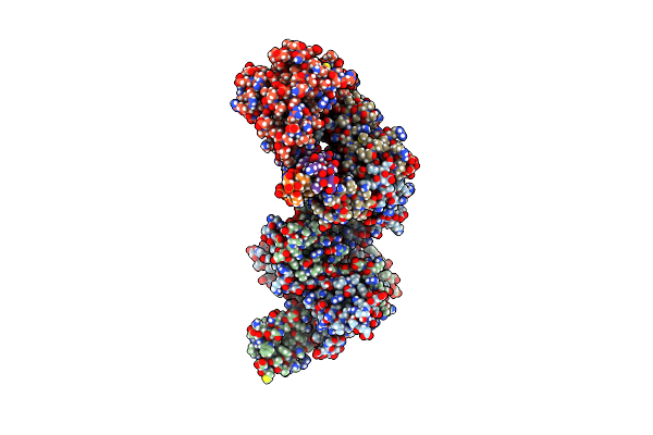 |
Mukef In Complex With The Phage Protein Gp5.9
Organism: Escherichia coli, Escherichia phage t7
Method: ELECTRON MICROSCOPY Release Date: 2025-03-26 Classification: DNA BINDING PROTEIN |
Organism: Escherichia coli, Escherichia phage t7
Method: ELECTRON MICROSCOPY
Release Date: 2025-03-26
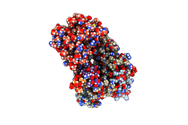 |
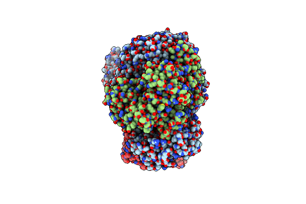
Mukef In Complex With The Phage Protein Gp5.9 (Focus)
Organism: Escherichia coli, Escherichia phage t7
Method: ELECTRON MICROSCOPY Release Date: 2025-03-26 Classification: DNA BINDING PROTEIN |
Organism: Escherichia coli, Escherichia phage t7
Method: ELECTRON MICROSCOPY
Release Date: 2025-03-26
 |
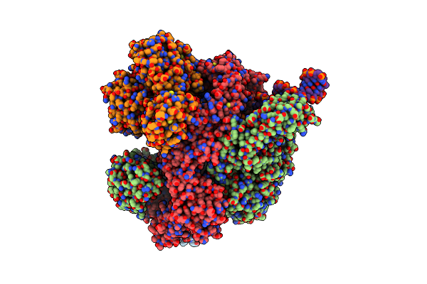
Recbcd In Complex With The Phage Protein Gp5.9
Organism: Escherichia coli, Escherichia phage t7
Method: ELECTRON MICROSCOPY Release Date: 2022-12-28 Classification: DNA BINDING PROTEIN Ligands: MG |
Organism: Escherichia coli, Escherichia phage t7
Method: ELECTRON MICROSCOPY
Release Date: 2022-12-28
Ligands: MG
 |
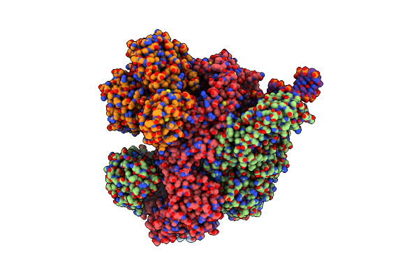
Recbcd-Dna In Complex With The Phage Protein Abc2
Organism: Escherichia coli, Salmonella phage p22, Synthetic construct
Method: ELECTRON MICROSCOPY Release Date: 2022-12-28 Classification: DNA BINDING PROTEIN Ligands: ANP, MG |
Organism: Escherichia coli, Salmonella phage p22, Synthetic construct
Method: ELECTRON MICROSCOPY
Release Date: 2022-12-28
Ligands: ANP, MG
 |
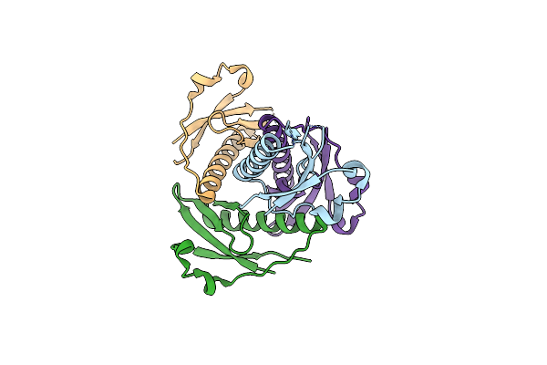
Recbcd-Dna In Complex With The Phage Protein Abc2 And Host Ppib
Organism: Escherichia coli, Salmonella phage p22, Synthetic construct
Method: ELECTRON MICROSCOPY Release Date: 2022-12-28 Classification: DNA BINDING PROTEIN Ligands: ANP, MG |
Organism: Escherichia coli, Salmonella phage p22, Synthetic construct
Method: ELECTRON MICROSCOPY
Release Date: 2022-12-28
Ligands: ANP, MG
 |
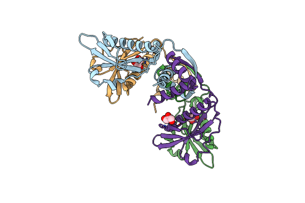
Crystal Structure Of The N-Terminal Domain Of Burkholderia Pseudomallei Antitoxin Hicb
Organism: Burkholderia pseudomallei k96243
Method: X-RAY DIFFRACTION Resolution:1.56 Å Release Date: 2018-10-31 Classification: ANTITOXIN |
Organism: Burkholderia pseudomallei k96243
Method: X-RAY DIFFRACTION
Release Date: 2018-10-31
 |
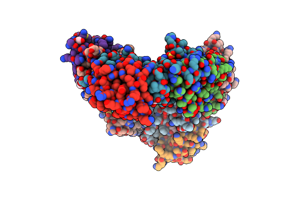
Crystal Structure Of The Burkholderia Pseudomallei Antitoxin Hicb
Organism: Burkholderia pseudomallei
Method: X-RAY DIFFRACTION Resolution:1.85 Å Release Date: 2018-10-31 Classification: ANTITOXIN Ligands: CL, GOL |
Organism: Burkholderia pseudomallei
Method: X-RAY DIFFRACTION
Release Date: 2018-10-31
Ligands: CL, GOL
 |
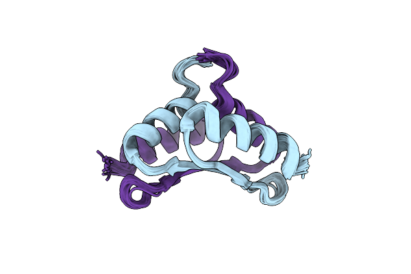
The Crystal Structure Of The Burkholderia Pseudomallei Hicab Complex
Organism: Burkholderia pseudomallei k96243
Method: X-RAY DIFFRACTION Resolution:2.49 Å Release Date: 2018-10-31 Classification: ANTITOXIN Ligands: SO4, EDO, PGE |
Organism: Burkholderia pseudomallei k96243
Method: X-RAY DIFFRACTION
Release Date: 2018-10-31
Ligands: SO4, EDO, PGE
 |
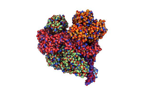
Solution Nmr Structure Of The C-Terminal Domain Of Parb (Spo0J)
Organism: Bacillus subtilis subsp. subtilis str. 168
Method: SOLUTION NMR Release Date: 2017-12-13 Classification: DNA BINDING PROTEIN |
Organism: Bacillus subtilis subsp. subtilis str. 168
Method: SOLUTION NMR
Release Date: 2017-12-13
 |
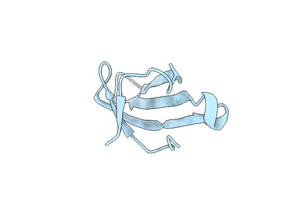
Cryo-Em Structure Of Lambda Phage Protein Gams Bound To Recbcd.
Organism: Escherichia coli, Enterobacteria phage lambda
Method: ELECTRON MICROSCOPY Release Date: 2017-01-11 Classification: DNA BINDING PROTEIN |
Organism: Escherichia coli, Enterobacteria phage lambda
Method: ELECTRON MICROSCOPY
Release Date: 2017-01-11
 |
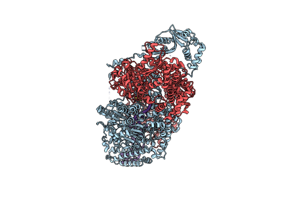
Crystal Structure Of C-Terminal Tudor Domain In Pcra/Uvrd Helicase
Organism: Geobacillus stearothermophilus
Method: X-RAY DIFFRACTION Resolution:1.53 Å Release Date: 2016-09-28 Classification: HYDROLASE |
Organism: Geobacillus stearothermophilus
Method: X-RAY DIFFRACTION
Release Date: 2016-09-28
 |
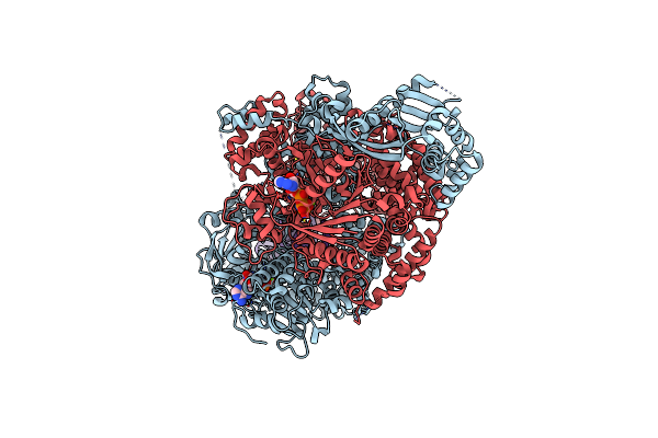
Crystal Structure Of Addab With A Forked Dna Substrate
Organism: Bacillus subtilis subsp. subtilis str. 168, Synthetic construct
Method: X-RAY DIFFRACTION Resolution:3.24 Å Release Date: 2014-03-12 Classification: HYDROLASE/DNA Ligands: SF4 |
Organism: Bacillus subtilis subsp. subtilis str. 168, Synthetic construct
Method: X-RAY DIFFRACTION
Release Date: 2014-03-12
Ligands: SF4
 |
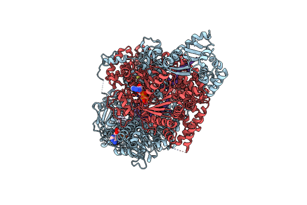
Crystal Structure Of Adpnp-Bound Addab With A Forked Dna Substrate
Organism: Bacillus subtilis subsp. subtilis str. 168, Synthetic construct
Method: X-RAY DIFFRACTION Resolution:2.80 Å Release Date: 2014-03-12 Classification: HYDROLASE/DNA Ligands: ANP, MG, SF4 |
Organism: Bacillus subtilis subsp. subtilis str. 168, Synthetic construct
Method: X-RAY DIFFRACTION
Release Date: 2014-03-12
Ligands: ANP, MG, SF4
 |
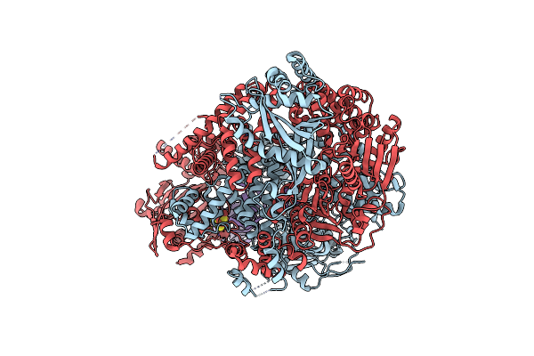
Crystal Structure Of Addab-Dna-Adpnp Complex At 3 Angstrom Resolution
Organism: Bacillus subtilis subsp. subtilis str. 168, Synthetic construct
Method: X-RAY DIFFRACTION Resolution:3.00 Å Release Date: 2014-03-12 Classification: HYDROLASE/DNA Ligands: ANP, MG, SF4 |
Organism: Bacillus subtilis subsp. subtilis str. 168, Synthetic construct
Method: X-RAY DIFFRACTION
Release Date: 2014-03-12
Ligands: ANP, MG, SF4
 |
Crystal Structure Of Addab-Dna Complex
Organism: Bacillus subtilis
Method: X-RAY DIFFRACTION Resolution:3.20 Å Release Date: 2012-03-21 Classification: HYDROLASE/DNA Ligands: SF4 |
Organism: Bacillus subtilis
Method: X-RAY DIFFRACTION
Release Date: 2012-03-21
Ligands: SF4