

Search Count: 20
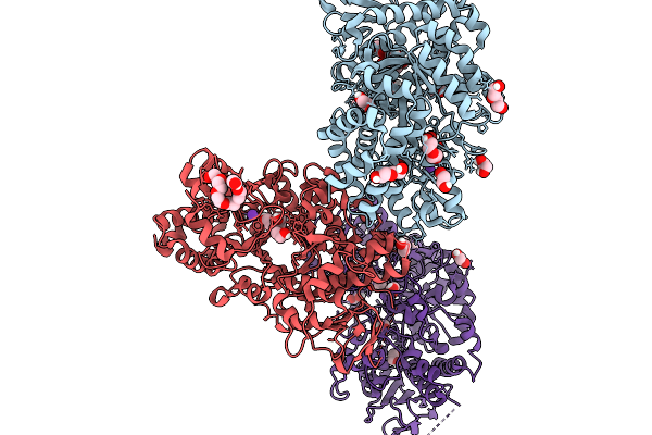 |
Sucrose Hydrolase Suxb From Xanthomonas Oryzae Pv. Oryzae
Organism: Xanthomonas oryzae pv. oryzae
Method: X-RAY DIFFRACTION Resolution:2.40 Å Release Date: 2026-03-25 Classification: HYDROLASE Ligands: EDO, PEG, K, P6G |
Organism: Xanthomonas oryzae pv. oryzae
Method: X-RAY DIFFRACTION
Release Date: 2026-03-25
Ligands: EDO, PEG, K, P6G
 |
Sucrose Hydrolase Suxb From Xanthomonas Oryzae Pv. Oryzae In Complex With Glucose
Organism: Xanthomonas oryzae pv. oryzae
Method: X-RAY DIFFRACTION Resolution:2.95 Å Release Date: 2026-03-25 Classification: HYDROLASE Ligands: K, GLC |
Organism: Xanthomonas oryzae pv. oryzae
Method: X-RAY DIFFRACTION
Release Date: 2026-03-25
Ligands: K, GLC
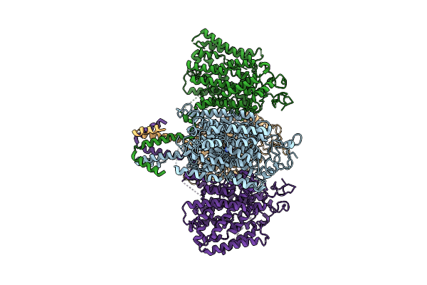 |
Zea Mays Ure Transporter Dur3 - Ure Bound
Organism: Zea mays
Method: ELECTRON MICROSCOPY Resolution:3.41 Å Release Date: 2025-11-05 Classification: CHOLINE-BINDING PROTEIN Ligands: URE |
Organism: Zea mays
Method: ELECTRON MICROSCOPY
Release Date: 2025-11-05
Ligands: URE
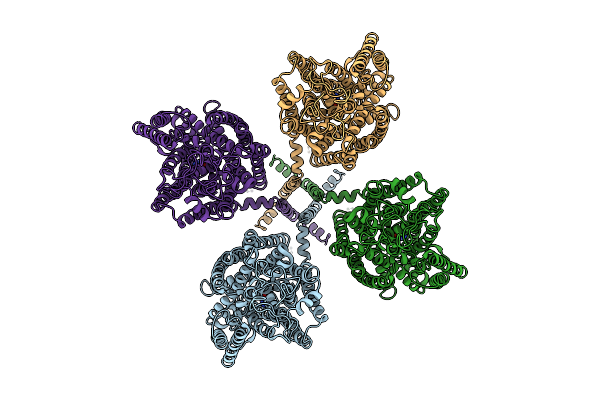 |
Arabidopsis Thaliana Ure Transporter Dur3 - Ure Bound
Organism: Arabidopsis thaliana
Method: ELECTRON MICROSCOPY Resolution:3.19 Å Release Date: 2025-11-05 Classification: PLANT PROTEIN Ligands: URE |
Organism: Arabidopsis thaliana
Method: ELECTRON MICROSCOPY
Release Date: 2025-11-05
Ligands: URE
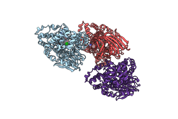 |
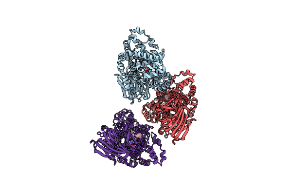
Synthesis And Evaluation Of Diaryl Ether Modulators Of The Leukotriene A4 Hydrolase Aminopeptidase Activity
Organism: Homo sapiens
Method: X-RAY DIFFRACTION Resolution:2.70 Å Release Date: 2024-08-28 Classification: HYDROLASE Ligands: VI7, ZN |
Organism: Homo sapiens
Method: X-RAY DIFFRACTION
Release Date: 2024-08-28
Ligands: VI7, ZN
 |
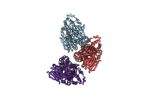
Substrate-Dependent Divergence Of Leukotriene A4 Hydrolase Aminopeptidase Activity
Organism: Homo sapiens
Method: X-RAY DIFFRACTION Resolution:2.85 Å Release Date: 2022-06-22 Classification: HYDROLASE Ligands: ZN, N0Y, 28T |
Organism: Homo sapiens
Method: X-RAY DIFFRACTION
Release Date: 2022-06-22
Ligands: ZN, N0Y, 28T
 |
Substrate-Dependent Divergence Of Leukotriene A4 Hydrolase Aminopeptidase Activity
Organism: Homo sapiens
Method: X-RAY DIFFRACTION Resolution:2.90 Å Release Date: 2022-06-15 Classification: HYDROLASE Ligands: ZN, N0Y, PGE |
Organism: Homo sapiens
Method: X-RAY DIFFRACTION
Release Date: 2022-06-15
Ligands: ZN, N0Y, PGE
 |
Structure Of Glutamate Receptor-Like Channel Glr3.4 Ligand-Binding Domain In Complex With Glutamate
Organism: Arabidopsis thaliana
Method: X-RAY DIFFRACTION Resolution:2.29 Å Release Date: 2021-07-28 Classification: MEMBRANE PROTEIN Ligands: GLU, CL, SO4, GOL |
Organism: Arabidopsis thaliana
Method: X-RAY DIFFRACTION
Release Date: 2021-07-28
Ligands: GLU, CL, SO4, GOL
 |
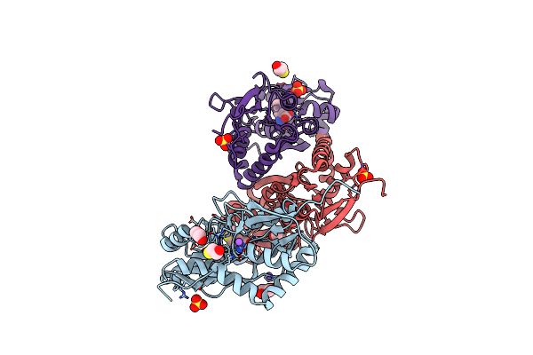
Structure Of Glutamate Receptor-Like Channel Glr3.4 Ligand-Binding Domain In Complex With Serine
Organism: Arabidopsis thaliana
Method: X-RAY DIFFRACTION Resolution:1.51 Å Release Date: 2021-07-28 Classification: MEMBRANE PROTEIN Ligands: SER, GOL, SO4 |
Organism: Arabidopsis thaliana
Method: X-RAY DIFFRACTION
Release Date: 2021-07-28
Ligands: SER, GOL, SO4
 |
Structure Of Glutamate Receptor-Like Channel Glr3.4 Ligand-Binding Domain In Complex With Methionine
Organism: Arabidopsis thaliana
Method: X-RAY DIFFRACTION Resolution:1.50 Å Release Date: 2021-07-28 Classification: MEMBRANE PROTEIN Ligands: MET, NA, SO4, GOL, BME, CL |
Organism: Arabidopsis thaliana
Method: X-RAY DIFFRACTION
Release Date: 2021-07-28
Ligands: MET, NA, SO4, GOL, BME, CL
 |
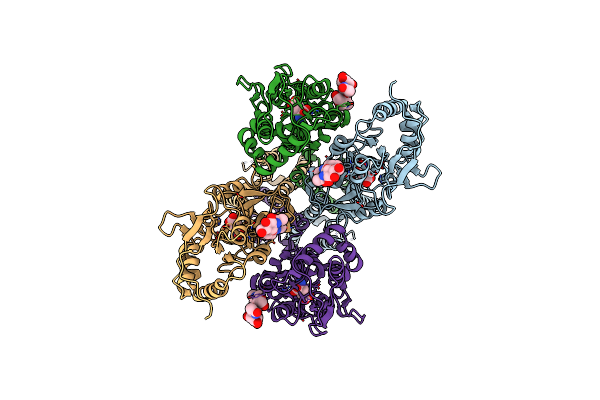
Structure Of The Glutamate Receptor-Like Channel Atglr3.4
Organism: Arabidopsis thaliana
Method: ELECTRON MICROSCOPY Release Date: 2021-07-28 Classification: TRANSPORT PROTEIN Ligands: GLU, NAG, GSH |
Organism: Arabidopsis thaliana
Method: ELECTRON MICROSCOPY
Release Date: 2021-07-28
Ligands: GLU, NAG, GSH
 |
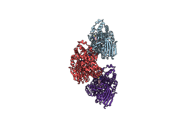
Structure Of The Glutamate Receptor-Like Channel Atglr3.4
Organism: Arabidopsis thaliana
Method: ELECTRON MICROSCOPY Release Date: 2021-07-28 Classification: TRANSPORT PROTEIN Ligands: GLU |
Organism: Arabidopsis thaliana
Method: ELECTRON MICROSCOPY
Release Date: 2021-07-28
Ligands: GLU
 |
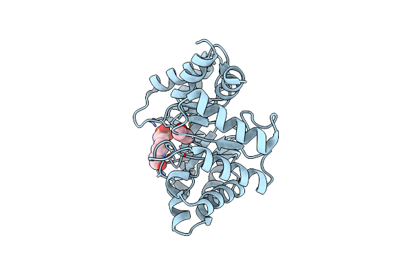
The Effect Of Modifier Structure On The Activation Of Leukotriene A4 Hydrolase Aminopeptidase Activity.
Organism: Homo sapiens
Method: X-RAY DIFFRACTION Resolution:2.84 Å Release Date: 2019-12-04 Classification: HYDROLASE Ligands: LNJ, ZN |
Organism: Homo sapiens
Method: X-RAY DIFFRACTION
Release Date: 2019-12-04
Ligands: LNJ, ZN
 |
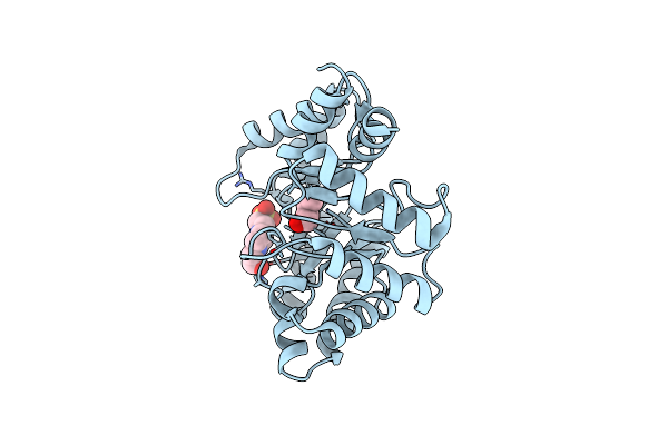
Crystal Structure Of Deoxyribose-Phosphate Aldolase From Escherichia Coli (K58E-Y96W Mutant)
Organism: Escherichia coli
Method: X-RAY DIFFRACTION Resolution:1.10 Å Release Date: 2016-05-04 Classification: LYASE Ligands: BU2, EPE |
Organism: Escherichia coli
Method: X-RAY DIFFRACTION
Release Date: 2016-05-04
Ligands: BU2, EPE
 |
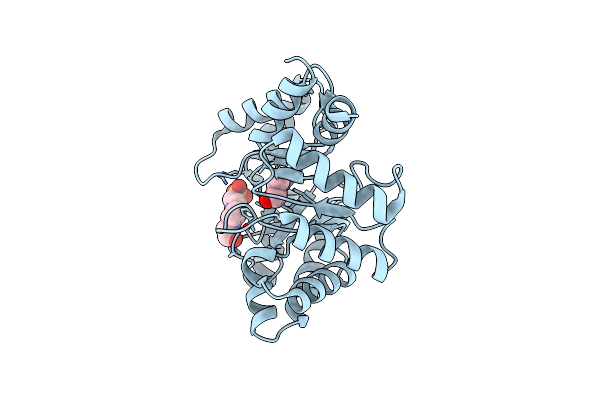
Crystal Structure Of Deoxyribose-Phosphate Aldolase From Escherichia Coli (K58E-Y96W Mutant) After Acetaldehyde Treatment
Organism: Escherichia coli k-12
Method: X-RAY DIFFRACTION Resolution:1.25 Å Release Date: 2016-05-04 Classification: LYASE Ligands: 1BO, EPE |
Organism: Escherichia coli k-12
Method: X-RAY DIFFRACTION
Release Date: 2016-05-04
Ligands: 1BO, EPE
 |
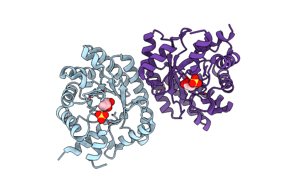
Crystal Structure Of Deoxyribose-Phosphate Aldolase From Escherichia Coli (K58E-Y96W Mutant) After Acetaldehyde Treatment And Heating
Organism: Escherichia coli k-12
Method: X-RAY DIFFRACTION Resolution:1.50 Å Release Date: 2016-05-04 Classification: LYASE Ligands: 1BO, EPE |
Organism: Escherichia coli k-12
Method: X-RAY DIFFRACTION
Release Date: 2016-05-04
Ligands: 1BO, EPE
 |
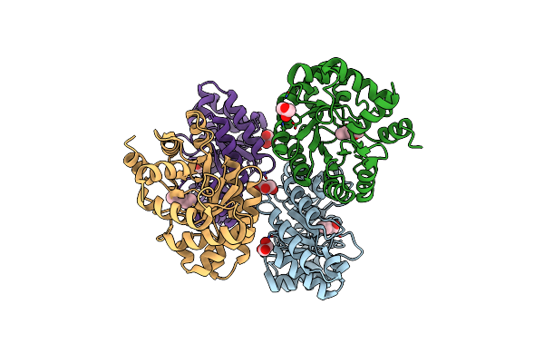
Crystal Structure Of Deoxyribose-Phosphate Aldolase From Colwellia Psychrerythraea (Tetragonal Form)
Organism: Colwellia psychrerythraea
Method: X-RAY DIFFRACTION Resolution:2.11 Å Release Date: 2016-02-03 Classification: LYASE Ligands: UNL, SO4, CO3 |
Organism: Colwellia psychrerythraea
Method: X-RAY DIFFRACTION
Release Date: 2016-02-03
Ligands: UNL, SO4, CO3
 |
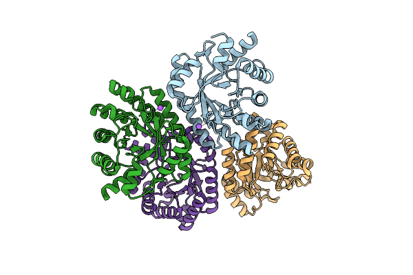
Crystal Structure Of Deoxyribose-Phosphate Aldolase From Colwellia Psychrerythraea (Hexagonal Form)
Organism: Colwellia psychrerythraea (strain 34h / atcc baa-681)
Method: X-RAY DIFFRACTION Resolution:2.10 Å Release Date: 2016-02-03 Classification: LYASE Ligands: UNL, GOL |
Organism: Colwellia psychrerythraea (strain 34h / atcc baa-681)
Method: X-RAY DIFFRACTION
Release Date: 2016-02-03
Ligands: UNL, GOL
 |
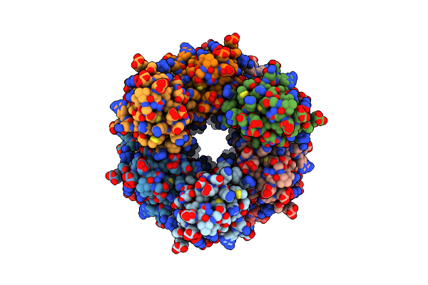
Crystal Structure Of Deoxyribose-Phosphate Aldolase From Shewanella Halifaxensis
Organism: Shewanella halifaxensis
Method: X-RAY DIFFRACTION Resolution:1.76 Å Release Date: 2016-02-03 Classification: LYASE |
Organism: Shewanella halifaxensis
Method: X-RAY DIFFRACTION
Release Date: 2016-02-03
 |
Structure And Assembly Of The Mouse Asc Filament By Combined Nmr Spectroscopy And Cryo-Electron Microscopy
Organism: Mus musculus
Method: SOLID-STATE NMR, ELECTRON MICROSCOPY Release Date: 2015-10-14 Classification: APOPTOSIS |
Organism: Mus musculus
Method: SOLID-STATE NMR, ELECTRON MICROSCOPY
Release Date: 2015-10-14