

Search Count: 36
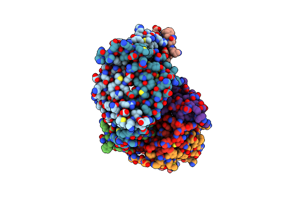 |
D-Protein Rfx-V2 Bound To The Vegfr1 Domain 3 Site On Vegf-A
Organism: Homo sapiens, Synthetic construct
Method: X-RAY DIFFRACTION Resolution:2.90 Å Release Date: 2021-03-03 Classification: BIOSYNTHETIC PROTEIN |
Organism: Homo sapiens, Synthetic construct
Method: X-RAY DIFFRACTION
Release Date: 2021-03-03
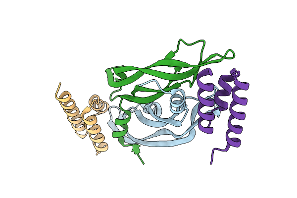 |
D-Protein Rfx-V1 Bound To The Vegfr1 Domain 2 Site On Vegf-A
Organism: Homo sapiens, Synthetic construct
Method: X-RAY DIFFRACTION Resolution:2.31 Å Release Date: 2021-02-17 Classification: BIOSYNTHETIC PROTEIN |
Organism: Homo sapiens, Synthetic construct
Method: X-RAY DIFFRACTION
Release Date: 2021-02-17
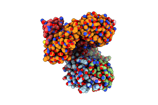 |
Context-Independent Anti-Hypusine Antibody Fabhpu98.61 In Complex With Hypusine
Organism: Oryctolagus cuniculus, Synthetic construct
Method: X-RAY DIFFRACTION Resolution:1.90 Å Release Date: 2016-02-17 Classification: IMMUNE SYSTEM Ligands: EDO, 5CT |
Organism: Oryctolagus cuniculus, Synthetic construct
Method: X-RAY DIFFRACTION
Release Date: 2016-02-17
Ligands: EDO, 5CT
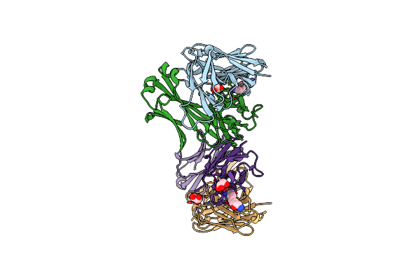 |
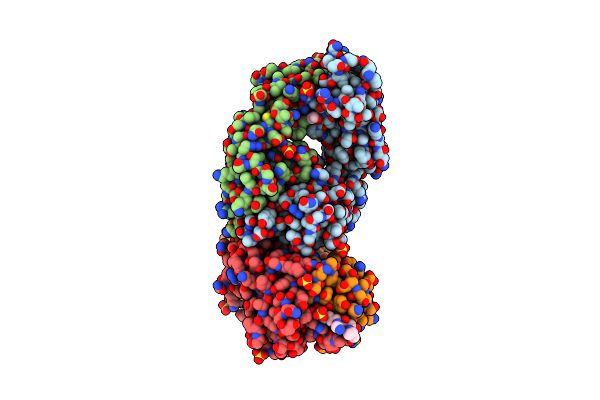
Context-Independent Anti-Hypusine Antibody Fabhpu24 In Complex With Hypusine
Organism: Oryctolagus cuniculus
Method: X-RAY DIFFRACTION Resolution:1.99 Å Release Date: 2016-01-20 Classification: IMMUNE SYSTEM Ligands: 5CT, GOL |
Organism: Oryctolagus cuniculus
Method: X-RAY DIFFRACTION
Release Date: 2016-01-20
Ligands: 5CT, GOL
 |
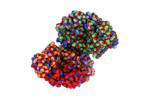
Context-Independent Anti-Hypusine Antibody Fabhpu98 In Complex With Hypusine
Organism: Oryctolagus cuniculus, Synthetic construct
Method: X-RAY DIFFRACTION Resolution:1.95 Å Release Date: 2016-01-20 Classification: IMMUNE SYSTEM Ligands: EDO, SO4 |
Organism: Oryctolagus cuniculus, Synthetic construct
Method: X-RAY DIFFRACTION
Release Date: 2016-01-20
Ligands: EDO, SO4
 |
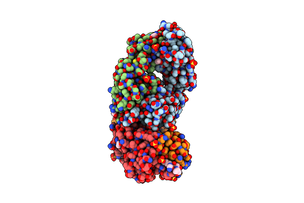
Context-Independent Anti-Hypusine Antibody Fabhpu24.B In Complex With Hypusine
Organism: Oryctolagus cuniculus, Synthetic construct
Method: X-RAY DIFFRACTION Resolution:2.40 Å Release Date: 2016-01-20 Classification: IMMUNE SYSTEM |
Organism: Oryctolagus cuniculus, Synthetic construct
Method: X-RAY DIFFRACTION
Release Date: 2016-01-20
 |
Context-Independent Anti-Hypusine Antibody Fabhpu98 In Complex With Deoxyhypusine
Organism: Oryctolagus cuniculus, Synthetic construct
Method: X-RAY DIFFRACTION Resolution:2.00 Å Release Date: 2016-01-20 Classification: IMMUNE SYSTEM Ligands: EDO, SO4 |
Organism: Oryctolagus cuniculus, Synthetic construct
Method: X-RAY DIFFRACTION
Release Date: 2016-01-20
Ligands: EDO, SO4
 |
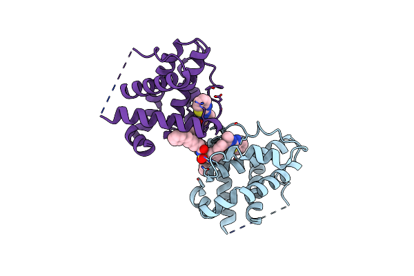
Discovery Of A Potent And Selective Bcl-Xl Inhibitor That Demonstrates Thrombocytopenia And Inhibits Tumor Growth In Vivo
Organism: Homo sapiens
Method: X-RAY DIFFRACTION Resolution:2.10 Å Release Date: 2015-07-22 Classification: APOPTOSIS/INHIBITOR Ligands: 3CQ |
Organism: Homo sapiens
Method: X-RAY DIFFRACTION
Release Date: 2015-07-22
Ligands: 3CQ
 |
Crystal Structure Of Bcl-Xl In Complex With Inhibitor (Compound 2).
Organism: Homo sapiens
Method: X-RAY DIFFRACTION Resolution:2.48 Å Release Date: 2013-04-24 Classification: APOPTOSIS Ligands: H1I |
Organism: Homo sapiens
Method: X-RAY DIFFRACTION
Release Date: 2013-04-24
Ligands: H1I
 |
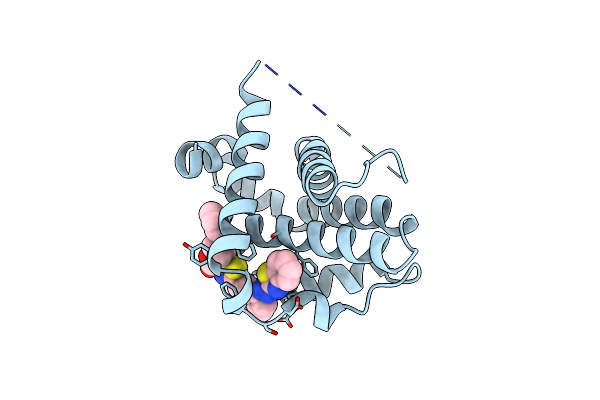
Crystal Structure Of Bcl-Xl In Complex With Inhibitor (Compound 3)
Organism: Homo sapiens
Method: X-RAY DIFFRACTION Resolution:2.29 Å Release Date: 2013-04-24 Classification: APOPTOSIS Ligands: H0Y, SO4, EDO |
Organism: Homo sapiens
Method: X-RAY DIFFRACTION
Release Date: 2013-04-24
Ligands: H0Y, SO4, EDO
 |
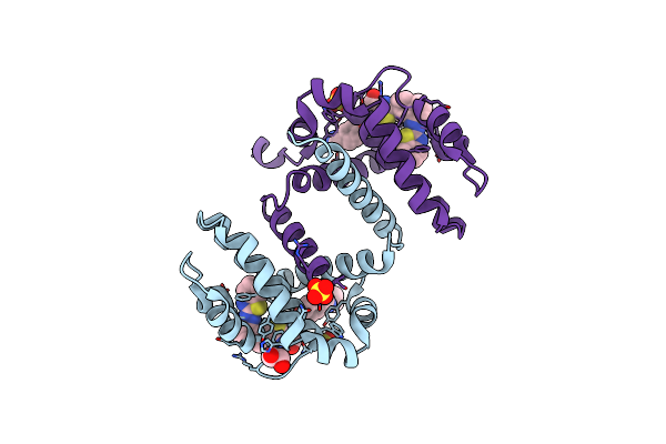
Crystal Structure Of Bcl-Xl In Complex With Inhibitor (Compound 6)
Organism: Homo sapiens
Method: X-RAY DIFFRACTION Resolution:2.60 Å Release Date: 2013-04-24 Classification: APOPTOSIS Ligands: X8U |
Organism: Homo sapiens
Method: X-RAY DIFFRACTION
Release Date: 2013-04-24
Ligands: X8U
 |
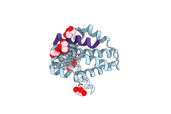
Crystal Structure Of Bcl-Xl In Complex With Inhibitor (Wehi-539)
Organism: Homo sapiens
Method: X-RAY DIFFRACTION Resolution:2.03 Å Release Date: 2013-04-24 Classification: APOPTOSIS Ligands: X0B, EDO, SO4 |
Organism: Homo sapiens
Method: X-RAY DIFFRACTION
Release Date: 2013-04-24
Ligands: X0B, EDO, SO4
 |
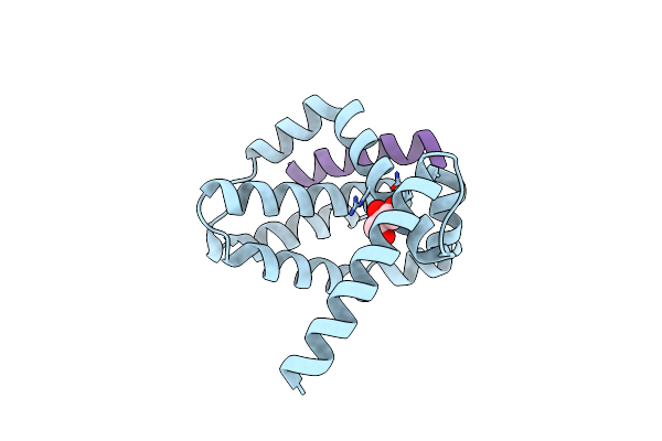
Structure Of Bcl-Xl Bound To Bimsahb
Organism: Homo sapiens
Method: X-RAY DIFFRACTION Resolution:1.80 Å Release Date: 2012-11-28 Classification: APOPTOSIS Ligands: GOL |
Organism: Homo sapiens
Method: X-RAY DIFFRACTION
Release Date: 2012-11-28
Ligands: GOL
 |
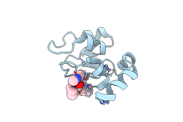
Structure Of Bcl-Xl Bound To Bimlock
Organism: Homo sapiens
Method: X-RAY DIFFRACTION Resolution:1.90 Å Release Date: 2012-11-28 Classification: APOPTOSIS Ligands: GOL |
Organism: Homo sapiens
Method: X-RAY DIFFRACTION
Release Date: 2012-11-28
Ligands: GOL
 |
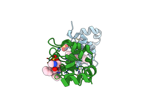
Crystal Structure Of Ciap1 Bir3 Bound To Gdc0152
Organism: Homo sapiens
Method: X-RAY DIFFRACTION Resolution:1.79 Å Release Date: 2012-02-22 Classification: Apoptosis Inhibitor Ligands: ZN |
Organism: Homo sapiens
Method: X-RAY DIFFRACTION
Release Date: 2012-02-22
Ligands: ZN
 |
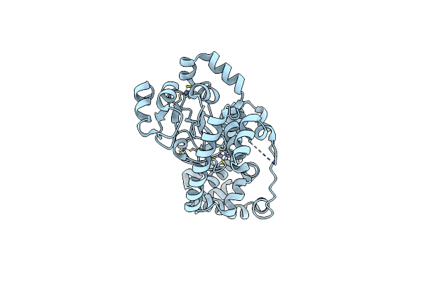
Crystal Structure Of The Bir Domain Of Mliap Bound To Gdc0152
Organism: Homo sapiens
Method: X-RAY DIFFRACTION Resolution:1.71 Å Release Date: 2012-02-22 Classification: Apoptosis Inhibitor Ligands: ZN, SO4 |
Organism: Homo sapiens
Method: X-RAY DIFFRACTION
Release Date: 2012-02-22
Ligands: ZN, SO4
 |
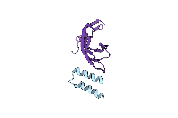
Iap Antagonist-Induced Conformational Change In Ciap1 Promotes E3 Ligase Activation Via Dimerization
Organism: Homo sapiens
Method: X-RAY DIFFRACTION Resolution:1.90 Å Release Date: 2011-11-02 Classification: APOPTOSIS Ligands: ZN |
Organism: Homo sapiens
Method: X-RAY DIFFRACTION
Release Date: 2011-11-02
Ligands: ZN
 |
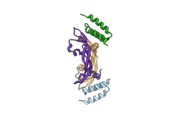
The Development Of Peptide-Based Tools For The Analysis Of Angiogenesis
Organism: Homo sapiens
Method: X-RAY DIFFRACTION Resolution:2.90 Å Release Date: 2011-08-17 Classification: SIGNALING PROTEIN |
Organism: Homo sapiens
Method: X-RAY DIFFRACTION
Release Date: 2011-08-17
 |
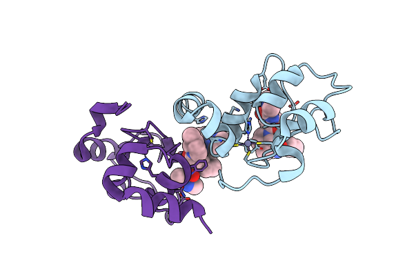
The Development Of Peptide-Based Tools For The Analysis Of Angiogenesis
Organism: Homo sapiens
Method: X-RAY DIFFRACTION Resolution:2.55 Å Release Date: 2011-08-17 Classification: SIGNALING PROTEIN |
Organism: Homo sapiens
Method: X-RAY DIFFRACTION
Release Date: 2011-08-17
 |
Crystal Structure Of Xiap Bir3 With Cs3
Organism: Homo sapiens
Method: X-RAY DIFFRACTION Resolution:1.80 Å Release Date: 2009-06-16 Classification: LIGASE Ligands: ZN, 9JZ |
Organism: Homo sapiens
Method: X-RAY DIFFRACTION
Release Date: 2009-06-16
Ligands: ZN, 9JZ