

Search Count: 19
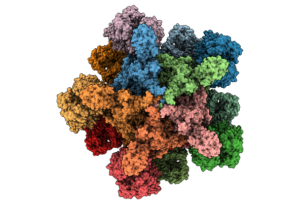 |
Yersinia Entomophaga Holotoxin Complex In Prepore Conformation
Organism: Yersinia entomophaga
Method: ELECTRON MICROSCOPY Release Date: 2025-12-31 Classification: TOXIN |
Organism: Yersinia entomophaga
Method: ELECTRON MICROSCOPY
Release Date: 2025-12-31
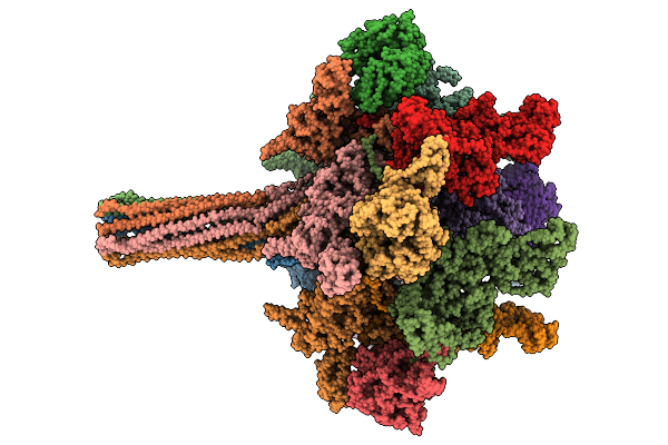 |
Yersinia Entomophaga Holotoxin Complex In Pore Conformation
Organism: Yersinia entomophaga
Method: ELECTRON MICROSCOPY Release Date: 2025-12-31 Classification: TOXIN |
Organism: Yersinia entomophaga
Method: ELECTRON MICROSCOPY
Release Date: 2025-12-31
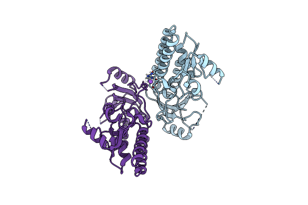 |
Crystal Structure Of Acinetobacter Baumannii Znua In The Metal-Free State
Organism: Acinetobacter baumannii
Method: X-RAY DIFFRACTION Resolution:2.13 Å Release Date: 2022-10-12 Classification: METAL BINDING PROTEIN |
Organism: Acinetobacter baumannii
Method: X-RAY DIFFRACTION
Release Date: 2022-10-12
 |
Crystal Structure Of The Molybdate-Binding Periplasmic Protein Moda From The Bacteria Pseudomonsa Aeruginosa In Ligand-Free Form
Organism: Pseudomonas aeruginosa pa1
Method: X-RAY DIFFRACTION Resolution:1.78 Å Release Date: 2022-05-04 Classification: METAL BINDING PROTEIN Ligands: GOL, NH4, SO4 |
Organism: Pseudomonas aeruginosa pa1
Method: X-RAY DIFFRACTION
Release Date: 2022-05-04
Ligands: GOL, NH4, SO4
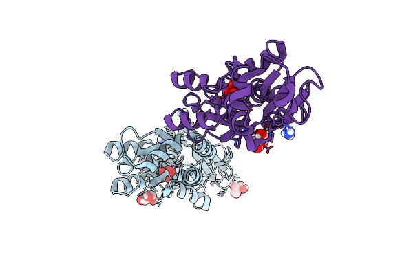 |
Crystal Structure Of The Molybdate-Binding Periplasmic Protein Moda From The Bacteria Pseudomonsa Aeruginosa In Chromate-Bound Form
Organism: Pseudomonas aeruginosa pa1
Method: X-RAY DIFFRACTION Resolution:1.90 Å Release Date: 2022-05-04 Classification: METAL BINDING PROTEIN Ligands: CQ4, NH4, GOL |
Organism: Pseudomonas aeruginosa pa1
Method: X-RAY DIFFRACTION
Release Date: 2022-05-04
Ligands: CQ4, NH4, GOL
 |
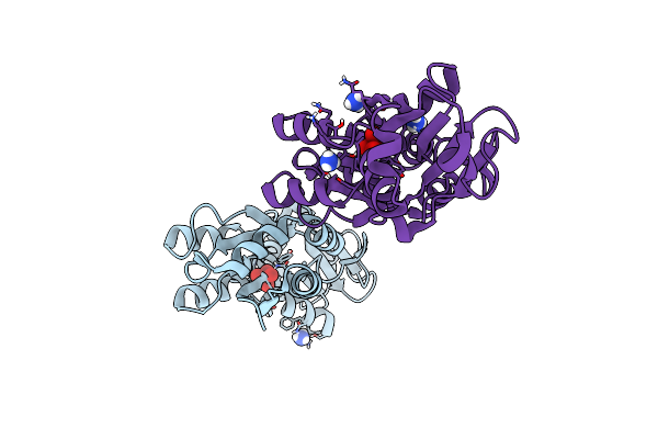
Crystal Structure Of The Molybdate-Binding Periplasmic Protein Moda From The Bacteria Pseudomonsa Aeruginosa In Molybdate-Bound Form
Organism: Pseudomonas aeruginosa pa1
Method: X-RAY DIFFRACTION Resolution:2.50 Å Release Date: 2022-05-04 Classification: METAL BINDING PROTEIN Ligands: NH4, MOO, GOL |
Organism: Pseudomonas aeruginosa pa1
Method: X-RAY DIFFRACTION
Release Date: 2022-05-04
Ligands: NH4, MOO, GOL
 |
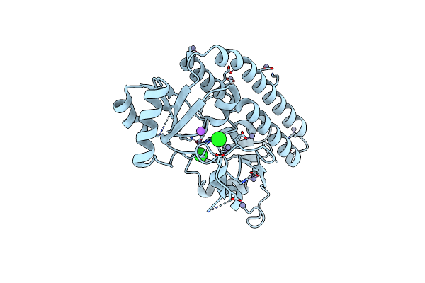
Crystal Structure Of The Molybdate-Binding Periplasmic Protein Moda From The Bacteria Pseudomonsa Aeruginosa In Tungstate-Bound Form
Organism: Pseudomonas aeruginosa pa1
Method: X-RAY DIFFRACTION Resolution:2.16 Å Release Date: 2022-05-04 Classification: METAL BINDING PROTEIN Ligands: WO4, NH4 |
Organism: Pseudomonas aeruginosa pa1
Method: X-RAY DIFFRACTION
Release Date: 2022-05-04
Ligands: WO4, NH4
 |
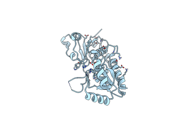
Crystal Structure Of The Zn(Ii)-Bound Adcaii H65A Mutant Variant Of Streptococcus Pneumoniae
Organism: Streptococcus pseudopneumoniae 5247
Method: X-RAY DIFFRACTION Resolution:2.40 Å Release Date: 2021-09-29 Classification: METAL BINDING PROTEIN Ligands: ZN, CL, NA |
Organism: Streptococcus pseudopneumoniae 5247
Method: X-RAY DIFFRACTION
Release Date: 2021-09-29
Ligands: ZN, CL, NA
 |
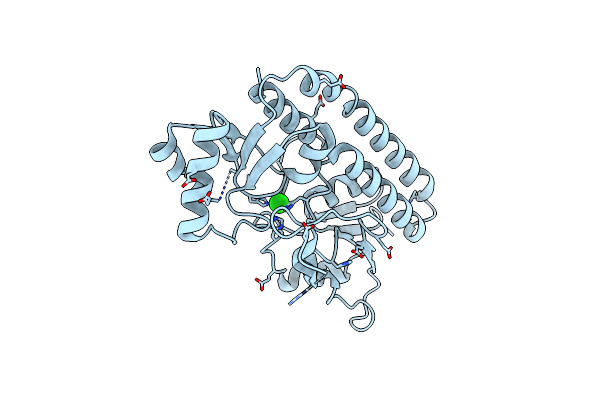
Crystal Structure Of The Zn(Ii)-Bound Adcaii H205L Mutant Variant Of Streptococcus Pneumoniae
Organism: Streptococcus pseudopneumoniae 5247
Method: X-RAY DIFFRACTION Resolution:3.37 Å Release Date: 2021-09-29 Classification: METAL BINDING PROTEIN Ligands: ZN |
Organism: Streptococcus pseudopneumoniae 5247
Method: X-RAY DIFFRACTION
Release Date: 2021-09-29
Ligands: ZN
 |
Crystal Structure Of The Zn(Ii)-Bound Adcaii E280Q Mutant Variant Of Streptococcus Pneumoniae
Organism: Streptococcus pseudopneumoniae 5247
Method: X-RAY DIFFRACTION Resolution:3.12 Å Release Date: 2021-09-29 Classification: METAL BINDING PROTEIN Ligands: ZN, CL |
Organism: Streptococcus pseudopneumoniae 5247
Method: X-RAY DIFFRACTION
Release Date: 2021-09-29
Ligands: ZN, CL
 |
Crystal Structure Of The Zn(Ii)-Bound Znua-Like Domain Of Streptococcus Pneumoniae Adca
Organism: Streptococcus pneumoniae (strain atcc baa-255 / r6)
Method: X-RAY DIFFRACTION Resolution:2.03 Å Release Date: 2020-12-23 Classification: METAL BINDING PROTEIN Ligands: ZN |
Organism: Streptococcus pneumoniae (strain atcc baa-255 / r6)
Method: X-RAY DIFFRACTION
Release Date: 2020-12-23
Ligands: ZN
 |
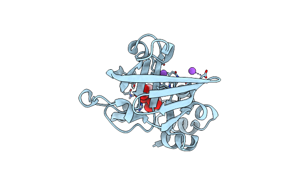
Crystal Structure Of Zn(Ii)-Bound Adca From Streptococcus Pneumoniae
Organism: Streptococcus pneumoniae (strain atcc baa-255 / r6)
Method: X-RAY DIFFRACTION Resolution:1.58 Å Release Date: 2020-12-23 Classification: METAL BINDING PROTEIN Ligands: ZN, CL |
Organism: Streptococcus pneumoniae (strain atcc baa-255 / r6)
Method: X-RAY DIFFRACTION
Release Date: 2020-12-23
Ligands: ZN, CL
 |
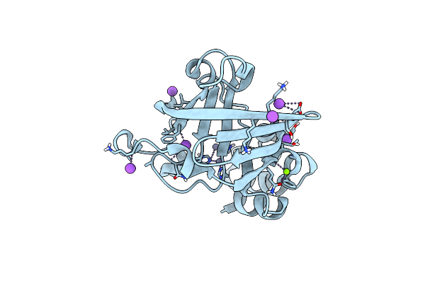
Crystal Structure Of The Zint-Like Domain Of Streptococcus Pneumoniae Adca In The Apo Form
Organism: Streptococcus pneumoniae (strain atcc baa-255 / r6)
Method: X-RAY DIFFRACTION Resolution:1.01 Å Release Date: 2020-12-23 Classification: METAL BINDING PROTEIN Ligands: EDT, NA |
Organism: Streptococcus pneumoniae (strain atcc baa-255 / r6)
Method: X-RAY DIFFRACTION
Release Date: 2020-12-23
Ligands: EDT, NA
 |
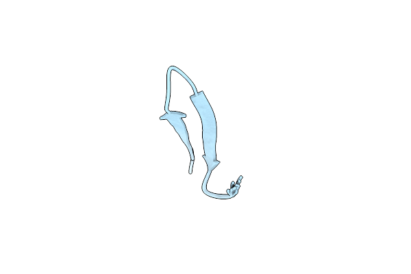
Crystal Structure Of Zn(Ii)-Bound Zint-Like Domain Of Streptococcus Pneumoniae Adca
Organism: Streptococcus pneumoniae (strain atcc baa-255 / r6)
Method: X-RAY DIFFRACTION Resolution:1.10 Å Release Date: 2020-12-23 Classification: METAL BINDING PROTEIN Ligands: ZN, NA, MG |
Organism: Streptococcus pneumoniae (strain atcc baa-255 / r6)
Method: X-RAY DIFFRACTION
Release Date: 2020-12-23
Ligands: ZN, NA, MG
 |
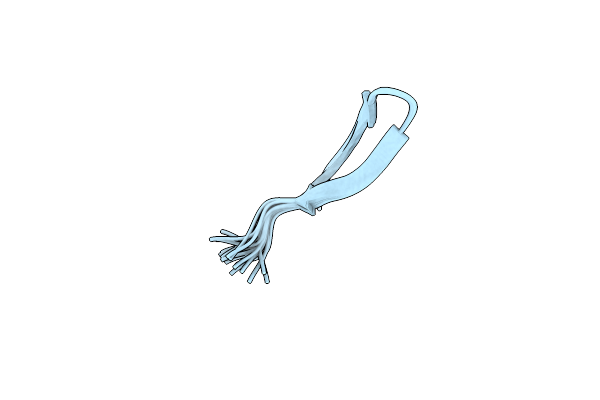
Solution Structure Of Gomesin At 278 K
Organism: Acanthoscurria gomesiana
Method: SOLUTION NMR Release Date: 2019-11-06 Classification: TOXIN |
Organism: Acanthoscurria gomesiana
Method: SOLUTION NMR
Release Date: 2019-11-06
 |
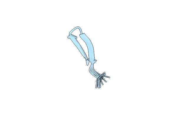
Solution Structure Of Gomesin At 298 K
Organism: Acanthoscurria gomesiana
Method: SOLUTION NMR Release Date: 2019-11-06 Classification: TOXIN |
Organism: Acanthoscurria gomesiana
Method: SOLUTION NMR
Release Date: 2019-11-06
 |
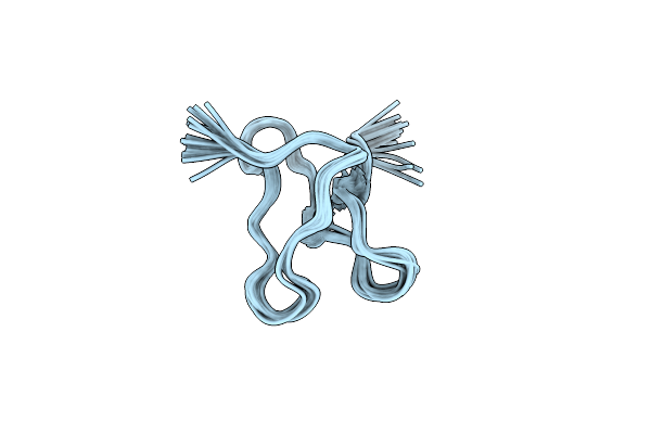
Solution Structure Of Gomesin At 310K
Organism: Acanthoscurria gomesiana
Method: SOLUTION NMR Release Date: 2019-11-06 Classification: TOXIN |
Organism: Acanthoscurria gomesiana
Method: SOLUTION NMR
Release Date: 2019-11-06
 |
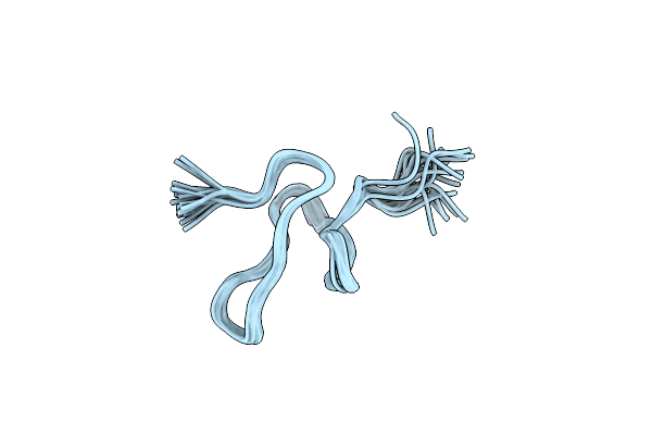
Solution Nmr Structure Of Ghwtx-Iv
|
Organism: Haplopelma schmidti
Method: SOLUTION NMR
Release Date: 2017-02-22
 |
Nmr Solution Structure Of Protx-Ii
|
Organism: Thrixopelma pruriens
Method: SOLUTION NMR
Release Date: 2016-07-06