

Search Count: 24
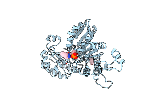 |
Crystal Structure Of Citrobacter Freundii Methionine Gamma-Lyase With C115H Replacement In The Complex With L-Norleucine
Organism: Citrobacter freundii
Method: X-RAY DIFFRACTION Resolution:1.45 Å Release Date: 2017-06-28 Classification: LYASE Ligands: PLP, NLE, PGE, PY6 |
Organism: Citrobacter freundii
Method: X-RAY DIFFRACTION
Release Date: 2017-06-28
Ligands: PLP, NLE, PGE, PY6
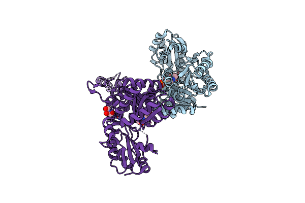 |
Crystal Structure Of O-Acetylhomoserine Sulfhydrolase From Brucella Melitensis At 2.0 A Resolution
Organism: Brucella melitensis biotype 1 (strain 16m / atcc 23456 / nctc 10094)
Method: X-RAY DIFFRACTION Resolution:2.00 Å Release Date: 2017-04-05 Classification: HYDROLASE Ligands: PLP, GOL |
Organism: Brucella melitensis biotype 1 (strain 16m / atcc 23456 / nctc 10094)
Method: X-RAY DIFFRACTION
Release Date: 2017-04-05
Ligands: PLP, GOL
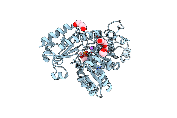 |
Crystal Structure Of Methionine Gamma-Lyase From Citrobacter Freundii With C115A Substitution
Organism: Citrobacter freundii
Method: X-RAY DIFFRACTION Resolution:2.27 Å Release Date: 2016-10-12 Classification: LYASE Ligands: 1PE, PEG, NA, PLP |
Organism: Citrobacter freundii
Method: X-RAY DIFFRACTION
Release Date: 2016-10-12
Ligands: 1PE, PEG, NA, PLP
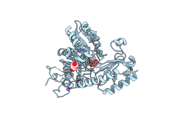 |
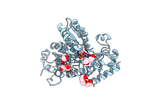
Crystal Structure Of Methionine Gamma-Lyase From Citrobacter Freundii, S339A Mutant
Organism: Citrobacter freundii
Method: X-RAY DIFFRACTION Resolution:1.70 Å Release Date: 2016-08-17 Classification: LYASE Ligands: PEG, NA, PLP |
Organism: Citrobacter freundii
Method: X-RAY DIFFRACTION
Release Date: 2016-08-17
Ligands: PEG, NA, PLP
 |
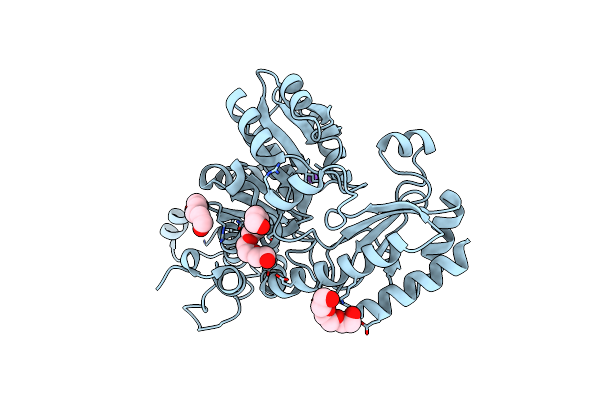
The Crystal Structure Of Methionine Gamma-Lyase From Citrobacter Freundii In Complex With L-Cycloserine Pyridoxal-5'-Phosphate
Organism: Citrobacter freundii
Method: X-RAY DIFFRACTION Resolution:1.60 Å Release Date: 2014-11-26 Classification: LYASE Ligands: LCS, PGE, PEG, CL |
Organism: Citrobacter freundii
Method: X-RAY DIFFRACTION
Release Date: 2014-11-26
Ligands: LCS, PGE, PEG, CL
 |
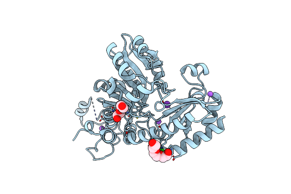
Crystal Structure Of L-Methionine Gamma-Lyase From Citrobacter Freundii Modified By Allicine
Organism: Citrobacter freundii
Method: X-RAY DIFFRACTION Resolution:1.85 Å Release Date: 2014-11-12 Classification: LYASE Ligands: 1PE, PGE, NA |
Organism: Citrobacter freundii
Method: X-RAY DIFFRACTION
Release Date: 2014-11-12
Ligands: 1PE, PGE, NA
 |
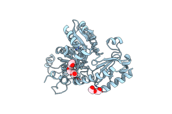
Crystal Structure Of C115A Mutant L-Methionine Gamma-Lyase From Citrobacter Freundii Modified By Allicine
Organism: Citrobacter freundii
Method: X-RAY DIFFRACTION Resolution:1.45 Å Release Date: 2014-11-12 Classification: LYASE Ligands: PGE, NA, CL, K |
Organism: Citrobacter freundii
Method: X-RAY DIFFRACTION
Release Date: 2014-11-12
Ligands: PGE, NA, CL, K
 |
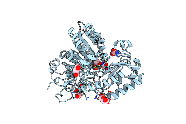
L-Methionine Gamma-Lyase From Citrobacter Freundii With Y58F Substitution
Organism: Citrobacter freundii gtc 09479
Method: X-RAY DIFFRACTION Resolution:1.96 Å Release Date: 2014-11-12 Classification: LYASE Ligands: PEG, 1PE |
Organism: Citrobacter freundii gtc 09479
Method: X-RAY DIFFRACTION
Release Date: 2014-11-12
Ligands: PEG, 1PE
 |
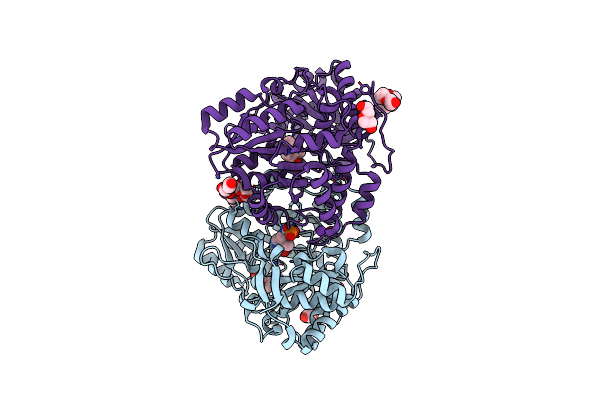
Crystal Structure Of L-Methionine Gamma-Lyase From Citrobacter Freundii With Glycine
Organism: Citrobacter freundii
Method: X-RAY DIFFRACTION Resolution:2.45 Å Release Date: 2013-11-06 Classification: LYASE Ligands: PLG, GLY, PEG |
Organism: Citrobacter freundii
Method: X-RAY DIFFRACTION
Release Date: 2013-11-06
Ligands: PLG, GLY, PEG
 |
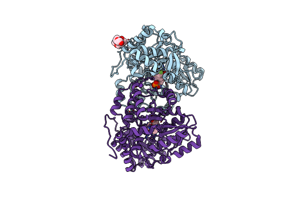
D214A Mutant Of Tyrosine Phenol-Lyase From Citrobacter Freundii
Organism: Citrobacter freundii
Method: X-RAY DIFFRACTION Resolution:1.91 Å Release Date: 2012-05-02 Classification: LYASE Ligands: K, PG4, PEG, PLP, P33 |
Organism: Citrobacter freundii
Method: X-RAY DIFFRACTION
Release Date: 2012-05-02
Ligands: K, PG4, PEG, PLP, P33
 |
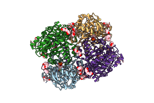
Y71F Mutant Of Tyrosine Phenol-Lyase From Citrobacter Freundii In Complex With Quinonoid Intermediate Formed With 3-Fluoro-L-Tyrosine
Organism: Citrobacter freundii
Method: X-RAY DIFFRACTION Resolution:2.04 Å Release Date: 2011-09-14 Classification: LYASE Ligands: K, P61, PG4, PEG |
Organism: Citrobacter freundii
Method: X-RAY DIFFRACTION
Release Date: 2011-09-14
Ligands: K, P61, PG4, PEG
 |
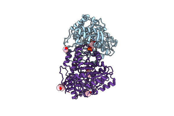
F448H Mutant Of Tyrosine Phenol-Lyase From Citrobacter Freundii In Complex With Quinonoid Intermediate Formed With 3-Fluoro-L-Tyrosine
Organism: Citrobacter freundii
Method: X-RAY DIFFRACTION Resolution:2.00 Å Release Date: 2011-09-14 Classification: LYASE Ligands: P61, PGE, PG4, K, EDO, 1PE, P33, PEG |
Organism: Citrobacter freundii
Method: X-RAY DIFFRACTION
Release Date: 2011-09-14
Ligands: P61, PGE, PG4, K, EDO, 1PE, P33, PEG
 |
Tyrosine Phenol-Lyase From Citrobacter Freundii In Complex With Pyridine N-Oxide And The Quinonoid Intermediate Formed With L-Alanine
Organism: Citrobacter freundii
Method: X-RAY DIFFRACTION Resolution:2.25 Å Release Date: 2011-09-14 Classification: LYASE Ligands: PLP, PLI, K, 9PO, PO4, P33 |
Organism: Citrobacter freundii
Method: X-RAY DIFFRACTION
Release Date: 2011-09-14
Ligands: PLP, PLI, K, 9PO, PO4, P33
 |
Crystal Structure Of L-Methionine Gamma-Lyase From Citrobacter Freundii With S-Ethyl-Cysteine
Organism: Citrobacter freundii
Method: X-RAY DIFFRACTION Resolution:1.80 Å Release Date: 2010-09-08 Classification: LYASE Ligands: ECX, PEG |
Organism: Citrobacter freundii
Method: X-RAY DIFFRACTION
Release Date: 2010-09-08
Ligands: ECX, PEG
 |
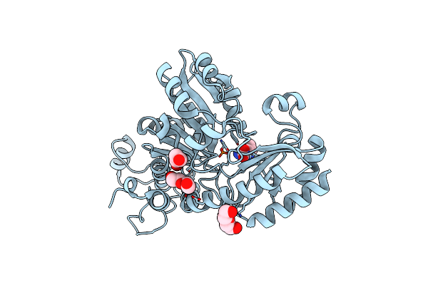
Crystal Structure Of L-Methionine Gamma-Lyase From Citrobacter Freundii With Methionine Phosphinate
Organism: Citrobacter freundii
Method: X-RAY DIFFRACTION Resolution:1.45 Å Release Date: 2010-09-08 Classification: LYASE Ligands: MPJ, PEG |
Organism: Citrobacter freundii
Method: X-RAY DIFFRACTION
Release Date: 2010-09-08
Ligands: MPJ, PEG
 |
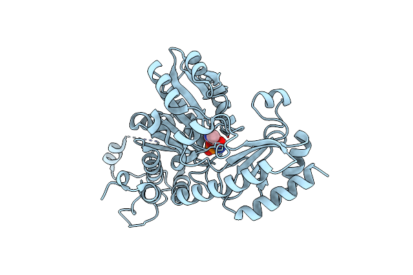
Crystal Structure Of L-Methionine Gamma-Lyase From Citrobacter Freundii With Norleucine
Organism: Citrobacter freundii
Method: X-RAY DIFFRACTION Resolution:1.63 Å Release Date: 2010-09-08 Classification: LYASE Ligands: NLE, PEG |
Organism: Citrobacter freundii
Method: X-RAY DIFFRACTION
Release Date: 2010-09-08
Ligands: NLE, PEG
 |
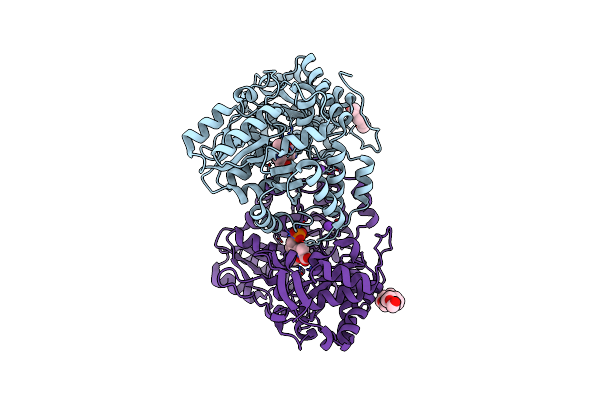
Methionine Gamma-Lyase From Citrobacter Freundii With Pyridoximine-5'-Phosphate
Organism: Citrobacter freundii
Method: X-RAY DIFFRACTION Resolution:1.65 Å Release Date: 2010-08-11 Classification: LYASE Ligands: PZP |
Organism: Citrobacter freundii
Method: X-RAY DIFFRACTION
Release Date: 2010-08-11
Ligands: PZP
 |
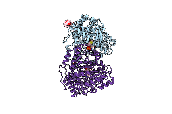
Quinonoid Intermediate Of Citrobacter Freundii Tyrosine Phenol-Lyase Formed With Alanine
Organism: Citrobacter freundii
Method: X-RAY DIFFRACTION Resolution:1.89 Å Release Date: 2008-08-19 Classification: LYASE Ligands: PLI, PGE, K, P33 |
Organism: Citrobacter freundii
Method: X-RAY DIFFRACTION
Release Date: 2008-08-19
Ligands: PLI, PGE, K, P33
 |
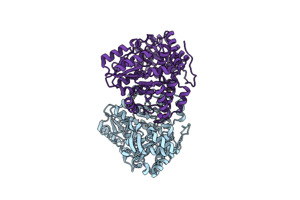
Quinonoid Intermediate Of Citrobacter Freundii Tyrosine Phenol-Lyase Formed With Methionine
Organism: Citrobacter freundii
Method: X-RAY DIFFRACTION Resolution:1.95 Å Release Date: 2008-08-19 Classification: LYASE Ligands: PM9, K, PGE, P33, PLP |
Organism: Citrobacter freundii
Method: X-RAY DIFFRACTION
Release Date: 2008-08-19
Ligands: PM9, K, PGE, P33, PLP
 |
Holo Tyrosine Phenol-Lyase From Citrobacter Freundii At Ph 8.0
Organism: Citrobacter freundii
Method: X-RAY DIFFRACTION Resolution:1.90 Å Release Date: 2006-07-25 Classification: LYASE Ligands: K |
Organism: Citrobacter freundii
Method: X-RAY DIFFRACTION
Release Date: 2006-07-25
Ligands: K