

Search Count: 171
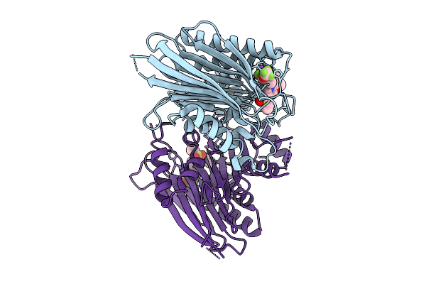 |
Co-Crystal Structure Of Circularly Permuted Human Taspase-1 Bound To Ligand Smdc1014883 (2R)-4-(Ethenesulfonyl)-1-{[3-Fluoro-4-(Trifluoromethoxy)Phenyl]Methyl}-2-[(Prop-2-Yn-1-Yloxy)Methyl]Piperazine
Organism: Homo sapiens
Method: X-RAY DIFFRACTION Resolution:2.15 Å Release Date: 2025-08-13 Classification: HYDROLASE Ligands: CL, NA, R1J |
Organism: Homo sapiens
Method: X-RAY DIFFRACTION
Release Date: 2025-08-13
Ligands: CL, NA, R1J
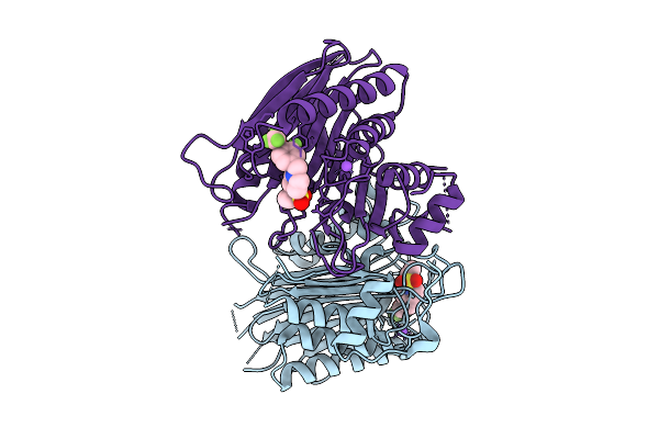 |
Co-Crystal Structure Of Circularly Permuted Human Taspase-1 Bound To Ligand Smdc1014689 1-(Ethenesulfonyl)-4-{[3-Fluoro-4-(Trifluoromethoxy)Phenyl]Methyl}Piperazine
Organism: Homo sapiens
Method: X-RAY DIFFRACTION Resolution:2.45 Å Release Date: 2025-08-13 Classification: HYDROLASE |
Organism: Homo sapiens
Method: X-RAY DIFFRACTION
Release Date: 2025-08-13
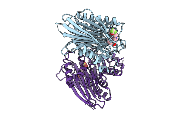 |
Co-Crystal Structure Of Circularly Permuted Human Taspase-1 Bound To Ligand Smdc994967 1-(Ethenesulfonyl)-4-{[4- (Trifluoromethoxy)Phenyl]Methyl}Piperazine
Organism: Homo sapiens
Method: X-RAY DIFFRACTION Resolution:2.10 Å Release Date: 2025-08-13 Classification: HYDROLASE |
Organism: Homo sapiens
Method: X-RAY DIFFRACTION
Release Date: 2025-08-13
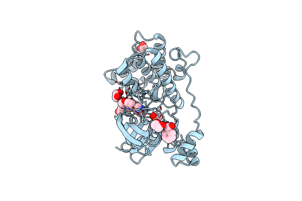 |
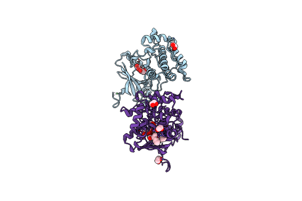
Crystal Structure Of Human Nuak1-Mark3 Kinase Domain Chimera Bound With Azepane (R)-#50 Small Molecule Inhibitor
Organism: Homo sapiens
Method: X-RAY DIFFRACTION Resolution:1.60 Å Release Date: 2024-11-06 Classification: TRANSFERASE/TRANSFERASE INHIBITOR Ligands: X5I, EDO, CL, BEZ |
Organism: Homo sapiens
Method: X-RAY DIFFRACTION
Release Date: 2024-11-06
Ligands: X5I, EDO, CL, BEZ
 |
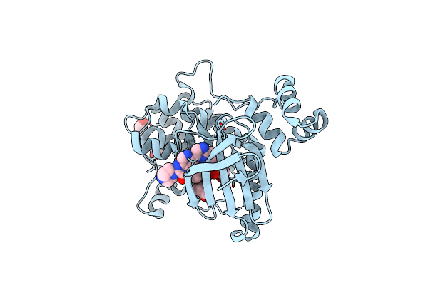
Crystal Structure Of Human Nuak1-Mark3 Kinase Domain Chimera Bound With Small Molecule Inhibitor #10
Organism: Homo sapiens
Method: X-RAY DIFFRACTION Resolution:2.15 Å Release Date: 2024-10-23 Classification: TRANSFERASE/TRANSFERASE INHIBITOR Ligands: EDO, NI, X4W |
Organism: Homo sapiens
Method: X-RAY DIFFRACTION
Release Date: 2024-10-23
Ligands: EDO, NI, X4W
 |
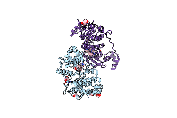
Crystal Structure Of Human Nuak1-Mark3 Kinase Domain Chimera Bound With Small Molecule Inhibitor #65
Organism: Homo sapiens
Method: X-RAY DIFFRACTION Resolution:1.80 Å Release Date: 2024-10-23 Classification: TRANSFERASE/TRANSFERASE INHIBITOR Ligands: X3Q, EDO |
Organism: Homo sapiens
Method: X-RAY DIFFRACTION
Release Date: 2024-10-23
Ligands: X3Q, EDO
 |
Crystal Structure Of Human Nuak1-Mark3 (7 Mutations) Kinase Domain Chimera Bound With Small Molecule Inhibitor #31
Organism: Homo sapiens
Method: X-RAY DIFFRACTION Resolution:1.85 Å Release Date: 2024-10-23 Classification: TRANSFERASE/TRANSFERASE INHIBITOR Ligands: X5N, EDO |
Organism: Homo sapiens
Method: X-RAY DIFFRACTION
Release Date: 2024-10-23
Ligands: X5N, EDO
 |
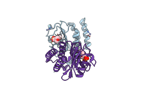
Crystal Structure Of Human Nuak1-Mark3 (6 Mutations) Kinase Domain Chimera Bound With Small Molecule Inhibitor #31
Organism: Homo sapiens
Method: X-RAY DIFFRACTION Resolution:1.90 Å Release Date: 2024-10-23 Classification: TRANSFERASE/TRANSFERASE INHIBITOR Ligands: X4H, EDO |
Organism: Homo sapiens
Method: X-RAY DIFFRACTION
Release Date: 2024-10-23
Ligands: X4H, EDO
 |
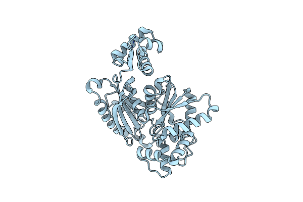
Crystal Structure Of Gaf Domain-Containing Protein, From Klebsiella Pneumoniae
Organism: Klebsiella pneumoniae subsp. pneumoniae hs11286
Method: X-RAY DIFFRACTION Resolution:1.90 Å Release Date: 2023-03-01 Classification: UNKNOWN FUNCTION Ligands: MES, ZN |
Organism: Klebsiella pneumoniae subsp. pneumoniae hs11286
Method: X-RAY DIFFRACTION
Release Date: 2023-03-01
Ligands: MES, ZN
 |
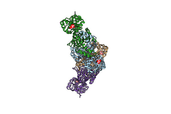
Crystal Structure Of Rok Family Protein From Burkholderia Vietnamiensis G4
Organism: Burkholderia vietnamiensis (strain g4 / lmg 22486)
Method: X-RAY DIFFRACTION Resolution:2.30 Å Release Date: 2021-11-17 Classification: TRANSCRIPTION |
Organism: Burkholderia vietnamiensis (strain g4 / lmg 22486)
Method: X-RAY DIFFRACTION
Release Date: 2021-11-17
 |
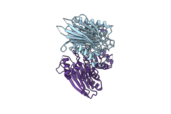
Crystal Structure Of Aspartate-Semialdehyde Dehydrogenase From Acinetobacter Baumannii
Organism: Acinetobacter baumannii
Method: X-RAY DIFFRACTION Resolution:1.80 Å Release Date: 2021-11-03 Classification: OXIDOREDUCTASE Ligands: TLA |
Organism: Acinetobacter baumannii
Method: X-RAY DIFFRACTION
Release Date: 2021-11-03
Ligands: TLA
 |
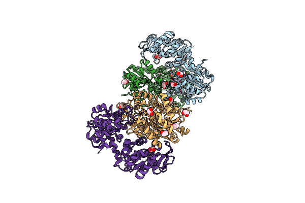
Crystal Structure Of Circularly Permuted Human Taspase-1
Organism: Homo sapiens
Method: X-RAY DIFFRACTION Resolution:2.15 Å Release Date: 2021-03-10 Classification: HYDROLASE |
Organism: Homo sapiens
Method: X-RAY DIFFRACTION
Release Date: 2021-03-10
 |
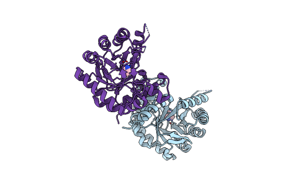
Crystal Structure Of Phosphoglycerate Kinase From Mycobacterium Avium
Organism: Mycobacterium avium (strain 104)
Method: X-RAY DIFFRACTION Resolution:1.60 Å Release Date: 2021-03-03 Classification: TRANSFERASE Ligands: EDO |
Organism: Mycobacterium avium (strain 104)
Method: X-RAY DIFFRACTION
Release Date: 2021-03-03
Ligands: EDO
 |
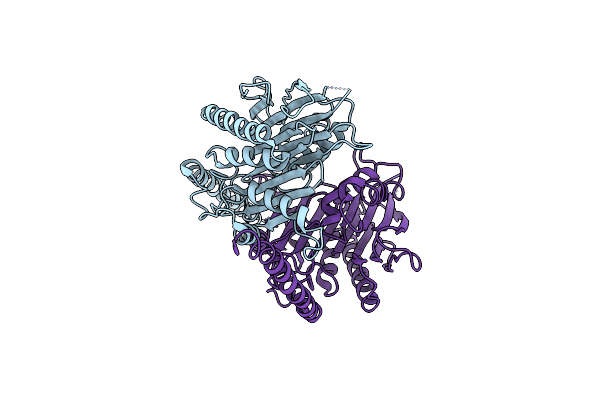
Crystal Structure Of Dihydropteroate Synthase From Stenotrophomonas Maltophilia With Active Site-Bound Imidazole
Organism: Stenotrophomonas maltophilia (strain k279a)
Method: X-RAY DIFFRACTION Resolution:2.35 Å Release Date: 2021-02-03 Classification: TRANSFERASE Ligands: IMD |
Organism: Stenotrophomonas maltophilia (strain k279a)
Method: X-RAY DIFFRACTION
Release Date: 2021-02-03
Ligands: IMD
 |
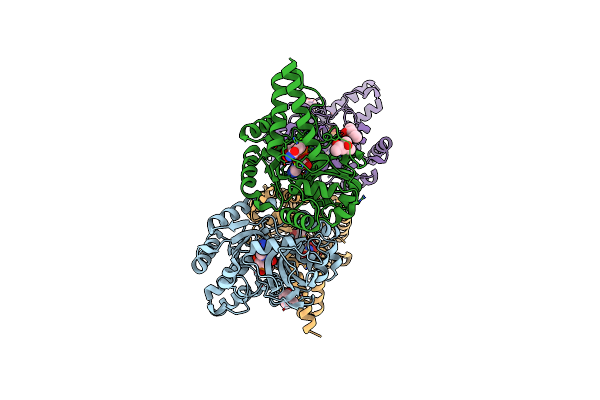
Crystallographic Structure Of The Circularly Permuted Human Taspase1 Protein
Organism: Homo sapiens
Method: X-RAY DIFFRACTION Resolution:3.04 Å Release Date: 2021-01-13 Classification: HYDROLASE |
Organism: Homo sapiens
Method: X-RAY DIFFRACTION
Release Date: 2021-01-13
 |
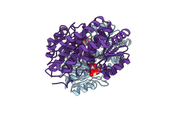
Structure Of Dna Ligase A From Mycobacterium Tuberculosis Bound To Nad
Organism: Mycobacterium tuberculosis (strain atcc 25618 / h37rv)
Method: X-RAY DIFFRACTION Resolution:2.05 Å Release Date: 2020-12-02 Classification: LIGASE Ligands: NAD, MG, CA, MPD |
Organism: Mycobacterium tuberculosis (strain atcc 25618 / h37rv)
Method: X-RAY DIFFRACTION
Release Date: 2020-12-02
Ligands: NAD, MG, CA, MPD
 |
Crystal Structure Of Glucosamine-6-Phosphate Deanimase From Strenotrophomonas Maltophilia
Organism: Stenotrophomonas maltophilia
Method: X-RAY DIFFRACTION Resolution:1.65 Å Release Date: 2020-11-25 Classification: HYDROLASE Ligands: CIT, GOL |
Organism: Stenotrophomonas maltophilia
Method: X-RAY DIFFRACTION
Release Date: 2020-11-25
Ligands: CIT, GOL
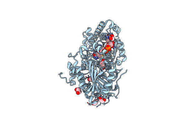 |
Structure Of Urocanate Hydratase From Legionella Pneumophila Bound To Nad
Organism: Legionella pneumophila subsp. pneumophila (strain philadelphia 1 / atcc 33152 / dsm 7513)
Method: X-RAY DIFFRACTION Resolution:1.70 Å Release Date: 2020-10-28 Classification: HYDROLASE Ligands: EDO, NAD, SIN |
Organism: Legionella pneumophila subsp. pneumophila (strain philadelphia 1 / atcc 33152 / dsm 7513)
Method: X-RAY DIFFRACTION
Release Date: 2020-10-28
Ligands: EDO, NAD, SIN
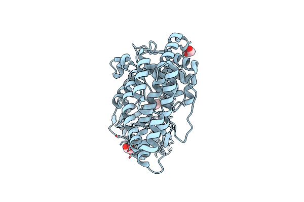 |
Structure Of Choline Kinase From Cryptococcus Neoformans Var. Grubii Serotype A
Organism: Cryptococcus neoformans var. grubii serotype a (strain h99 / atcc 208821 / cbs 10515 / fgsc 9487)
Method: X-RAY DIFFRACTION Resolution:2.25 Å Release Date: 2020-04-22 Classification: TRANSFERASE Ligands: EDO, CL |
Organism: Cryptococcus neoformans var. grubii serotype a (strain h99 / atcc 208821 / cbs 10515 / fgsc 9487)
Method: X-RAY DIFFRACTION
Release Date: 2020-04-22
Ligands: EDO, CL
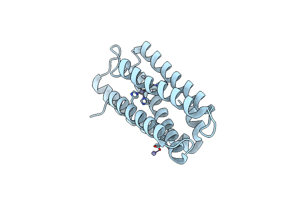 |
Crystal Structure Of Zinc-Bound Hemerythrin Hhe Cation Binding Domain-Containing Protein (Soak): Rv2633C Homolog From Mycobacterium Kansasii
Organism: Mycobacterium kansasii
Method: X-RAY DIFFRACTION Resolution:1.60 Å Release Date: 2020-01-22 Classification: PROTEIN BINDING Ligands: ZN |
Organism: Mycobacterium kansasii
Method: X-RAY DIFFRACTION
Release Date: 2020-01-22
Ligands: ZN