

Search Count: 30
 |
Neutron Structure Of Cellulase Cel6A From Phanerochaete Chrysosporium At Room Temperature
Organism: Phanerodontia chrysosporium
Method: X-RAY DIFFRACTION, NEUTRON DIFFRACTION Resolution:1.36 Å, 1.86 Å Release Date: 2025-03-12 Classification: HYDROLASE |
Organism: Phanerodontia chrysosporium
Method: X-RAY DIFFRACTION, NEUTRON DIFFRACTION
Release Date: 2025-03-12
 |
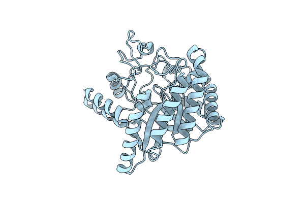
Neutron Structure Of Cellulase Cel6A From Phanerochaete Chrysosporium At Room Temperature, Enzyme-Product Complex
Organism: Phanerodontia chrysosporium
Method: X-RAY DIFFRACTION, NEUTRON DIFFRACTION Resolution:1.40 Å, 1.8 Å Release Date: 2025-03-12 Classification: HYDROLASE Ligands: BGC, NA |
Organism: Phanerodontia chrysosporium
Method: X-RAY DIFFRACTION, NEUTRON DIFFRACTION
Release Date: 2025-03-12
Ligands: BGC, NA
 |
Neutron Structure Of Cellulase Cel6A From Phanerochaete Chrysosporium At Room Temperature, Low-D2O-Solvent
Organism: Phanerodontia chrysosporium
Method: X-RAY DIFFRACTION, NEUTRON DIFFRACTION Resolution:1.40 Å, 2.15 Å Release Date: 2025-03-12 Classification: HYDROLASE |
Organism: Phanerodontia chrysosporium
Method: X-RAY DIFFRACTION, NEUTRON DIFFRACTION
Release Date: 2025-03-12
 |
Neutron Structure Of Cellulase Cel6A From Phanerochaete Chrysosporium At Room Temperature, Enzyme-Product Complex, H2O Solvent
Organism: Phanerodontia chrysosporium
Method: X-RAY DIFFRACTION, NEUTRON DIFFRACTION Resolution:1.80 Å, 2.59 Å Release Date: 2025-03-12 Classification: HYDROLASE Ligands: BGC, NA |
Organism: Phanerodontia chrysosporium
Method: X-RAY DIFFRACTION, NEUTRON DIFFRACTION
Release Date: 2025-03-12
Ligands: BGC, NA
 |
High-Resolution X-Ray Structure Of Cellulase Cel6A From Phanerochaete Chrysosporium At Cryogenic Temperature
Organism: Phanerodontia chrysosporium
Method: X-RAY DIFFRACTION Resolution:0.80 Å Release Date: 2025-03-12 Classification: HYDROLASE Ligands: MPD, ACT |
Organism: Phanerodontia chrysosporium
Method: X-RAY DIFFRACTION
Release Date: 2025-03-12
Ligands: MPD, ACT
 |
High-Resolution X-Ray Structure Of Cellulase Cel6A From Phanerochaete Chrysosporium At Cryogenic Temperature, Enzyme-Product Complex
Organism: Phanerodontia chrysosporium
Method: X-RAY DIFFRACTION Resolution:0.85 Å Release Date: 2025-03-12 Classification: HYDROLASE Ligands: BGC, NA, CL, PEG |
Organism: Phanerodontia chrysosporium
Method: X-RAY DIFFRACTION
Release Date: 2025-03-12
Ligands: BGC, NA, CL, PEG
 |
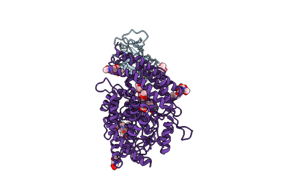
Structure Of The Sars-Cov-2 Eg.5.1 Spike Glycoprotein In Complex With Ace2 (1-Up State)
Organism: Severe acute respiratory syndrome coronavirus 2, Homo sapiens
Method: ELECTRON MICROSCOPY Release Date: 2024-05-01 Classification: VIRAL PROTEIN/PROTEIN BINDING Ligands: NAG |
Organism: Severe acute respiratory syndrome coronavirus 2, Homo sapiens
Method: ELECTRON MICROSCOPY
Release Date: 2024-05-01
Ligands: NAG
 |
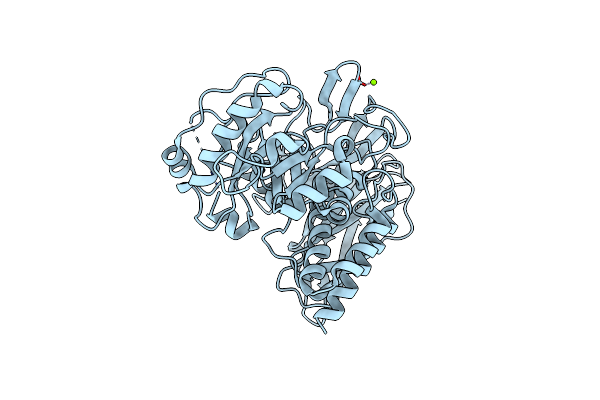
Structure Of The Sars-Cov-2 Eg.5.1 Spike Rbd In Complex With Ace2
Organism: Homo sapiens, Severe acute respiratory syndrome coronavirus 2
Method: ELECTRON MICROSCOPY Release Date: 2024-05-01 Classification: VIRAL PROTEIN/PROTEIN BINDING Ligands: NAG |
Organism: Homo sapiens, Severe acute respiratory syndrome coronavirus 2
Method: ELECTRON MICROSCOPY
Release Date: 2024-05-01
Ligands: NAG
 |
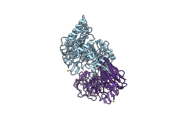
Crystal Structure Of Group 1 Oligosaccharide-Releasing Beta-N-Acetylgalactosaminidase Ngaca From Cohnella Abietis, Apo 1 Form
Organism: Cohnella abietis
Method: X-RAY DIFFRACTION Resolution:1.75 Å Release Date: 2024-04-24 Classification: HYDROLASE Ligands: MG |
Organism: Cohnella abietis
Method: X-RAY DIFFRACTION
Release Date: 2024-04-24
Ligands: MG
 |
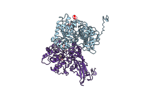
Crystal Structure Of Group 1 Oligosaccharide-Releasing Beta-N-Acetylgalactosaminidase Ngaca From Cohnella Abietis, Apo 2 Form
Organism: Cohnella abietis
Method: X-RAY DIFFRACTION Resolution:1.75 Å Release Date: 2024-04-24 Classification: HYDROLASE Ligands: MG |
Organism: Cohnella abietis
Method: X-RAY DIFFRACTION
Release Date: 2024-04-24
Ligands: MG
 |
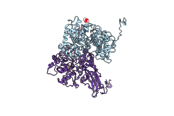
Crystal Structure Of Group 2Oligosaccharide/Monosaccharide-Releasing Beta-N-Acetylhexosaminidase Ngaat From Arabidopsis Thaliana In Complex With Galnac-Thiazoline
Organism: Arabidopsis thaliana
Method: X-RAY DIFFRACTION Resolution:2.20 Å Release Date: 2024-04-24 Classification: HYDROLASE Ligands: GNL, PEG, CL |
Organism: Arabidopsis thaliana
Method: X-RAY DIFFRACTION
Release Date: 2024-04-24
Ligands: GNL, PEG, CL
 |
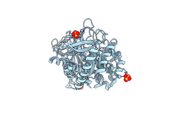
Crystal Structure Of Group 2 Oligosaccharide/Monosaccharide-Releasing Beta-N-Acetylhexosaminidase Ngaat From Arabidopsis Thaliana In Complex With Glcnac-Thiazoline
Organism: Arabidopsis thaliana x arabidopsis lyrata
Method: X-RAY DIFFRACTION Resolution:2.50 Å Release Date: 2024-04-24 Classification: HYDROLASE Ligands: PEG, NGT, CL |
Organism: Arabidopsis thaliana x arabidopsis lyrata
Method: X-RAY DIFFRACTION
Release Date: 2024-04-24
Ligands: PEG, NGT, CL
 |
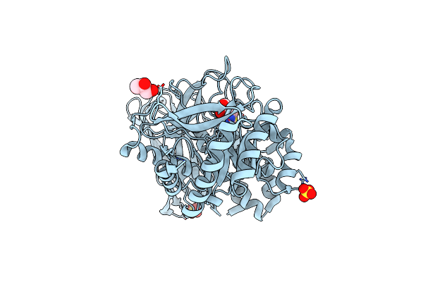
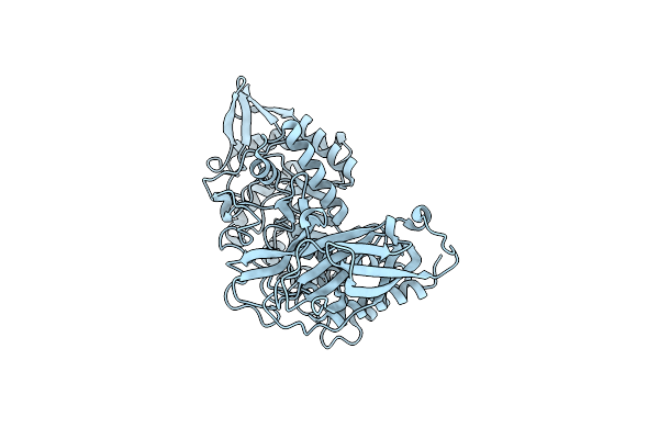
Crystal Structure Of Group 3 Oligosaccharide/Monosaccharide-Releasing Beta-N-Acetylgalactosaminidase Ngadssm, Apo Form
Organism: Metagenome
Method: X-RAY DIFFRACTION Resolution:1.76 Å Release Date: 2024-04-24 Classification: HYDROLASE Ligands: SO4 |
Organism: Metagenome
Method: X-RAY DIFFRACTION
Release Date: 2024-04-24
Ligands: SO4
 |
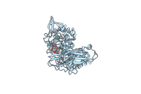
Crystal Structure Of Group 3 Oligosaccharide/Monosaccharide-Releasing Beta-N-Acetylgalactosaminidase Ngadssm In Complex With Galnac-Thiazoline
Organism: Metagenome
Method: X-RAY DIFFRACTION Resolution:1.75 Å Release Date: 2024-04-24 Classification: HYDROLASE Ligands: GNL, GOL, SO4, CL |
Organism: Metagenome
Method: X-RAY DIFFRACTION
Release Date: 2024-04-24
Ligands: GNL, GOL, SO4, CL
 |
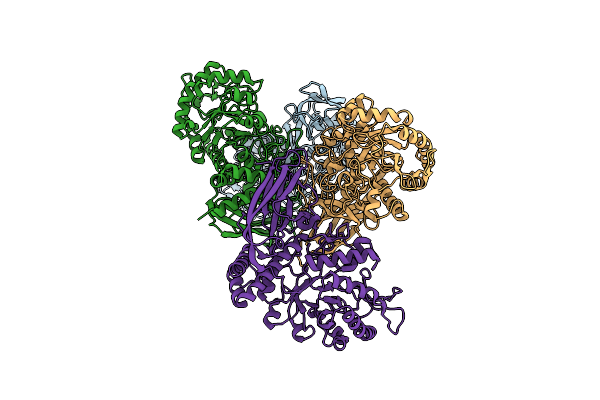
Crystal Structure Of Group 4 Monosaccharide-Releasing Beta-N-Acetylgalactosaminidase Ngap2 From Paenibacillus Sp. Ts12, Apo Form
Organism: Paenibacillus sp. ts12
Method: X-RAY DIFFRACTION Resolution:1.95 Å Release Date: 2024-04-24 Classification: HYDROLASE Ligands: BR |
Organism: Paenibacillus sp. ts12
Method: X-RAY DIFFRACTION
Release Date: 2024-04-24
Ligands: BR
 |
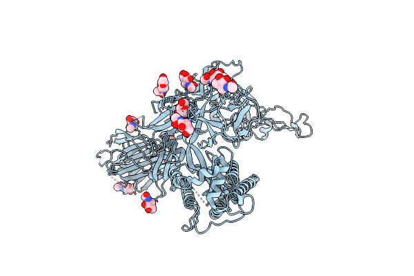
Crystal Structure Of Group 4 Monosaccharide-Releasing Beta-N-Acetylgalactosaminidase Ngap2 From Paenibacillus Sp. Ts12 In Complex With Galnac-Thiazoline
Organism: Paenibacillus sp. ts12
Method: X-RAY DIFFRACTION Resolution:1.65 Å Release Date: 2024-04-24 Classification: HYDROLASE Ligands: GNL, BR |
Organism: Paenibacillus sp. ts12
Method: X-RAY DIFFRACTION
Release Date: 2024-04-24
Ligands: GNL, BR
 |
Crystal Structure Of Group 4 Monosaccharide-Releasing Beta-N-Acetylgalactosaminidase Ngaly From Lacticaseibacillus Yichunensis, Apo Form
Organism: Lacticaseibacillus yichunensis
Method: X-RAY DIFFRACTION Resolution:2.50 Å Release Date: 2024-04-24 Classification: HYDROLASE |
Organism: Lacticaseibacillus yichunensis
Method: X-RAY DIFFRACTION
Release Date: 2024-04-24
 |
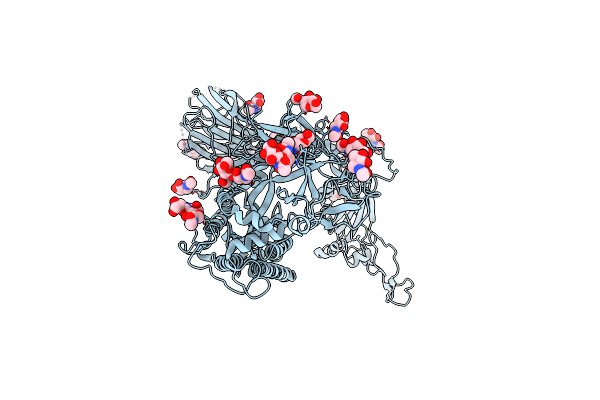
Structure Of The Sars-Cov-2 Eg.5.1 Spike Glycoprotein (Closed-2 State)
Organism: Severe acute respiratory syndrome coronavirus 2
Method: ELECTRON MICROSCOPY Release Date: 2024-04-24 Classification: VIRAL PROTEIN Ligands: NAG |
Organism: Severe acute respiratory syndrome coronavirus 2
Method: ELECTRON MICROSCOPY
Release Date: 2024-04-24
Ligands: NAG
 |
Structure Of The Sars-Cov-2 Eg.5.1 Spike Glycoprotein (Closed-1 State)
Organism: Severe acute respiratory syndrome coronavirus 2
Method: ELECTRON MICROSCOPY Release Date: 2024-04-24 Classification: VIRAL PROTEIN Ligands: NAG |
Organism: Severe acute respiratory syndrome coronavirus 2
Method: ELECTRON MICROSCOPY
Release Date: 2024-04-24
Ligands: NAG
 |
Structure Of The Sars-Cov-2 Xbb.1.5 Spike Glycoprotein (Closed State 1)
Organism: Severe acute respiratory syndrome coronavirus 2
Method: ELECTRON MICROSCOPY Release Date: 2024-01-03 Classification: VIRAL PROTEIN Ligands: NAG |
Organism: Severe acute respiratory syndrome coronavirus 2
Method: ELECTRON MICROSCOPY
Release Date: 2024-01-03
Ligands: NAG