

Search Count: 58
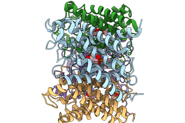 |
Cryo-Em Structure Of Aquaporin 3 At Ph 8.0
Organism: Homo sapiens
Method: ELECTRON MICROSCOPY Resolution:3.20 Å Release Date: 2026-02-18 Classification: MEMBRANE PROTEIN Ligands: GOL, A1H8K |
Organism: Homo sapiens
Method: ELECTRON MICROSCOPY
Release Date: 2026-02-18
Ligands: GOL, A1H8K
 |
Cryo-Em Structure Of Aquaporin 3 At Ph 5.5
Organism: Homo sapiens
Method: ELECTRON MICROSCOPY Resolution:3.20 Å Release Date: 2026-02-18 Classification: MEMBRANE PROTEIN |
Organism: Homo sapiens
Method: ELECTRON MICROSCOPY
Release Date: 2026-02-18
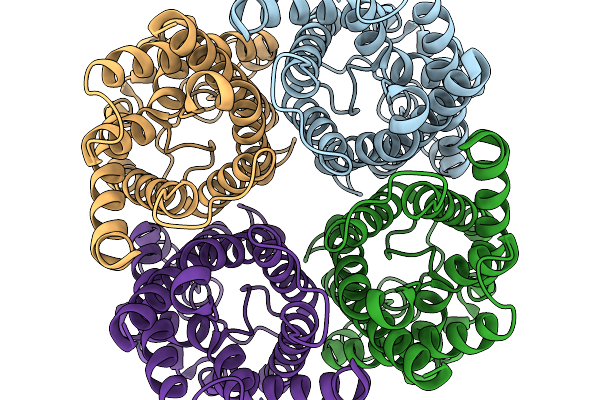 |
Cryo-Em Structure Of Aquaporin 3 At Ph 8.0 With Hydrogen Peroxide
Organism: Homo sapiens
Method: ELECTRON MICROSCOPY Resolution:3.00 Å Release Date: 2026-02-18 Classification: MEMBRANE PROTEIN Ligands: PEO |
Organism: Homo sapiens
Method: ELECTRON MICROSCOPY
Release Date: 2026-02-18
Ligands: PEO
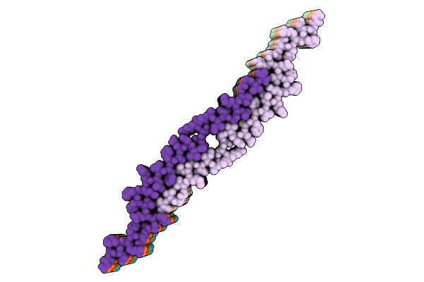 |
The L1 Amyloid-Beta(1-40)Fibril In The Presence Of Anle138B (Post-Treatment)
Organism: Homo sapiens
Method: ELECTRON MICROSCOPY Release Date: 2025-10-15 Classification: PROTEIN FIBRIL |
Organism: Homo sapiens
Method: ELECTRON MICROSCOPY
Release Date: 2025-10-15
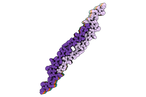 |
The L1 Amyloid-Beta(1-40)Fibril In The Presence Of Anle138B (Pre-Treatment)
Organism: Homo sapiens
Method: ELECTRON MICROSCOPY Release Date: 2025-10-15 Classification: PROTEIN FIBRIL |
Organism: Homo sapiens
Method: ELECTRON MICROSCOPY
Release Date: 2025-10-15
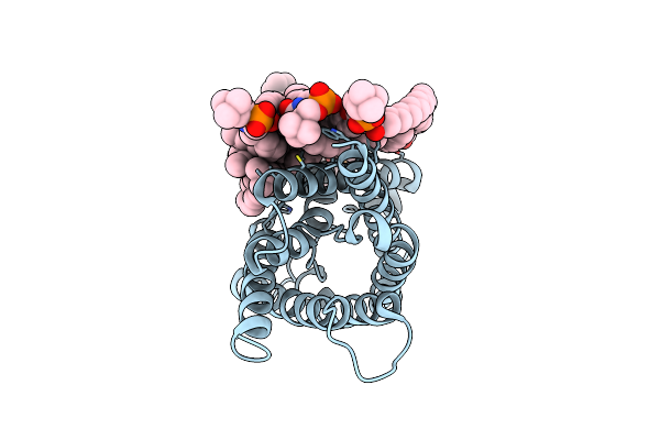 |
Structure Of Lens Aquaporin-0 Array In Sphingomyelin/Cholesterol Bilayer (2Sm:1Chol)
Organism: Ovis aries
Method: ELECTRON CRYSTALLOGRAPHY Resolution:2.50 Å Release Date: 2024-04-24 Classification: MEMBRANE PROTEIN Ligands: HWP, CLR |
Organism: Ovis aries
Method: ELECTRON CRYSTALLOGRAPHY
Release Date: 2024-04-24
Ligands: HWP, CLR
 |
Structure Of Lens Aquaporin-0 Array In Sphingomyelin/Cholesterol Bilayer (1Sm:2Chol)
Organism: Ovis aries
Method: ELECTRON CRYSTALLOGRAPHY Resolution:2.35 Å Release Date: 2024-04-24 Classification: MEMBRANE PROTEIN Ligands: HWP, CLR |
Organism: Ovis aries
Method: ELECTRON CRYSTALLOGRAPHY
Release Date: 2024-04-24
Ligands: HWP, CLR
 |
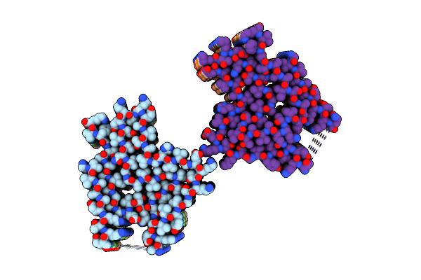
Aqp7_Inhibitor
Organism: Homo sapiens
Method: ELECTRON MICROSCOPY Release Date: 2024-01-31 Classification: MEMBRANE PROTEIN Ligands: T60 |
Organism: Homo sapiens
Method: ELECTRON MICROSCOPY
Release Date: 2024-01-31
Ligands: T60
 |
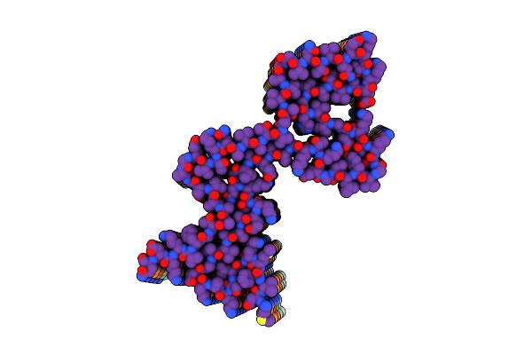
Lipidic Alpha-Synuclein Fibril - Polymorph L2B
Organism: Homo sapiens
Method: ELECTRON MICROSCOPY Release Date: 2022-10-12 Classification: PROTEIN FIBRIL |
Organism: Homo sapiens
Method: ELECTRON MICROSCOPY
Release Date: 2022-10-12
 |
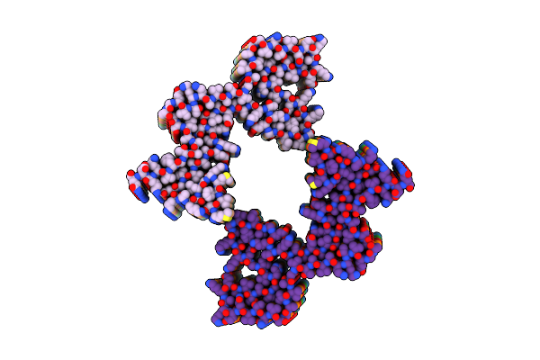
Lipidic Alpha-Synuclein Fibril - Polymorph L1A
Organism: Homo sapiens
Method: ELECTRON MICROSCOPY Release Date: 2022-10-12 Classification: PROTEIN FIBRIL |
Organism: Homo sapiens
Method: ELECTRON MICROSCOPY
Release Date: 2022-10-12
 |
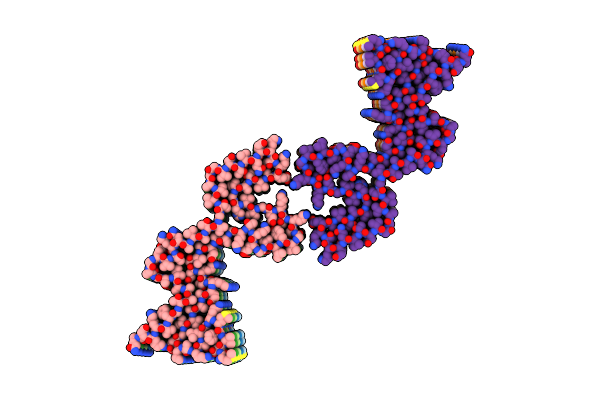
Lipidic Alpha-Synuclein Fibril - Polymorph L1B
Organism: Homo sapiens
Method: ELECTRON MICROSCOPY Release Date: 2022-10-12 Classification: PROTEIN FIBRIL |
Organism: Homo sapiens
Method: ELECTRON MICROSCOPY
Release Date: 2022-10-12
 |
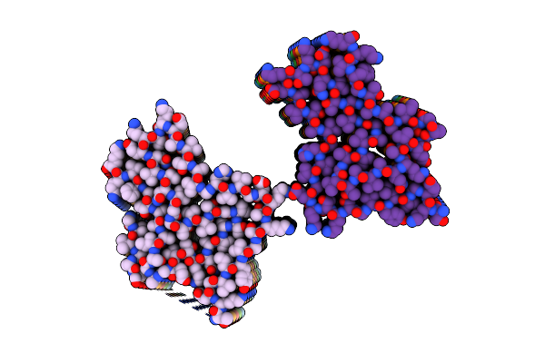
Lipidic Alpha-Synuclein Fibril - Polymorph L1C
Organism: Homo sapiens
Method: ELECTRON MICROSCOPY Release Date: 2022-10-12 Classification: PROTEIN FIBRIL |
Organism: Homo sapiens
Method: ELECTRON MICROSCOPY
Release Date: 2022-10-12
 |
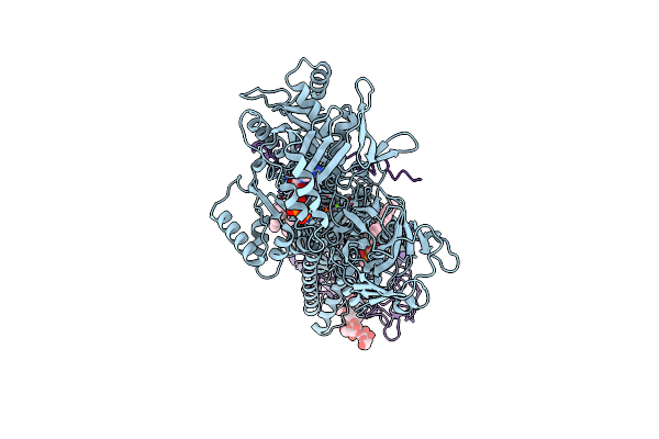
Lipidic Alpha-Synuclein Fibril - Polymorph L3A
Organism: Homo sapiens
Method: ELECTRON MICROSCOPY Release Date: 2022-10-12 Classification: PROTEIN FIBRIL |
Organism: Homo sapiens
Method: ELECTRON MICROSCOPY
Release Date: 2022-10-12
 |
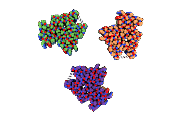
Cryo-Em Structure Of Phophorylated Drs2P-Cdc50P In A Ps And Atp-Bound E2P State
Organism: Saccharomyces cerevisiae (strain atcc 204508 / s288c)
Method: ELECTRON MICROSCOPY Release Date: 2022-08-24 Classification: MEMBRANE PROTEIN Ligands: 2Y5, MG, ATP, Q3G |
Organism: Saccharomyces cerevisiae (strain atcc 204508 / s288c)
Method: ELECTRON MICROSCOPY
Release Date: 2022-08-24
Ligands: 2Y5, MG, ATP, Q3G
 |
Lipidic Alpha-Synuclein Fibril - Polymorph L2A
Organism: Homo sapiens
Method: ELECTRON MICROSCOPY Release Date: 2022-08-17 Classification: PROTEIN FIBRIL |
Organism: Homo sapiens
Method: ELECTRON MICROSCOPY
Release Date: 2022-08-17
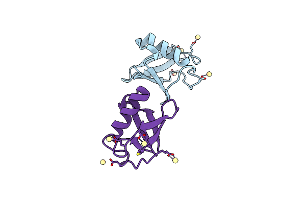 |
Structure Of D-Thr53 Ubiquitin
Organism: Homo sapiens
Method: X-RAY DIFFRACTION Resolution:2.60 Å Release Date: 2022-05-18 Classification: PROTEIN BINDING Ligands: CD |
Organism: Homo sapiens
Method: X-RAY DIFFRACTION
Release Date: 2022-05-18
Ligands: CD
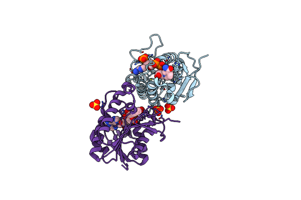 |
Crystal Structure Of Human Biliverdin Ix-Beta Reductase B
Organism: Homo sapiens
Method: X-RAY DIFFRACTION Resolution:1.60 Å Release Date: 2022-01-19 Classification: OXIDOREDUCTASE Ligands: NAP, SO4 |
Organism: Homo sapiens
Method: X-RAY DIFFRACTION
Release Date: 2022-01-19
Ligands: NAP, SO4
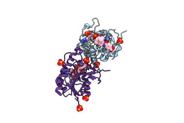 |
Crystal Structure Of Hyman Biliverdin Ix-Beta Reductase B With Tamibarotene (A80)
Organism: Homo sapiens
Method: X-RAY DIFFRACTION Resolution:1.70 Å Release Date: 2022-01-19 Classification: OXIDOREDUCTASE Ligands: NAP, A80, SO4 |
Organism: Homo sapiens
Method: X-RAY DIFFRACTION
Release Date: 2022-01-19
Ligands: NAP, A80, SO4
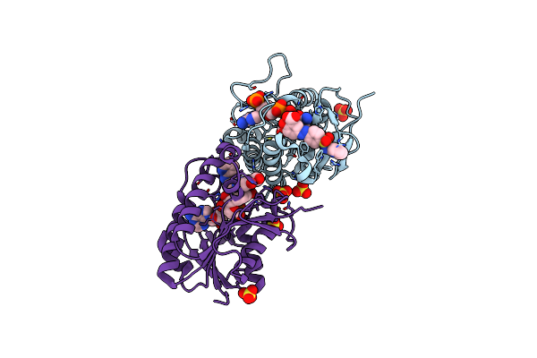 |
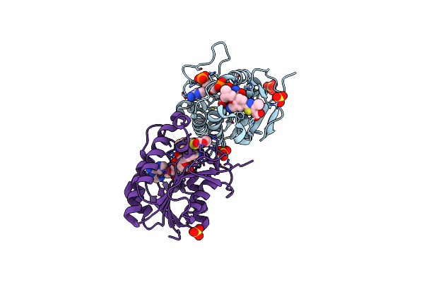
Crystal Structure Of Human Biliverdin Ix-Beta Reductase B With Sulfasalazine (Sas)
Organism: Homo sapiens
Method: X-RAY DIFFRACTION Resolution:1.45 Å Release Date: 2022-01-19 Classification: OXIDOREDUCTASE Ligands: NAP, SAS, SO4 |
Organism: Homo sapiens
Method: X-RAY DIFFRACTION
Release Date: 2022-01-19
Ligands: NAP, SAS, SO4
 |
Crystal Structure Of Human Biliverdin Ix-Beta Reductase B With Febuxostat (Tei)
Organism: Homo sapiens
Method: X-RAY DIFFRACTION Resolution:1.45 Å Release Date: 2022-01-19 Classification: OXIDOREDUCTASE Ligands: NAP, TEI, SO4 |
Organism: Homo sapiens
Method: X-RAY DIFFRACTION
Release Date: 2022-01-19
Ligands: NAP, TEI, SO4