

Search Count: 29
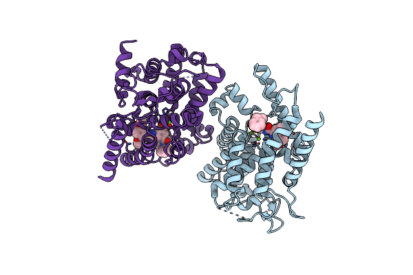 |
Crystal Structure Of Pde3B In Complex With Gsk4394835A
Organism: Homo sapiens
Method: X-RAY DIFFRACTION Resolution:2.70 Å Release Date: 2024-02-07 Classification: HYDROLASE Ligands: MG, X5W |
Organism: Homo sapiens
Method: X-RAY DIFFRACTION
Release Date: 2024-02-07
Ligands: MG, X5W
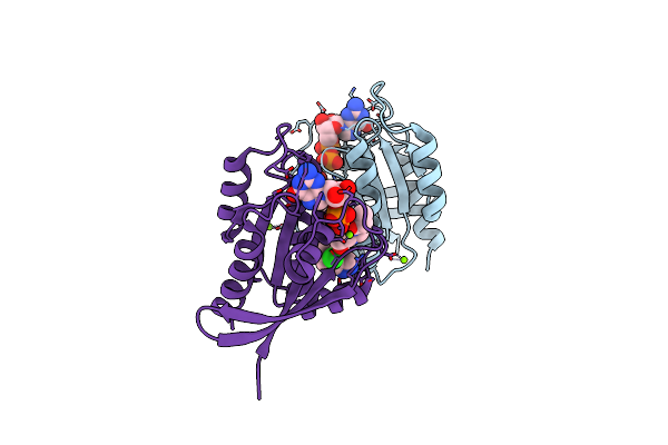 |
Krasg12C Ligand Complex
Organism: Homo sapiens
Method: X-RAY DIFFRACTION Resolution:1.70 Å Release Date: 2023-07-12 Classification: SIGNALING PROTEIN Ligands: MG, GDP, PQI |
Organism: Homo sapiens
Method: X-RAY DIFFRACTION
Release Date: 2023-07-12
Ligands: MG, GDP, PQI
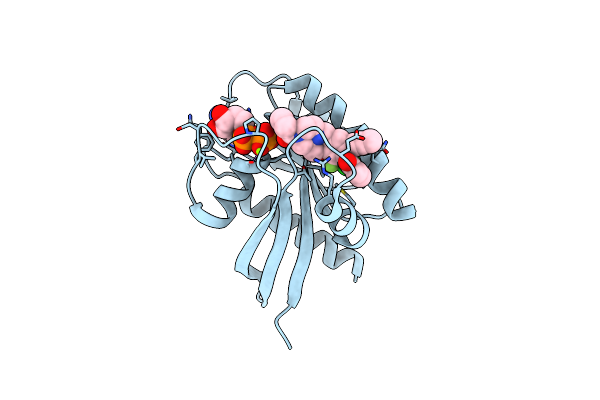 |
Krasg12C Ligand Complex
Organism: Homo sapiens
Method: X-RAY DIFFRACTION Resolution:1.11 Å Release Date: 2023-07-12 Classification: SIGNALING PROTEIN Ligands: MG, GDP, PUO |
Organism: Homo sapiens
Method: X-RAY DIFFRACTION
Release Date: 2023-07-12
Ligands: MG, GDP, PUO
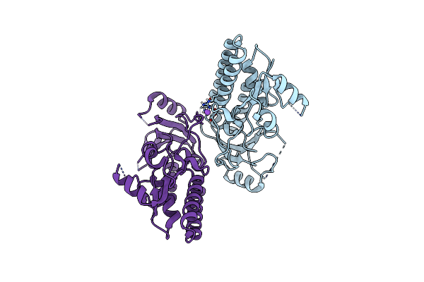 |
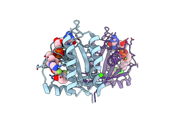
Crystal Structure Of Acinetobacter Baumannii Znua In The Metal-Free State
Organism: Acinetobacter baumannii
Method: X-RAY DIFFRACTION Resolution:2.13 Å Release Date: 2022-10-12 Classification: METAL BINDING PROTEIN |
Organism: Acinetobacter baumannii
Method: X-RAY DIFFRACTION
Release Date: 2022-10-12
 |
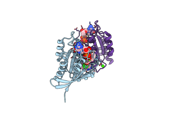
Krasg12C Ligand Complex
Organism: Homo sapiens
Method: X-RAY DIFFRACTION Resolution:1.18 Å Release Date: 2022-04-20 Classification: SIGNALING PROTEIN Ligands: V4T, MG, GDP, CA |
Organism: Homo sapiens
Method: X-RAY DIFFRACTION
Release Date: 2022-04-20
Ligands: V4T, MG, GDP, CA
 |
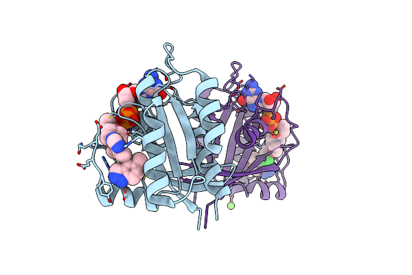
Krasg12C Ligand Complex
Organism: Homo sapiens
Method: X-RAY DIFFRACTION Resolution:2.38 Å Release Date: 2022-04-20 Classification: SIGNALING PROTEIN Ligands: MG, GDP, V52, CA |
Organism: Homo sapiens
Method: X-RAY DIFFRACTION
Release Date: 2022-04-20
Ligands: MG, GDP, V52, CA
 |
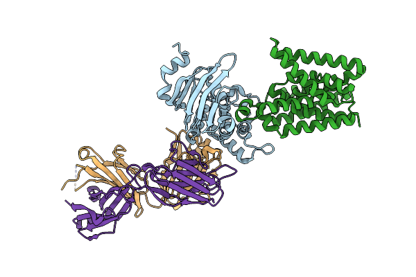
Krasg12C Ligand Complex
Organism: Homo sapiens
Method: X-RAY DIFFRACTION Resolution:1.48 Å Release Date: 2022-04-20 Classification: SIGNALING PROTEIN Ligands: MG, GDP, VLE, CA |
Organism: Homo sapiens
Method: X-RAY DIFFRACTION
Release Date: 2022-04-20
Ligands: MG, GDP, VLE, CA
 |
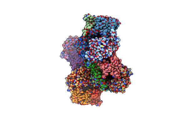
Psabc From Streptococcus Pneumoniae In Complex With Fab
Organism: Streptococcus pneumoniae serotype 2 (strain d39 / nctc 7466), Mus musculus
Method: X-RAY DIFFRACTION Resolution:2.85 Å Release Date: 2021-08-25 Classification: TRANSPORT PROTEIN/IMMUNE SYSTEM |
 |
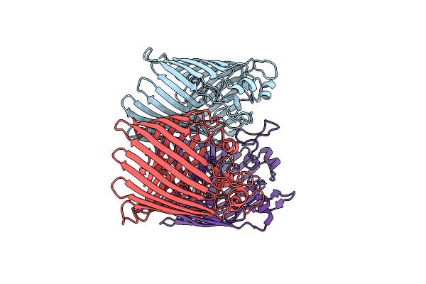
Psabc From Streptococcus Pneumoniae In Complex With Fab
Organism: Streptococcus pneumoniae serotype 2 (strain d39 / nctc 7466)
Method: X-RAY DIFFRACTION Resolution:2.90 Å Release Date: 2021-08-25 Classification: TRANSPORT PROTEIN Ligands: PO4, MA4 |
Organism: Streptococcus pneumoniae serotype 2 (strain d39 / nctc 7466)
Method: X-RAY DIFFRACTION
Release Date: 2021-08-25
Ligands: PO4, MA4
 |
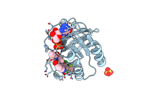
Ompk37 Porin
Organism: Klebsiella pneumoniae
Method: X-RAY DIFFRACTION Resolution:2.60 Å Release Date: 2020-04-01 Classification: MEMBRANE PROTEIN |
Organism: Klebsiella pneumoniae
Method: X-RAY DIFFRACTION
Release Date: 2020-04-01
 |
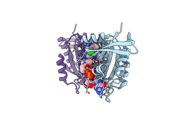
Krasg12C Ligand Complex
Organism: Homo sapiens
Method: X-RAY DIFFRACTION Resolution:1.37 Å Release Date: 2020-02-26 Classification: SIGNALING PROTEIN Ligands: MG, GDP, O7K, SO4 |
Organism: Homo sapiens
Method: X-RAY DIFFRACTION
Release Date: 2020-02-26
Ligands: MG, GDP, O7K, SO4
 |
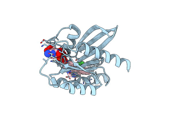
Krasg12C Ligand Complex
Organism: Homo sapiens
Method: X-RAY DIFFRACTION Resolution:1.72 Å Release Date: 2020-02-19 Classification: SIGNALING PROTEIN Ligands: MG, GDP, MKW |
Organism: Homo sapiens
Method: X-RAY DIFFRACTION
Release Date: 2020-02-19
Ligands: MG, GDP, MKW
 |
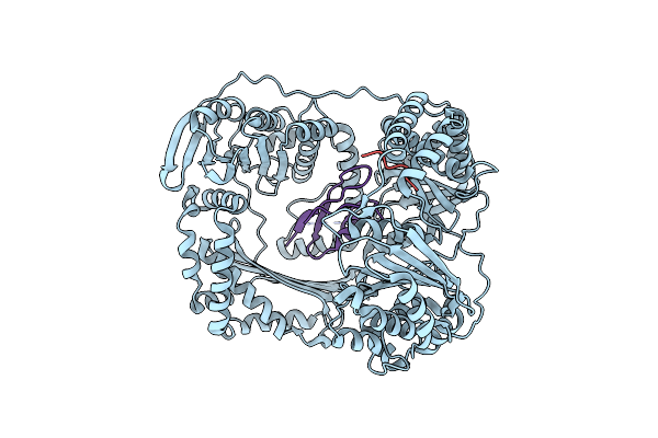
Krasg12C Ligand Complex
Organism: Homo sapiens
Method: X-RAY DIFFRACTION Resolution:1.31 Å Release Date: 2020-02-19 Classification: SIGNALING PROTEIN Ligands: MG, GDP, MKZ |
Organism: Homo sapiens
Method: X-RAY DIFFRACTION
Release Date: 2020-02-19
Ligands: MG, GDP, MKZ
 |
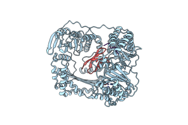
The Crystal Structure Of The Ferredoxin Protease Fusc In Complex With Its Substrate Plant Ferredoxin
Organism: Pectobacterium atrosepticum scri1043, Arabidopsis thaliana
Method: X-RAY DIFFRACTION Resolution:2.70 Å Release Date: 2018-06-20 Classification: HYDROLASE |
Organism: Pectobacterium atrosepticum scri1043, Arabidopsis thaliana
Method: X-RAY DIFFRACTION
Release Date: 2018-06-20
 |
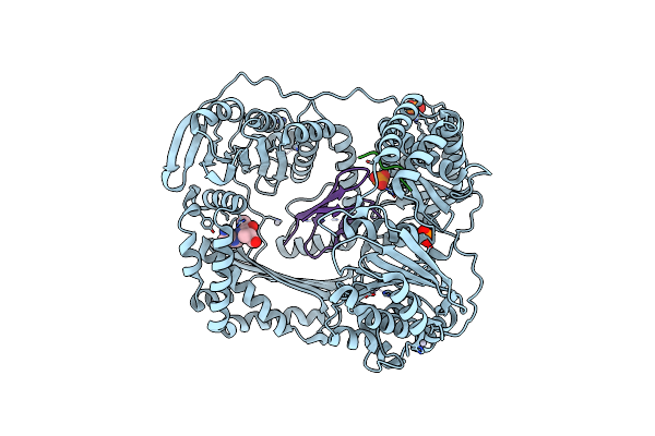
The Crystal Structure Of The Ferredoxin Protease Fusc E83A Mutant In Complex With Arabidopsis Ferredoxin
Organism: Pectobacterium atrosepticum (strain scri 1043 / atcc baa-672), Arabidopsis thaliana
Method: X-RAY DIFFRACTION Resolution:1.90 Å Release Date: 2018-06-20 Classification: HYDROLASE Ligands: ZN |
Organism: Pectobacterium atrosepticum (strain scri 1043 / atcc baa-672), Arabidopsis thaliana
Method: X-RAY DIFFRACTION
Release Date: 2018-06-20
Ligands: ZN
 |
The Crystal Structure Of The Ferredoxin Protease Fusc In Complex With Arabidopsis Ferredoxin, Ethylmercury Phosphate Soaked Dataset
Organism: Pectobacterium atrosepticum (strain scri 1043 / atcc baa-672), Arabidopsis thaliana
Method: X-RAY DIFFRACTION Resolution:2.30 Å Release Date: 2018-06-20 Classification: HYDROLASE Ligands: PO4, HG |
Organism: Pectobacterium atrosepticum (strain scri 1043 / atcc baa-672), Arabidopsis thaliana
Method: X-RAY DIFFRACTION
Release Date: 2018-06-20
Ligands: PO4, HG
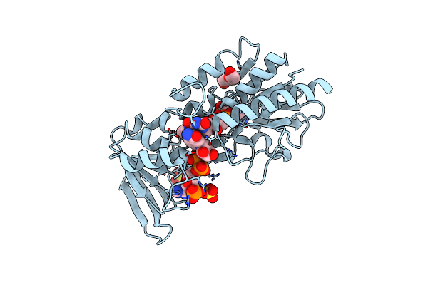 |
Crystal Structure Of Lactococcus Lactis Thioredoxin Reductase (Fr Conformation)
Organism: Lactococcus lactis subsp. cremoris
Method: X-RAY DIFFRACTION Resolution:2.14 Å Release Date: 2017-04-12 Classification: OXIDOREDUCTASE Ligands: FAD, NAP, PEG, SO4 |
Organism: Lactococcus lactis subsp. cremoris
Method: X-RAY DIFFRACTION
Release Date: 2017-04-12
Ligands: FAD, NAP, PEG, SO4
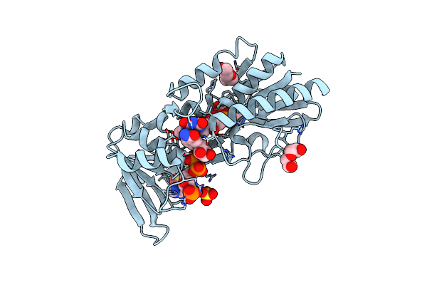 |
Crystal Structure Of Lactococcus Lactis Thioredoxin Reductase Exposed To Visible Light (30 Min)
Organism: Lactococcus lactis subsp. cremoris
Method: X-RAY DIFFRACTION Resolution:2.00 Å Release Date: 2017-04-12 Classification: OXIDOREDUCTASE Ligands: FAD, NAP, PEG, SO4 |
Organism: Lactococcus lactis subsp. cremoris
Method: X-RAY DIFFRACTION
Release Date: 2017-04-12
Ligands: FAD, NAP, PEG, SO4
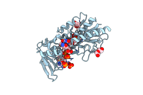 |
Crystal Structure Of Lactococcus Lactis Thioredoxin Reductase Exposed To Visible Light (60 Min)
Organism: Lactococcus lactis subsp. cremoris
Method: X-RAY DIFFRACTION Resolution:1.92 Å Release Date: 2017-04-12 Classification: OXIDOREDUCTASE Ligands: FAD, NAP, PEG, SO4 |
Organism: Lactococcus lactis subsp. cremoris
Method: X-RAY DIFFRACTION
Release Date: 2017-04-12
Ligands: FAD, NAP, PEG, SO4
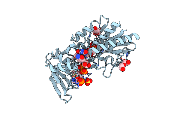 |
Crystal Structure Of Lactococcus Lactis Thioredoxin Reductase Exposed To Visible Light (120 Min)
Organism: Lactococcus lactis subsp. cremoris
Method: X-RAY DIFFRACTION Resolution:2.00 Å Release Date: 2017-04-12 Classification: OXIDOREDUCTASE Ligands: FAD, NAP, PEG, SO4 |
Organism: Lactococcus lactis subsp. cremoris
Method: X-RAY DIFFRACTION
Release Date: 2017-04-12
Ligands: FAD, NAP, PEG, SO4