

Search Count: 9
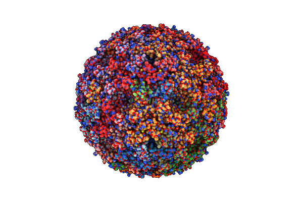 |
Structure Of A Synthetic Beta-Carboxysome Shell, T=3
Organism: Halothece sp. (strain pcc 7418)
Method: ELECTRON MICROSCOPY Release Date: 2019-09-25 Classification: STRUCTURAL PROTEIN |
Organism: Halothece sp. (strain pcc 7418)
Method: ELECTRON MICROSCOPY
Release Date: 2019-09-25
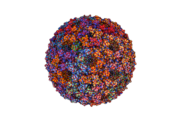 |
Structure Of A Synthetic Beta-Carboxysome Shell, T=4
Organism: Halothece sp. (strain pcc 7418)
Method: ELECTRON MICROSCOPY Release Date: 2019-09-25 Classification: STRUCTURAL PROTEIN |
Organism: Halothece sp. (strain pcc 7418)
Method: ELECTRON MICROSCOPY
Release Date: 2019-09-25
 |
T.Elongatus Ndh (Data-Set 1)
Organism: Thermosynechococcus elongatus (strain bp-1)
Method: ELECTRON MICROSCOPY Release Date: 2019-02-27 Classification: OXIDOREDUCTASE Ligands: SF4 |
Organism: Thermosynechococcus elongatus (strain bp-1)
Method: ELECTRON MICROSCOPY
Release Date: 2019-02-27
Ligands: SF4
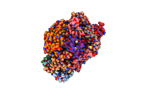 |
T.Elongatus Ndh (Data-Set 2)
Organism: Thermosynechococcus elongatus (strain bp-1)
Method: ELECTRON MICROSCOPY Release Date: 2019-02-27 Classification: OXIDOREDUCTASE Ligands: SF4 |
Organism: Thermosynechococcus elongatus (strain bp-1)
Method: ELECTRON MICROSCOPY
Release Date: 2019-02-27
Ligands: SF4
 |
T.Elongatus Ndh (Composite Model)
Organism: Thermosynechococcus elongatus bp-1
Method: ELECTRON MICROSCOPY Resolution:3.10 Å Release Date: 2019-02-27 Classification: OXIDOREDUCTASE Ligands: SF4 |
Organism: Thermosynechococcus elongatus bp-1
Method: ELECTRON MICROSCOPY
Release Date: 2019-02-27
Ligands: SF4
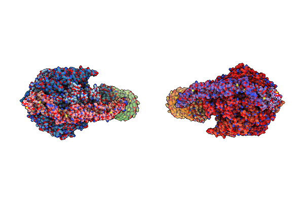 |
Model Of The Yeast F1Fo-Atp Synthase Dimer Based On Subtomogram Average
Organism: Saccharomyces cerevisiae, Bos taurus
Method: ELECTRON MICROSCOPY Resolution:37.00 Å Release Date: 2012-08-29 Classification: HYDROLASE Ligands: ATP, MG, ADP |
Organism: Saccharomyces cerevisiae, Bos taurus
Method: ELECTRON MICROSCOPY
Release Date: 2012-08-29
Ligands: ATP, MG, ADP
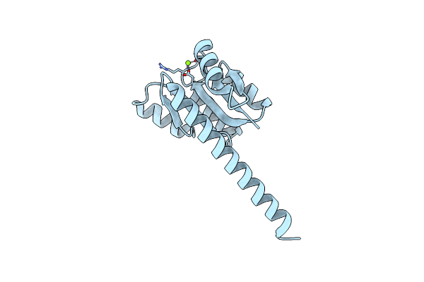 |
Crystal Structure Of The Wild-Type Hupr Receiver Domain
Organism: Rhodobacter capsulatus
Method: X-RAY DIFFRACTION Resolution:2.10 Å Release Date: 2008-11-11 Classification: DNA-BINDING Ligands: MG |
Organism: Rhodobacter capsulatus
Method: X-RAY DIFFRACTION
Release Date: 2008-11-11
Ligands: MG
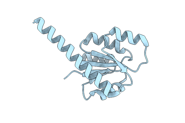 |
Crystal Structure Of The D55E Mutant Of The Hupr Receiver Domain
Organism: Rhodobacter capsulatus
Method: X-RAY DIFFRACTION Resolution:2.50 Å Release Date: 2008-11-11 Classification: DNA-BINDING |
Organism: Rhodobacter capsulatus
Method: X-RAY DIFFRACTION
Release Date: 2008-11-11
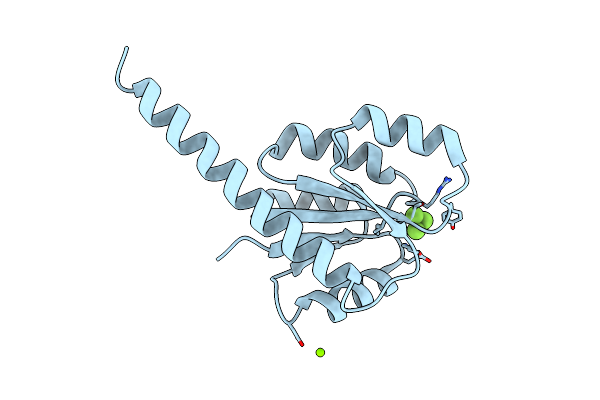 |
Crystal Structure Of The Hupr Receiver Domain In Inhibitory Phospho- State
Organism: Rhodobacter capsulatus
Method: X-RAY DIFFRACTION Resolution:2.90 Å Release Date: 2008-11-11 Classification: DNA-BINDING Ligands: BEF, MG |
Organism: Rhodobacter capsulatus
Method: X-RAY DIFFRACTION
Release Date: 2008-11-11
Ligands: BEF, MG