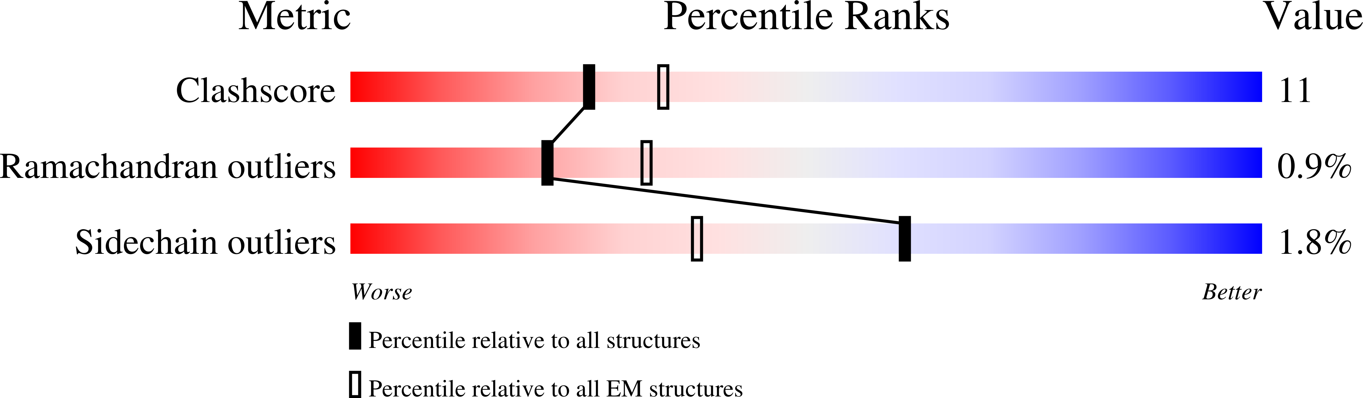
Deposition Date
2012-07-17
Release Date
2012-08-29
Last Version Date
2024-05-08
Entry Detail
PDB ID:
4B2Q
Keywords:
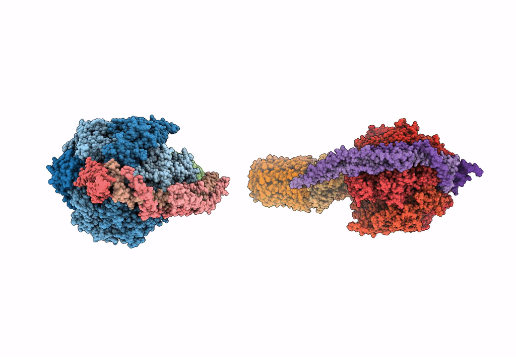
Title:
Model of the yeast F1Fo-ATP synthase dimer based on subtomogram average
Biological Source:
Source Organism(s):
SACCHAROMYCES CEREVISIAE (Taxon ID: 4932)
BOS TAURUS (Taxon ID: 9913)
BOS TAURUS (Taxon ID: 9913)
Method Details:
Experimental Method:
Resolution:
37.00 Å
Aggregation State:
CELL
Reconstruction Method:
TOMOGRAPHY