

Search Count: 38
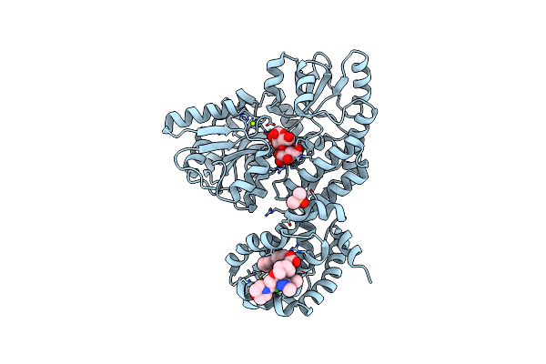 |
Crystal Structure Of Human Mbp-Myeloid Cell Leukemia 1 (Mcl-1) In Complex With Brd810 Inhibitor
Organism: Escherichia coli k-12, Homo sapiens
Method: X-RAY DIFFRACTION Resolution:1.56 Å Release Date: 2024-06-19 Classification: APOPTOSIS Ligands: MG, YI7, DMS, PGE |
Organism: Escherichia coli k-12, Homo sapiens
Method: X-RAY DIFFRACTION
Release Date: 2024-06-19
Ligands: MG, YI7, DMS, PGE
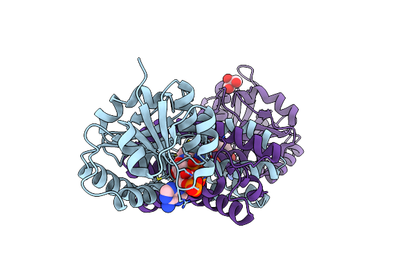 |
Structure Of Imine Reductase From Pseudomonas Sp.
Organism: Pseudomonas putida
Method: X-RAY DIFFRACTION Resolution:1.59 Å Release Date: 2021-08-25 Classification: OXIDOREDUCTASE Ligands: NDP, EDO, GOL |
Organism: Pseudomonas putida
Method: X-RAY DIFFRACTION
Release Date: 2021-08-25
Ligands: NDP, EDO, GOL
 |
N-Terminal Bromodomain Of Human Brd4 With Ibet-Bd1 (Gsk778)
Organism: Homo sapiens
Method: X-RAY DIFFRACTION Resolution:1.28 Å Release Date: 2020-04-01 Classification: TRANSCRIPTION Ligands: GLU, LWB, EDO |
Organism: Homo sapiens
Method: X-RAY DIFFRACTION
Release Date: 2020-04-01
Ligands: GLU, LWB, EDO
 |
C-Terminal Bromodomain Of Human Brd2 With Ibet-Bd1 (Gsk778)
Organism: Homo sapiens
Method: X-RAY DIFFRACTION Resolution:1.60 Å Release Date: 2020-04-01 Classification: TRANSCRIPTION Ligands: LWB, EDO |
Organism: Homo sapiens
Method: X-RAY DIFFRACTION
Release Date: 2020-04-01
Ligands: LWB, EDO
 |
C-Terminal Bromodomain Of Human Brd2 With Ibet-Bd2 (Gsk046)
Organism: Homo sapiens
Method: X-RAY DIFFRACTION Resolution:1.60 Å Release Date: 2020-04-01 Classification: TRANSCRIPTION Ligands: EDO, LW5 |
Organism: Homo sapiens
Method: X-RAY DIFFRACTION
Release Date: 2020-04-01
Ligands: EDO, LW5
 |
N-Terminal Bromodomain Of Human Brd4 With Ibet-Bd2 (Gsk046)
Organism: Homo sapiens
Method: X-RAY DIFFRACTION Resolution:1.60 Å Release Date: 2020-04-01 Classification: TRANSCRIPTION Ligands: EDO, LW5 |
Organism: Homo sapiens
Method: X-RAY DIFFRACTION
Release Date: 2020-04-01
Ligands: EDO, LW5
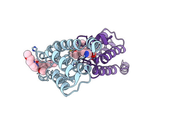 |
Crystal Structure Of Human Brd9 Bromodomain In Complex With A Protac
Organism: Homo sapiens
Method: X-RAY DIFFRACTION Resolution:2.40 Å Release Date: 2019-01-16 Classification: TRANSCRIPTION Ligands: GBW |
Organism: Homo sapiens
Method: X-RAY DIFFRACTION
Release Date: 2019-01-16
Ligands: GBW
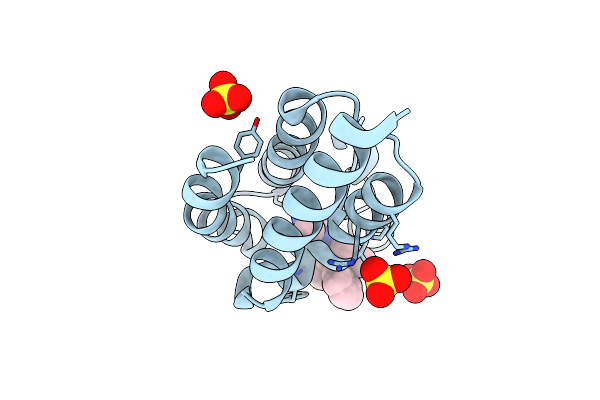 |
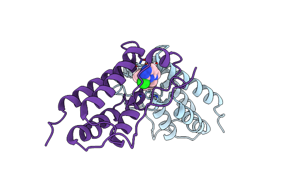
Bromodomain From Plasmodium Faciparum Gcn5, Complexed With Compound
Organism: Plasmodium falciparum (isolate 3d7)
Method: X-RAY DIFFRACTION Resolution:2.10 Å Release Date: 2017-01-04 Classification: TRANSFERASE Ligands: CL, SO4, 7H7 |
Organism: Plasmodium falciparum (isolate 3d7)
Method: X-RAY DIFFRACTION
Release Date: 2017-01-04
Ligands: CL, SO4, 7H7
 |
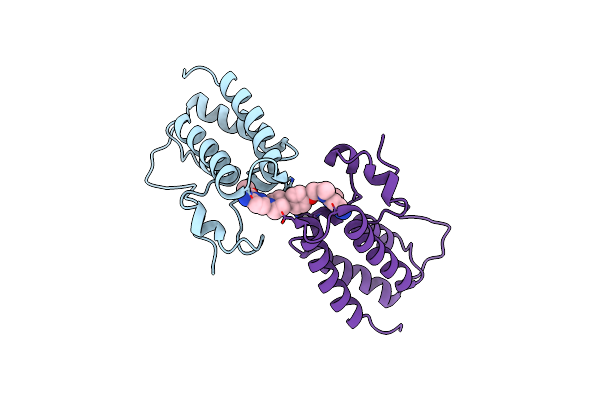
Bivalent Binding To Bet Bromodomains
Organism: Homo sapiens
Method: X-RAY DIFFRACTION Resolution:2.01 Å Release Date: 2016-09-28 Classification: TRANSCRIPTION Ligands: ETU |
Organism: Homo sapiens
Method: X-RAY DIFFRACTION
Release Date: 2016-09-28
Ligands: ETU
 |
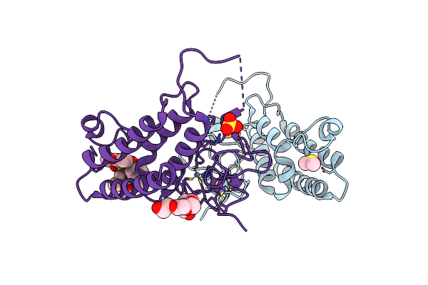
Bivalent Binding To Bet Bromodomains
Organism: Homo sapiens
Method: X-RAY DIFFRACTION Resolution:1.49 Å Release Date: 2016-09-28 Classification: TRANSCRIPTION Ligands: K6K |
Organism: Homo sapiens
Method: X-RAY DIFFRACTION
Release Date: 2016-09-28
Ligands: K6K
 |
Crystal Structure Of Trim24 With 3,4-Dimethoxy-N-(6-(4-Methoxyphenoxy)-1,3-Dimethyl-2-Oxo-2,3-Dihydro-1H-Benzo[D]Imidazol-5-Yl)Benzenesulfonamide Inhibitor
Organism: Homo sapiens
Method: X-RAY DIFFRACTION Resolution:1.79 Å Release Date: 2015-06-17 Classification: LIGASE Ligands: ZN, SO4, DMS, 4QH, 1PE |
Organism: Homo sapiens
Method: X-RAY DIFFRACTION
Release Date: 2015-06-17
Ligands: ZN, SO4, DMS, 4QH, 1PE
 |
Crystal Structure Of Brd9 Bromodomain In Complex With A Valerolactam Quinolone Ligand
Organism: Homo sapiens
Method: X-RAY DIFFRACTION Resolution:1.80 Å Release Date: 2015-05-20 Classification: TRANSCRIPTION Ligands: 4L2 |
Organism: Homo sapiens
Method: X-RAY DIFFRACTION
Release Date: 2015-05-20
Ligands: 4L2
 |
Crystal Structure Of Brd9 Bromodomain In Complex With A Substituted Valerolactam Quinolone Ligand
Organism: Homo sapiens
Method: X-RAY DIFFRACTION Resolution:1.95 Å Release Date: 2015-05-20 Classification: TRANSCRIPTION Ligands: 4L3 |
Organism: Homo sapiens
Method: X-RAY DIFFRACTION
Release Date: 2015-05-20
Ligands: 4L3
 |
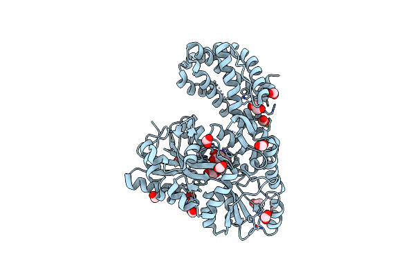
Structure Of Mcl1 Bound To Brd Inhibitor Ligand 1 At 1.7A
Organism: Homo sapiens
Method: X-RAY DIFFRACTION Resolution:1.70 Å Release Date: 2015-05-06 Classification: Apoptosis/Inhibitor Ligands: ZN, 865, POP |
Organism: Homo sapiens
Method: X-RAY DIFFRACTION
Release Date: 2015-05-06
Ligands: ZN, 865, POP
 |
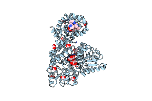
Structure Of Apo Mbp-Mcl1 At 1.9A
Organism: Escherichia coli, Homo sapiens
Method: X-RAY DIFFRACTION Resolution:1.90 Å Release Date: 2015-05-06 Classification: APOPTOSIS Ligands: MG, EDO, FMT |
Organism: Escherichia coli, Homo sapiens
Method: X-RAY DIFFRACTION
Release Date: 2015-05-06
Ligands: MG, EDO, FMT
 |
Structure Of Mbp-Mcl1 Bound To Ligand 1 At 2.35A
Organism: Escherichia coli, Homo sapiens
Method: X-RAY DIFFRACTION Resolution:2.35 Å Release Date: 2015-05-06 Classification: APOPTOSIS Ligands: 865, EDO, FMT |
Organism: Escherichia coli, Homo sapiens
Method: X-RAY DIFFRACTION
Release Date: 2015-05-06
Ligands: 865, EDO, FMT
 |
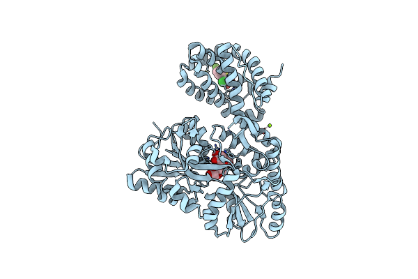
Structure Of Mbp-Mcl1 Bound To Ligand 2 At 1.55A
Organism: Escherichia coli, Homo sapiens
Method: X-RAY DIFFRACTION Resolution:1.55 Å Release Date: 2015-05-06 Classification: APOPTOSIS Ligands: 19H, MG, FMT, NA, EDO |
Organism: Escherichia coli, Homo sapiens
Method: X-RAY DIFFRACTION
Release Date: 2015-05-06
Ligands: 19H, MG, FMT, NA, EDO
 |
Structure Of Mbp-Mcl1 Bound To Ligand 4 At 2.4A
Organism: Escherichia coli, Homo sapiens
Method: X-RAY DIFFRACTION Resolution:2.40 Å Release Date: 2015-05-06 Classification: APOPTOSIS Ligands: CL, MG, 3R4 |
Organism: Escherichia coli, Homo sapiens
Method: X-RAY DIFFRACTION
Release Date: 2015-05-06
Ligands: CL, MG, 3R4
 |
The Structure Of Mbp-Mcl1 Bound To Ligand 5 At 1.9A
Organism: Escherichia coli, Homo sapiens
Method: X-RAY DIFFRACTION Resolution:1.90 Å Release Date: 2015-05-06 Classification: APOPTOSIS Ligands: MG, EDO, FMT, 3R6 |
Organism: Escherichia coli, Homo sapiens
Method: X-RAY DIFFRACTION
Release Date: 2015-05-06
Ligands: MG, EDO, FMT, 3R6
 |
The Structure Of Mbp-Mcl1 Bound To Ligand 6 At 2.0A
Organism: Escherichia coli, Homo sapiens
Method: X-RAY DIFFRACTION Resolution:2.00 Å Release Date: 2015-05-06 Classification: APOPTOSIS Ligands: 3R7, FMT, MG |
Organism: Escherichia coli, Homo sapiens
Method: X-RAY DIFFRACTION
Release Date: 2015-05-06
Ligands: 3R7, FMT, MG