
Search Count: 69
All
Selected
 |
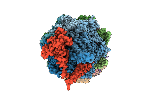
Crystal Structure Of Glycosyltransferase Ugt73C1 In Complex With Udp And Quercetin
Organism: Arabidopsis thaliana
Method: X-RAY DIFFRACTION Resolution:2.08 Å Release Date: 2026-04-29 Classification: PLANT PROTEIN Ligands: UDP, GOL, QUE |
 |
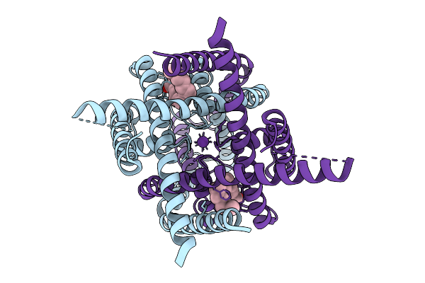
Organism: Mus musculus
Method: ELECTRON MICROSCOPY Release Date: 2026-04-08 Classification: MEMBRANE PROTEIN |
 |
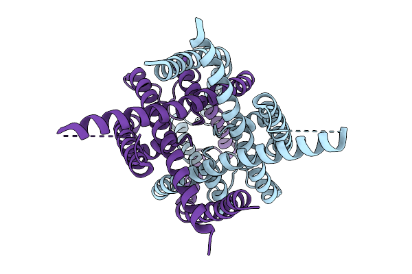
Organism: Mus musculus
Method: ELECTRON MICROSCOPY Release Date: 2026-04-08 Classification: MEMBRANE PROTEIN |
 |
Organism: Mus musculus
Method: ELECTRON MICROSCOPY Release Date: 2026-04-08 Classification: MEMBRANE PROTEIN Ligands: DSN |
 |
Organism: Mus musculus
Method: ELECTRON MICROSCOPY Release Date: 2026-04-08 Classification: MEMBRANE PROTEIN |
 |
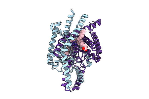
Structure Of Atd Truncated Glutamate Receptor Mglud1 Complexed With D-Serine
Organism: Mus musculus
Method: ELECTRON MICROSCOPY Release Date: 2026-04-08 Classification: MEMBRANE PROTEIN Ligands: DSN |
 |
Structure Of Atd Truncated Glutamate Receptor Mglud1 Complexed With Gaba And Calcium
Organism: Mus musculus
Method: ELECTRON MICROSCOPY Release Date: 2026-04-08 Classification: MEMBRANE PROTEIN |
 |
Organism: Homo sapiens, Escherichia coli str. k-12 substr. mg1655, Synthetic construct
Method: ELECTRON MICROSCOPY Resolution:3.42 Å Release Date: 2026-04-01 Classification: HYDROLASE Ligands: ZN, ATP, MG, ADP, LDZ |
 |
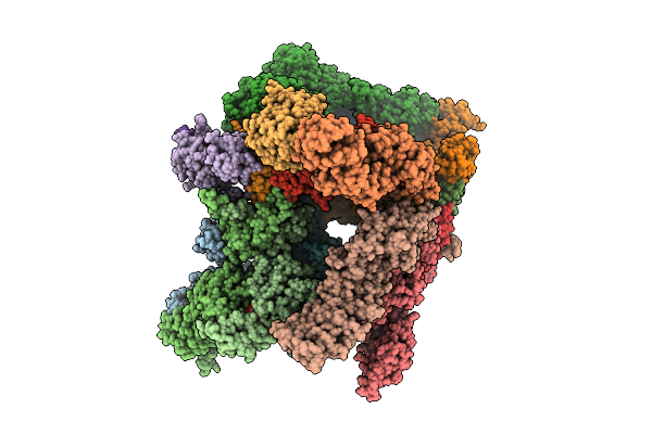
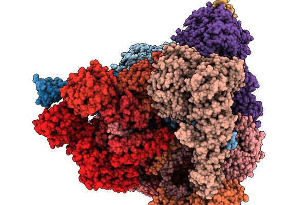
Substrate-Free Human 26S Proteasome Purified By Midnolin, 20S Proteasome, Rpts And Rpn11 Part
Organism: Homo sapiens
Method: ELECTRON MICROSCOPY Release Date: 2026-04-01 Classification: HYDROLASE Ligands: ZN, ATP, MG, ADP, LDZ |
 |
Organism: Homo sapiens
Method: ELECTRON MICROSCOPY Release Date: 2026-04-01 Classification: MEMBRANE PROTEIN Ligands: ACD, A1ENQ |
 |
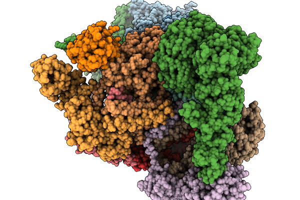
Structure Of Human 26S Proteasome Complexed With Midnolin, 19S Proteasome With Ubl Bound
Organism: Escherichia coli k-12, Homo sapiens, Purpureocillium lilacinum
Method: ELECTRON MICROSCOPY Resolution:3.65 Å Release Date: 2026-04-01 Classification: HYDROLASE Ligands: ADP, ATP, ZN, MG |
 |
Structure Of Human 26S Proteasome Complexed With Midnolin, 19S Proteasome With Ubl And Catch Domain Resolved
Organism: Escherichia coli k-12, Homo sapiens, Pseudotamlana agarivorans
Method: ELECTRON MICROSCOPY Release Date: 2026-04-01 Classification: HYDROLASE Ligands: ADP, ATP, ZN, MG |
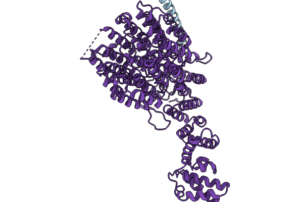 |
Focused Refinement Of Rpn1 And The C-Terminal Helix Of Midnolin In The Substrate-Engaged Human 26S Proteasome
Organism: Homo sapiens
Method: ELECTRON MICROSCOPY Release Date: 2026-03-25 Classification: HYDROLASE |
 |
Substrate-Engaged Human 26S Proteasome Bound To Midnolin With Rpt1 At Top Of Spiral Staircase
Organism: Homo sapiens
Method: ELECTRON MICROSCOPY Release Date: 2026-03-25 Classification: HYDROLASE Ligands: ZN, ATP, MG, ADP, LDZ |
 |
Substrate-Engaged Human 26S Proteasome Bound To Midnolin With Rpt5 At Top Of Spiral Staircase
Organism: Homo sapiens
Method: ELECTRON MICROSCOPY Release Date: 2026-03-25 Classification: HYDROLASE Ligands: ZN, ATP, MG, ADP, LDZ |
 |
Substrate-Engaged Human 26S Proteasome Bound To Midnolin With Rpt2 At Top Of Spiral Staircase
Organism: Homo sapiens
Method: ELECTRON MICROSCOPY Release Date: 2026-03-25 Classification: HYDROLASE Ligands: ADP, ATP, MG, LDZ, ZN |
 |
Focused Refinement Of 19S In The Substrate-Engaged Human 26S Proteasome Bound To Midnolin With Rpt6 At Top Of Spiral Staircase
Organism: Homo sapiens
Method: ELECTRON MICROSCOPY Release Date: 2026-03-25 Classification: HYDROLASE Ligands: ADP, ZN, ATP, MG |
 |
Organism: Homo sapiens
Method: ELECTRON MICROSCOPY Release Date: 2026-03-18 Classification: MEMBRANE PROTEIN Ligands: ACD, K |
 |
Organism: Homo sapiens
Method: ELECTRON MICROSCOPY Release Date: 2026-03-18 Classification: MEMBRANE PROTEIN Ligands: HLT |
 |
Organism: Homo sapiens
Method: ELECTRON MICROSCOPY Release Date: 2026-03-18 Classification: MEMBRANE PROTEIN |