

Search Count: 20
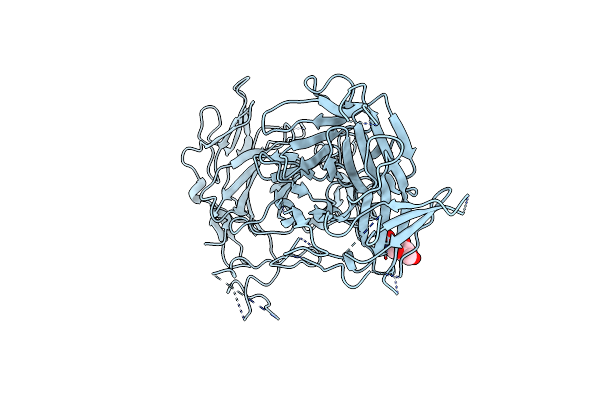 |
N-Terminal 5 Domains Of Ci-Mpr
Organism: Homo sapiens
Method: X-RAY DIFFRACTION Resolution:2.46 Å Release Date: 2020-09-30 Classification: SUGAR BINDING PROTEIN Ligands: NAG |
Organism: Homo sapiens
Method: X-RAY DIFFRACTION
Release Date: 2020-09-30
Ligands: NAG
 |
N-Terminal 5 Domains Of Igfiir
Organism: Homo sapiens
Method: X-RAY DIFFRACTION Resolution:2.54 Å Release Date: 2020-06-24 Classification: SUGAR BINDING PROTEIN |
Organism: Homo sapiens
Method: X-RAY DIFFRACTION
Release Date: 2020-06-24
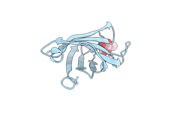 |
Crystal Structure Of The Mrh Domain Of Glucosidase Ii Beta Bound To Mannose
Organism: Schizosaccharomyces pombe (strain 972 / atcc 24843)
Method: X-RAY DIFFRACTION Resolution:1.63 Å Release Date: 2015-07-22 Classification: HYDROLASE Ligands: MAN |
Organism: Schizosaccharomyces pombe (strain 972 / atcc 24843)
Method: X-RAY DIFFRACTION
Release Date: 2015-07-22
Ligands: MAN
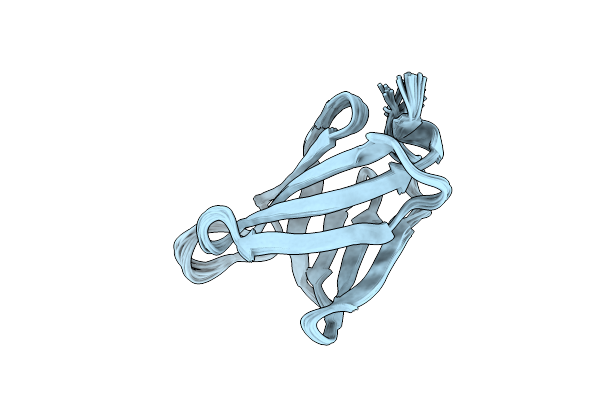 |
Mrh Domain Of The Glucosidase Ii Beta Subunit From S. Pombe
Organism: Schizosaccharomyces pombe
Method: SOLUTION NMR Release Date: 2013-05-01 Classification: HYDROLASE, sugar binding protein |
Organism: Schizosaccharomyces pombe
Method: SOLUTION NMR
Release Date: 2013-05-01
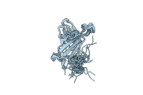 |
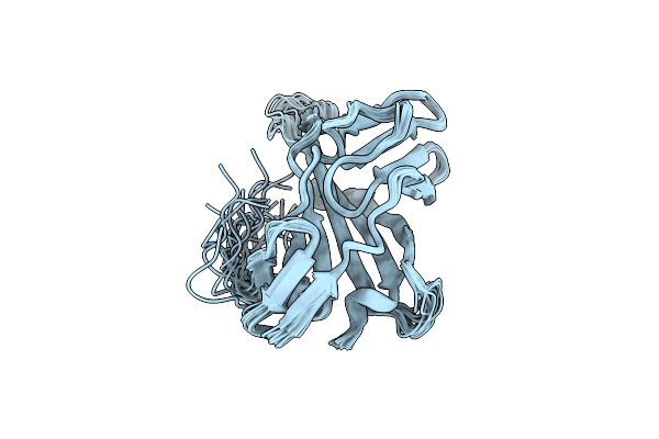
Solution Structure Of Ci-Mpr Ligand-Free Domain 5
Organism: Bos taurus
Method: SOLUTION NMR Release Date: 2010-07-07 Classification: PROTEIN TRANSPORT |
Organism: Bos taurus
Method: SOLUTION NMR
Release Date: 2010-07-07
 |
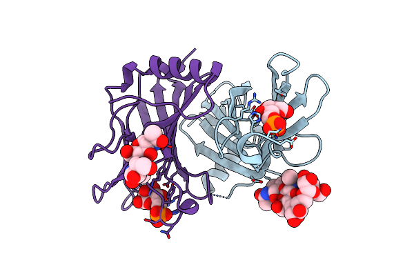
Solution Structure Of Ci-Mpr Domain 5 Bound To N-Acetylglucosaminyl 6-Phosphomethylmannoside
Organism: Bos taurus
Method: SOLUTION NMR Release Date: 2010-07-07 Classification: PROTEIN TRANSPORT |
Organism: Bos taurus
Method: SOLUTION NMR
Release Date: 2010-07-07
 |
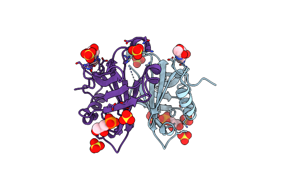
Crystal Structure Of Scd-Mpr Mutant E19Q/K137M Bound To Man-6-P
Organism: Bos taurus
Method: X-RAY DIFFRACTION Resolution:1.90 Å Release Date: 2009-11-24 Classification: Protein transport, sugar binding protein Ligands: M6D |
Organism: Bos taurus
Method: X-RAY DIFFRACTION
Release Date: 2009-11-24
Ligands: M6D
 |
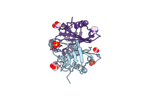
Crystal Structure Of Scd-Mpr Mutant E19Q/K137M Ph 7.0
Organism: Bos taurus
Method: X-RAY DIFFRACTION Resolution:2.30 Å Release Date: 2009-11-24 Classification: protein transport, sugar binding protein Ligands: NAG, 1GP, SO4 |
Organism: Bos taurus
Method: X-RAY DIFFRACTION
Release Date: 2009-11-24
Ligands: NAG, 1GP, SO4
 |
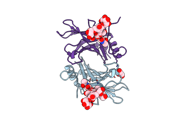
Crystal Structure Of Scd-Mpr Mutant E19Q/K137M Ph 6.5
Organism: Bos taurus
Method: X-RAY DIFFRACTION Resolution:2.00 Å Release Date: 2009-11-24 Classification: protein transport, sugar binding protein Ligands: NAG, SO4, ACT, IMD |
Organism: Bos taurus
Method: X-RAY DIFFRACTION
Release Date: 2009-11-24
Ligands: NAG, SO4, ACT, IMD
 |
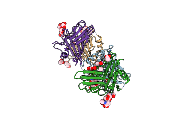
Crystal Structure Cation-Dependent Mannose 6-Phosphate Receptor At Ph 7.4
Organism: Bos taurus
Method: X-RAY DIFFRACTION Resolution:1.51 Å Release Date: 2008-05-13 Classification: PROTEIN TRANSPORT, SUGAR BINDING PROTEIN Ligands: GOL |
Organism: Bos taurus
Method: X-RAY DIFFRACTION
Release Date: 2008-05-13
Ligands: GOL
 |
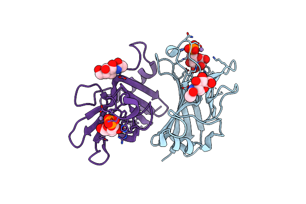
Crystal Structure Cation-Dependent Mannose 6-Phosphate Receptor At Ph 4.8
Organism: Bos taurus
Method: X-RAY DIFFRACTION Resolution:2.00 Å Release Date: 2008-02-12 Classification: Protein transport, sugar binding protein Ligands: GOL, ACT, CAC |
Organism: Bos taurus
Method: X-RAY DIFFRACTION
Release Date: 2008-02-12
Ligands: GOL, ACT, CAC
 |
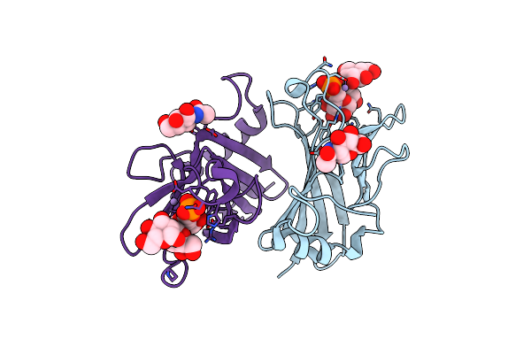
Crystal Structure Cation-Dependent Mannose 6-Phosphate Receptor At Ph 6.5 Bound To M6P
Organism: Bos taurus
Method: X-RAY DIFFRACTION Resolution:1.45 Å Release Date: 2008-02-12 Classification: Protein transport, sugar binding protein Ligands: NAG, MN, M6D |
Organism: Bos taurus
Method: X-RAY DIFFRACTION
Release Date: 2008-02-12
Ligands: NAG, MN, M6D
 |
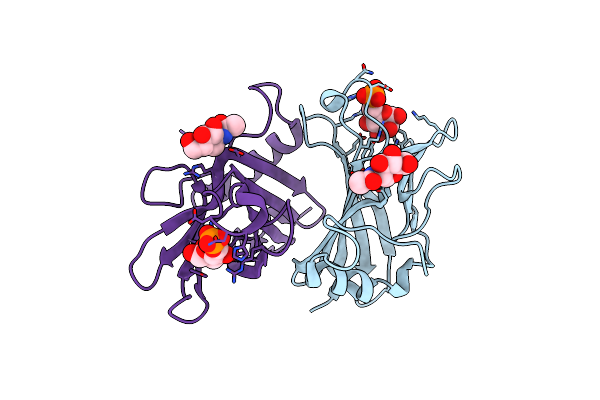
Crystal Structure Cation-Dependent Mannose 6-Phosphate Receptor At Ph 6.5 Bound To Trimannoside
Organism: Bos taurus
Method: X-RAY DIFFRACTION Resolution:2.40 Å Release Date: 2008-02-12 Classification: Protein transport, sugar binding protein Ligands: NAG, MN |
Organism: Bos taurus
Method: X-RAY DIFFRACTION
Release Date: 2008-02-12
Ligands: NAG, MN
 |
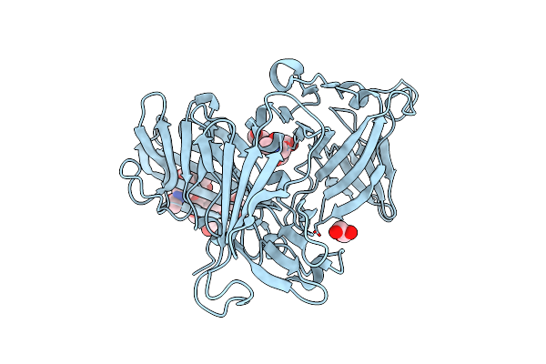
Crystal Structure Cation-Dependent Mannose 6-Phosphate Receptor At Ph 6.5 Bound To M6P In Absence Of Mn
Organism: Bos taurus
Method: X-RAY DIFFRACTION Resolution:1.75 Å Release Date: 2008-02-12 Classification: Protein transport, sugar binding protein Ligands: NAG, M6D |
Organism: Bos taurus
Method: X-RAY DIFFRACTION
Release Date: 2008-02-12
Ligands: NAG, M6D
 |
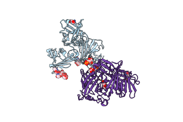
Crystal Structure Of N-Terminal 3 Domains Of Ci-Mpr
Organism: Bos taurus
Method: X-RAY DIFFRACTION Resolution:1.80 Å Release Date: 2004-06-29 Classification: PROTEIN TRANSPORT,SUGAR BINDING PROTEIN Ligands: GOL |
Organism: Bos taurus
Method: X-RAY DIFFRACTION
Release Date: 2004-06-29
Ligands: GOL
 |
N-Terminal 3 Domains Of Ci-Mpr Bound To Mannose 6-Phosphate
Organism: Bos taurus
Method: X-RAY DIFFRACTION Resolution:2.20 Å Release Date: 2004-06-29 Classification: PROTEIN TRANSPORT,SUGAR BINDING PROTEIN Ligands: NAG, M6P, GOL |
Organism: Bos taurus
Method: X-RAY DIFFRACTION
Release Date: 2004-06-29
Ligands: NAG, M6P, GOL
 |
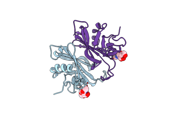
N-Terminal 3 Domains Of Ci-Mpr Bound To Mannose 6-Phosphate
Organism: Bos taurus
Method: X-RAY DIFFRACTION Resolution:2.10 Å Release Date: 2004-06-29 Classification: PROTEIN TRANSPORT,SUGAR BINDING PROTEIN Ligands: NAG, OS, M6P |
Organism: Bos taurus
Method: X-RAY DIFFRACTION
Release Date: 2004-06-29
Ligands: NAG, OS, M6P
 |
Twists And Turns Of The Cd-Mpr: Ligand-Bound Versus Ligand-Free Receptor
Organism: Bos taurus
Method: X-RAY DIFFRACTION Resolution:2.20 Å Release Date: 2002-01-23 Classification: SUGAR BINDING PROTEIN Ligands: NAG |
Organism: Bos taurus
Method: X-RAY DIFFRACTION
Release Date: 2002-01-23
Ligands: NAG
 |
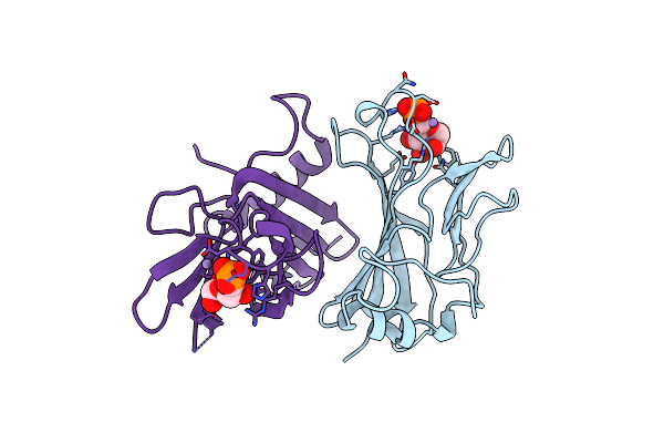
Structure Of Cation-Dependent Mannose 6-Phosphate Receptor Bound To Pentamannosyl Phosphate
Organism: Bos taurus
Method: X-RAY DIFFRACTION Resolution:1.85 Å Release Date: 2000-01-14 Classification: SIGNALING PROTEIN Ligands: MN |
Organism: Bos taurus
Method: X-RAY DIFFRACTION
Release Date: 2000-01-14
Ligands: MN
 |
Extracytoplasmic Domain Of Bovine Cation-Dependent Mannose 6-Phosphate Receptor
Organism: Bos taurus
Method: X-RAY DIFFRACTION Resolution:1.80 Å Release Date: 1999-04-27 Classification: RECEPTOR Ligands: MN, M6P |
Organism: Bos taurus
Method: X-RAY DIFFRACTION
Release Date: 1999-04-27
Ligands: MN, M6P