

Search Count: 16
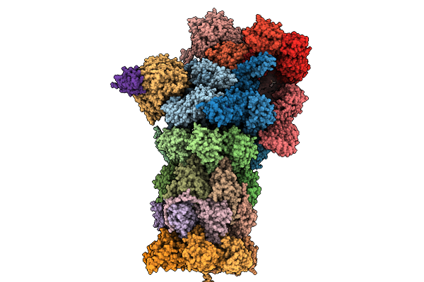 |
Co-Chaperone Bag1-Bound Human 26S Proteasome In Sbag1 State
Organism: Homo sapiens
Method: ELECTRON MICROSCOPY Release Date: 2025-12-31 Classification: HYDROLASE Ligands: ATP, ADP |
Organism: Homo sapiens
Method: ELECTRON MICROSCOPY
Release Date: 2025-12-31
Ligands: ATP, ADP
 |
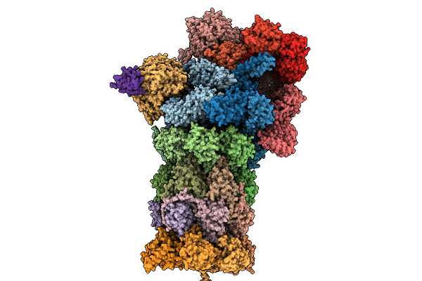
Co-Chaperone Bag1-Bound Human 26S Proteasome In Sbag1.2 State
Organism: Homo sapiens
Method: ELECTRON MICROSCOPY Release Date: 2025-12-31 Classification: HYDROLASE Ligands: ATP, ADP |
Organism: Homo sapiens
Method: ELECTRON MICROSCOPY
Release Date: 2025-12-31
Ligands: ATP, ADP
 |
Co-Chaperone Bag1-Bound Human 26S Proteasome In Sbag1.3 State
Organism: Homo sapiens
Method: ELECTRON MICROSCOPY Release Date: 2025-12-31 Classification: HYDROLASE Ligands: ATP, ADP |
Organism: Homo sapiens
Method: ELECTRON MICROSCOPY
Release Date: 2025-12-31
Ligands: ATP, ADP
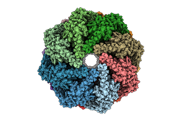 |
Ancestral Group Ii Chaperonin (Acii) Double-Ring In Closed Conformation
Organism: Synthetic construct
Method: ELECTRON MICROSCOPY Release Date: 2025-12-10 Classification: CHAPERONE Ligands: ADP, MG |
Organism: Synthetic construct
Method: ELECTRON MICROSCOPY
Release Date: 2025-12-10
Ligands: ADP, MG
 |
Ancestral Group Iii Chaperonin (Aciii) Double-Ring In Open Conformation Including The Equatorial And Intermediate Domains (Residues 12-200 And 356-507)
Organism: Synthetic construct
Method: ELECTRON MICROSCOPY Release Date: 2025-12-10 Classification: CHAPERONE |
Organism: Synthetic construct
Method: ELECTRON MICROSCOPY
Release Date: 2025-12-10
 |
Ancestral Fibrobacteres-Chlorobi-Bacteroidetes Group Chaperonin (Afcb) Double-Ring In Open Conformation
Organism: Synthetic construct
Method: ELECTRON MICROSCOPY Release Date: 2025-12-10 Classification: CHAPERONE |
Organism: Synthetic construct
Method: ELECTRON MICROSCOPY
Release Date: 2025-12-10
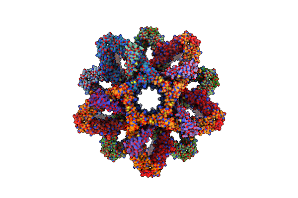 |
3D Reconstruction Of The Cylindrical Assembly Of Dnaja2 Delta G/F By Imposing D5 Symmetry
Organism: Saccharomyces cerevisiae s288c, Homo sapiens
Method: ELECTRON MICROSCOPY Resolution:6.90 Å Release Date: 2023-07-26 Classification: CHAPERONE Ligands: ZN |
Organism: Saccharomyces cerevisiae s288c, Homo sapiens
Method: ELECTRON MICROSCOPY
Release Date: 2023-07-26
Ligands: ZN
 |
Partial Structure Of Tyrosine Hydroxylase Lacking The First 35 Residues In Complex With Dopamine.
Organism: Homo sapiens
Method: ELECTRON MICROSCOPY Release Date: 2021-12-22 Classification: OXIDOREDUCTASE Ligands: LDP, FE |
Organism: Homo sapiens
Method: ELECTRON MICROSCOPY
Release Date: 2021-12-22
Ligands: LDP, FE
 |
Partial Structure Of Tyrosine Hydroxylase In Complex With Dopamine Showing The Catalytic Domain And An Alpha-Helix From The Regulatory Domain Involved In Dopamine Binding.
Organism: Homo sapiens
Method: ELECTRON MICROSCOPY Release Date: 2021-12-08 Classification: OXIDOREDUCTASE Ligands: LDP, FE |
Organism: Homo sapiens
Method: ELECTRON MICROSCOPY
Release Date: 2021-12-08
Ligands: LDP, FE
 |
Full-Length Structure Of The Substrate-Free Tyrosine Hydroxylase (Apo-Th).
Organism: Homo sapiens
Method: ELECTRON MICROSCOPY Release Date: 2021-12-01 Classification: OXIDOREDUCTASE Ligands: FE |
Organism: Homo sapiens
Method: ELECTRON MICROSCOPY
Release Date: 2021-12-01
Ligands: FE
 |
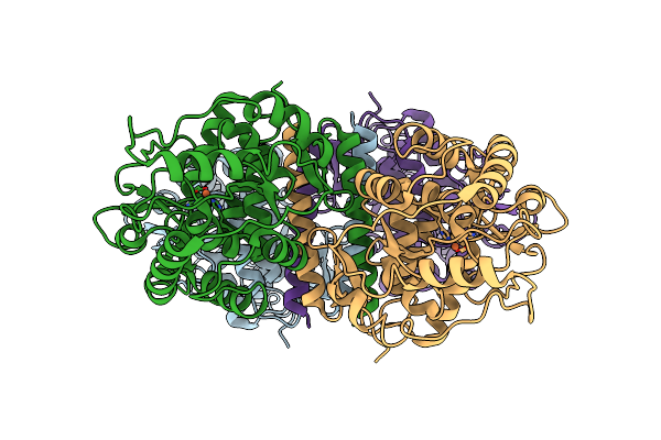
Atomic Model Of The Em-Based Structure Of The Full-Length Tyrosine Hydroxylase In Complex With Dopamine (Residues 40-497) In Which The Regulatory Domain (Residues 40-165) Has Been Included Only With The Backbone Atoms
Organism: Homo sapiens
Method: ELECTRON MICROSCOPY Release Date: 2021-11-17 Classification: OXIDOREDUCTASE Ligands: FE, LDP |
Organism: Homo sapiens
Method: ELECTRON MICROSCOPY
Release Date: 2021-11-17
Ligands: FE, LDP
 |
Partial Structure Of The Substrate-Free Tyrosine Hydroxylase (Apo-Th).
Organism: Homo sapiens
Method: ELECTRON MICROSCOPY Release Date: 2021-11-17 Classification: OXIDOREDUCTASE Ligands: FE |
Organism: Homo sapiens
Method: ELECTRON MICROSCOPY
Release Date: 2021-11-17
Ligands: FE
 |
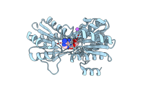
Atomic-Resolution Structure Of The Coiled-Coil Dimerisation Domain Of Human Arc
Organism: Homo sapiens
Method: X-RAY DIFFRACTION Resolution:0.95 Å Release Date: 2021-03-03 Classification: SIGNALING PROTEIN Ligands: MPD, CL |
Organism: Homo sapiens
Method: X-RAY DIFFRACTION
Release Date: 2021-03-03
Ligands: MPD, CL
 |
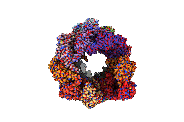
Borrelia Burgdorferi Bmpd Nucleoside Binding Protein Bound To Adenosine
Organism: Borrelia burgdorferi (strain jd1)
Method: X-RAY DIFFRACTION Resolution:1.43 Å Release Date: 2020-02-12 Classification: TRANSPORT PROTEIN Ligands: ADN, CL, NA |
Organism: Borrelia burgdorferi (strain jd1)
Method: X-RAY DIFFRACTION
Release Date: 2020-02-12
Ligands: ADN, CL, NA
 |
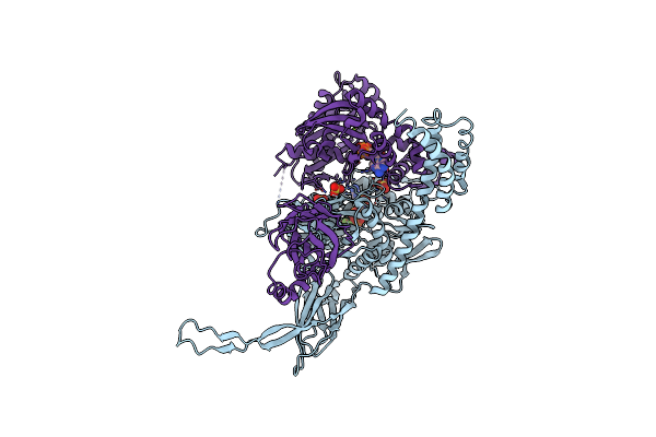
Human Cct:Mlst8 Complex
Organism: Homo sapiens
Method: ELECTRON MICROSCOPY Release Date: 2019-07-03 Classification: CHAPERONE Ligands: ADP |
Organism: Homo sapiens
Method: ELECTRON MICROSCOPY
Release Date: 2019-07-03
Ligands: ADP
 |
Structure Of The Hsp110:Hsc70 Nucleotide Exchange Complex
Organism: Saccharomyces cerevisiae, Bos taurus
Method: X-RAY DIFFRACTION Resolution:3.12 Å Release Date: 2008-05-27 Classification: CHAPERONE/CHAPERONE Ligands: MG, SO4, BEF, ADP, CL |
Organism: Saccharomyces cerevisiae, Bos taurus
Method: X-RAY DIFFRACTION
Release Date: 2008-05-27
Ligands: MG, SO4, BEF, ADP, CL