

Search Count: 18
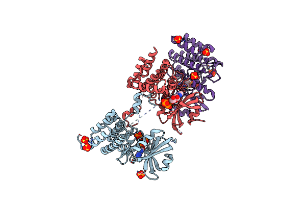 |
Crystal Structure Of Unphosphorylated Human Pkr Kinase Domain In Complex With Adp
Organism: Homo sapiens
Method: X-RAY DIFFRACTION Resolution:2.60 Å Release Date: 2019-07-10 Classification: TRANSFERASE Ligands: ADP, PO4, MG, SO4 |
Organism: Homo sapiens
Method: X-RAY DIFFRACTION
Release Date: 2019-07-10
Ligands: ADP, PO4, MG, SO4
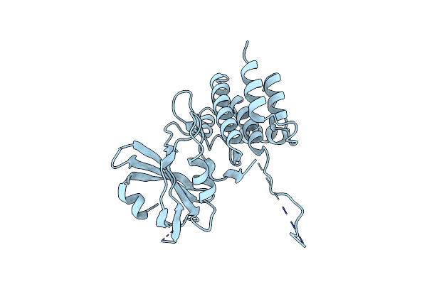 |
Crystal Structure Of Unphosphorylated Human Pkr
Organism: Homo sapiens
Method: X-RAY DIFFRACTION Resolution:3.10 Å Release Date: 2019-07-10 Classification: TRANSFERASE |
Organism: Homo sapiens
Method: X-RAY DIFFRACTION
Release Date: 2019-07-10
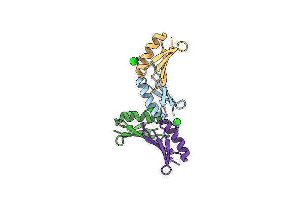 |
Crystal Structure Of Mutant B1 Immunoglobulin-Binding Domain Of Streptococcal Protein G (T16F, T18A, V21E, T25L, K28Y, V29I, K31R, Q32H, Y33L, N35K, D36H, N37Q)
Organism: Streptococcus
Method: X-RAY DIFFRACTION Resolution:1.40 Å Release Date: 2019-01-23 Classification: DE NOVO PROTEIN Ligands: ZN, CL |
Organism: Streptococcus
Method: X-RAY DIFFRACTION
Release Date: 2019-01-23
Ligands: ZN, CL
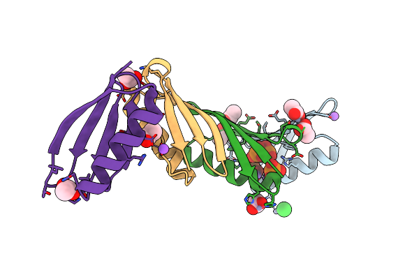 |
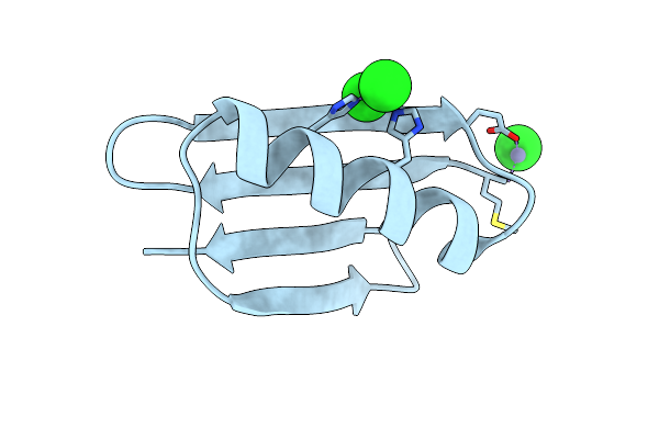
Crystal Structure Of B1 Immunoglobulin-Binding Domain Of Streptococcal Protein G (T16F, T18A, V21H, T25H, K28Y, V29I, K31R, Q32A, Y33L, N35K, D36A, N37Q)
Organism: Streptococcus
Method: X-RAY DIFFRACTION Resolution:1.40 Å Release Date: 2019-01-23 Classification: METAL BINDING PROTEIN Ligands: ZN, ACT, NA, CL, PO4, DPO |
Organism: Streptococcus
Method: X-RAY DIFFRACTION
Release Date: 2019-01-23
Ligands: ZN, ACT, NA, CL, PO4, DPO
 |
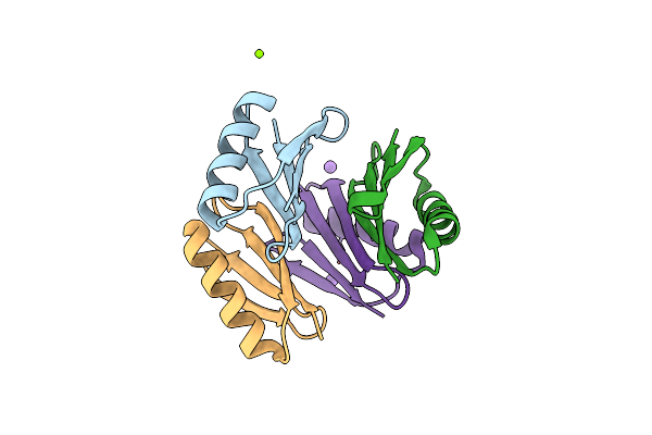
Crystal Structure Of De Novo Designed Metal-Controlled Dimer Of Mutant B1 Immunoglobulin-Binding Domain Of Streptococcal Protein G (L12H, T16L, V29H, Y33H, N37L)-Zinc
Organism: Streptococcus
Method: X-RAY DIFFRACTION Resolution:1.50 Å Release Date: 2019-01-23 Classification: METAL BINDING PROTEIN Ligands: ZN, CL |
Organism: Streptococcus
Method: X-RAY DIFFRACTION
Release Date: 2019-01-23
Ligands: ZN, CL
 |
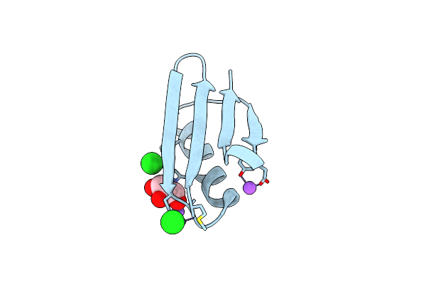
Crystal Structure Of De Novo Designed Metal-Controlled Dimer Of Mutant B1 Immunoglobulin-Binding Domain Of Streptococcal Protein G (L12H, T16L, V29H, Y33H, N37L)-Apo
Organism: Streptococcus
Method: X-RAY DIFFRACTION Resolution:1.70 Å Release Date: 2019-01-23 Classification: DE NOVO PROTEIN Ligands: MG, NA |
Organism: Streptococcus
Method: X-RAY DIFFRACTION
Release Date: 2019-01-23
Ligands: MG, NA
 |
Crystal Structure Of De Novo Designed Metal-Controlled Dimer Of B1 Immunoglobulin-Binding Domain Of Streptococcal Protein G (L12H, E15V, T16L, T18I, V29H, Y33H, N37L)-Zinc
Organism: Streptococcus
Method: X-RAY DIFFRACTION Resolution:1.34 Å Release Date: 2019-01-23 Classification: DE NOVO PROTEIN Ligands: ZN, CL, GOL, NA |
Organism: Streptococcus
Method: X-RAY DIFFRACTION
Release Date: 2019-01-23
Ligands: ZN, CL, GOL, NA
 |
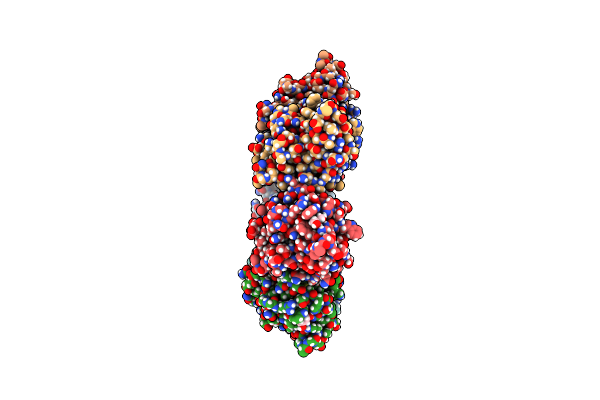
Crystal Structure Of De Novo Designed Metal-Controlled Dimer Of Mutant B1 Immunoglobulin-Binding Domain Of Streptococcal Protein G (L12H, E15V, T16L, T18I, V29H, Y33H, N37L)-Apo
Organism: Streptococcus
Method: X-RAY DIFFRACTION Resolution:2.30 Å Release Date: 2019-01-23 Classification: METAL BINDING PROTEIN Ligands: MG |
Organism: Streptococcus
Method: X-RAY DIFFRACTION
Release Date: 2019-01-23
Ligands: MG
 |
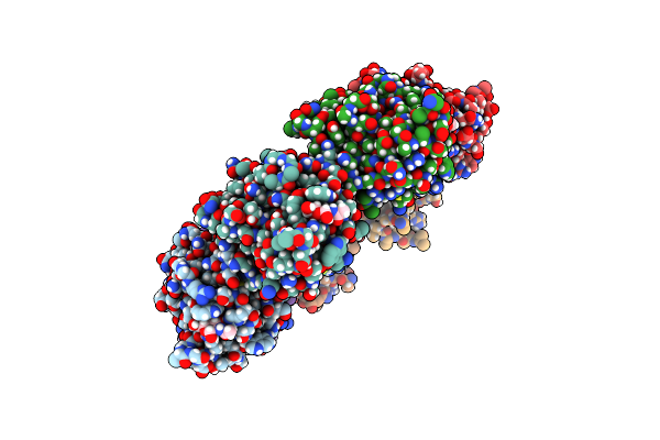
Crystal Structure Of The Vibrio Vulnificus Hemolysin/Cytolysin Beta-Trefoil Lectin
Organism: Vibrio vulnificus
Method: X-RAY DIFFRACTION Resolution:2.00 Å Release Date: 2014-05-28 Classification: TOXIN Ligands: GOL |
Organism: Vibrio vulnificus
Method: X-RAY DIFFRACTION
Release Date: 2014-05-28
Ligands: GOL
 |
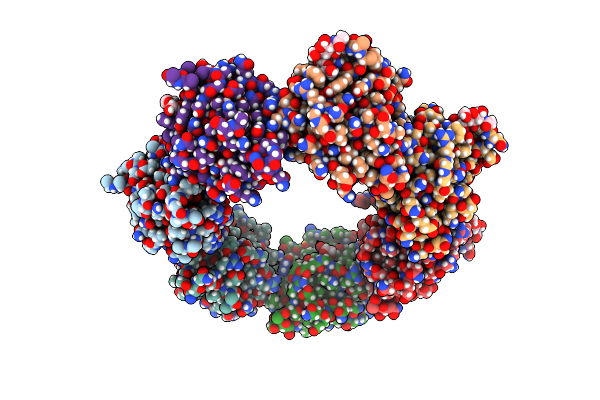
Crystal Structure Of The Vibrio Vulnificus Hemolysin/Cytolysin Beta-Trefoil Lectin With N-Acetyl-D-Galactosamine Bound
Organism: Vibrio vulnificus
Method: X-RAY DIFFRACTION Resolution:2.00 Å Release Date: 2014-05-28 Classification: TOXIN Ligands: NGA, GOL |
Organism: Vibrio vulnificus
Method: X-RAY DIFFRACTION
Release Date: 2014-05-28
Ligands: NGA, GOL
 |
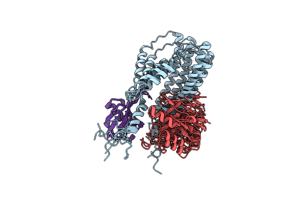
Crystal Structure Of The Vibrio Vulnificus Hemolysin/Cytolysin Beta-Trefoil Lectin With N-Acetyl-D-Lactosamine Bound
Organism: Vibrio vulnificus
Method: X-RAY DIFFRACTION Resolution:2.10 Å Release Date: 2014-05-28 Classification: TOXIN Ligands: GOL, GAL |
Organism: Vibrio vulnificus
Method: X-RAY DIFFRACTION
Release Date: 2014-05-28
Ligands: GOL, GAL
 |
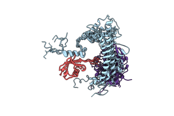
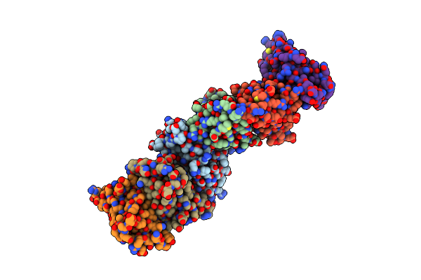
Nmr Structure Of Major S5A (196-306):K48 Linked Diubiquitin Species
Organism: Homo sapiens, Xenopus laevis
Method: SOLUTION NMR Release Date: 2009-09-01 Classification: PROTEIN BINDING |
Organism: Homo sapiens, Xenopus laevis
Method: SOLUTION NMR
Release Date: 2009-09-01
 |
Nmr Structure Of Minor S5A (196-306):K48 Linked Diubiquitin Species
Organism: Homo sapiens
Method: SOLUTION NMR Release Date: 2009-09-01 Classification: PROTEIN BINDING |
Organism: Homo sapiens
Method: SOLUTION NMR
Release Date: 2009-09-01
 |
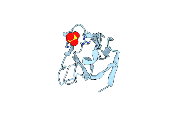
Crystal Structure Of Cd84: Insite Into Slam Family Function
Organism: Homo sapiens
Method: X-RAY DIFFRACTION Resolution:2.04 Å Release Date: 2007-06-26 Classification: SIGNALING PROTEIN Ligands: CL |
Organism: Homo sapiens
Method: X-RAY DIFFRACTION
Release Date: 2007-06-26
Ligands: CL
 |
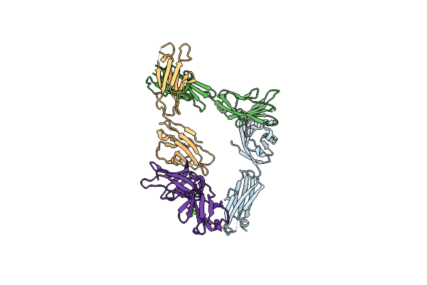
T Cell Immunoglobulin Mucin-3 Crystal Structure Revealed A Galectin-9-Independent Binding Surface
Organism: Mus musculus
Method: X-RAY DIFFRACTION Resolution:1.95 Å Release Date: 2007-04-10 Classification: SIGNALING PROTEIN Ligands: SO4 |
Organism: Mus musculus
Method: X-RAY DIFFRACTION
Release Date: 2007-04-10
Ligands: SO4
 |
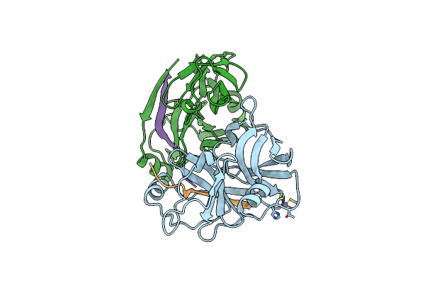
Crystal Structure Of Ntb-A
Organism: Homo sapiens
Method: X-RAY DIFFRACTION Resolution:3.00 Å Release Date: 2006-10-17 Classification: IMMUNE SYSTEM Ligands: CA, CL |
Organism: Homo sapiens
Method: X-RAY DIFFRACTION
Release Date: 2006-10-17
Ligands: CA, CL
 |
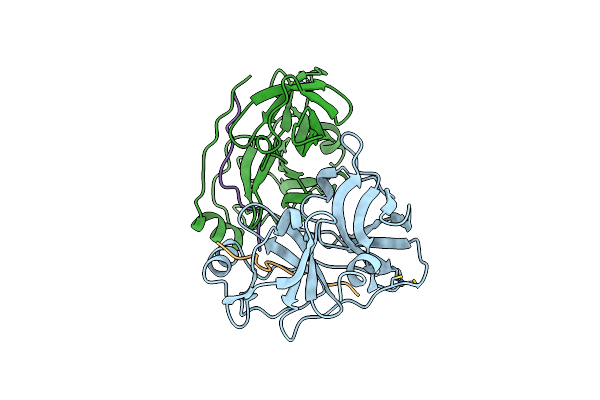
Structure Of Hcv Protease (Bk Strain)
Organism: Hepatitis c virus
Method: X-RAY DIFFRACTION Resolution:2.80 Å Release Date: 1998-04-08 Classification: COMPLEX (HYDROLASE/PEPTIDE) Ligands: ZN |
Organism: Hepatitis c virus
Method: X-RAY DIFFRACTION
Release Date: 1998-04-08
Ligands: ZN
 |
Bk Strain Hepatitis C Virus (Hcv) Ns3-Ns4A
Organism: Hepatitis c virus (isolate bk)
Method: X-RAY DIFFRACTION Resolution:2.20 Å Release Date: 1998-01-14 Classification: Viral protein complex Ligands: ZN |
Organism: Hepatitis c virus (isolate bk)
Method: X-RAY DIFFRACTION
Release Date: 1998-01-14
Ligands: ZN