

Search Count: 16
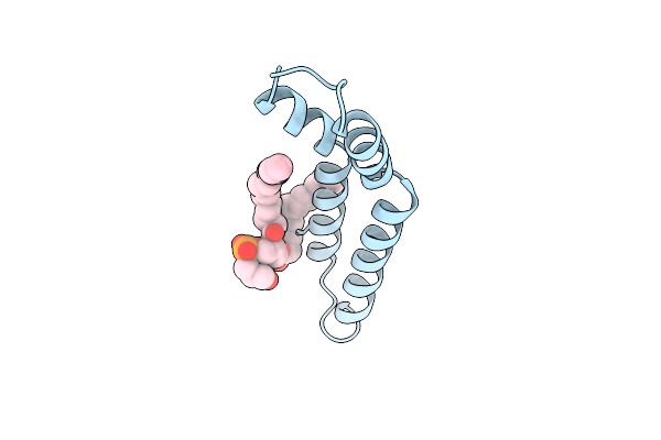 |
Cryoem Structure Of Cyclised H-Pilus
Organism: Salmonella
Method: ELECTRON MICROSCOPY Resolution:2.24 Å Release Date: 2025-03-26 Classification: PROTEIN FIBRIL Ligands: LHG |
Organism: Salmonella
Method: ELECTRON MICROSCOPY
Release Date: 2025-03-26
Ligands: LHG
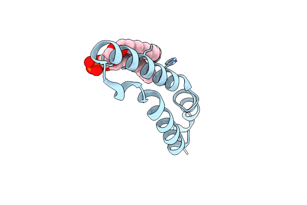 |
Cryo-Em Of F-Pilus
Organism: Escherichia coli
Method: ELECTRON MICROSCOPY Release Date: 2025-01-15 Classification: PROTEIN FIBRIL Ligands: X41 |
Organism: Escherichia coli
Method: ELECTRON MICROSCOPY
Release Date: 2025-01-15
Ligands: X41
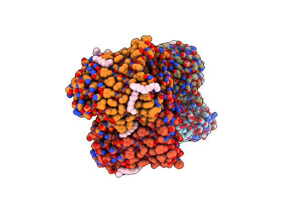 |
Crystal Structure Of The Ompk36 Td Insertion Chimera From Klebsiella Pneumonia
Organism: Klebsiella pneumoniae
Method: X-RAY DIFFRACTION Resolution:1.50 Å Release Date: 2022-07-06 Classification: MEMBRANE PROTEIN Ligands: LI, C14 |
Organism: Klebsiella pneumoniae
Method: X-RAY DIFFRACTION
Release Date: 2022-07-06
Ligands: LI, C14
 |
Crystal Structure Of The Ompk36 D Insertion Chimera From Klebsiella Pneumonia
Organism: Klebsiella pneumoniae
Method: X-RAY DIFFRACTION Resolution:1.79 Å Release Date: 2022-07-06 Classification: MEMBRANE PROTEIN Ligands: C8E, LI |
Organism: Klebsiella pneumoniae
Method: X-RAY DIFFRACTION
Release Date: 2022-07-06
Ligands: C8E, LI
 |
Crystal Structure Of The Ompk36 Gd Insertion Chimera From Klebsiella Pneumonia
Organism: Klebsiella pneumoniae
Method: X-RAY DIFFRACTION Resolution:2.03 Å Release Date: 2019-09-11 Classification: MEMBRANE PROTEIN Ligands: LDA, C8E, P6L |
Organism: Klebsiella pneumoniae
Method: X-RAY DIFFRACTION
Release Date: 2019-09-11
Ligands: LDA, C8E, P6L
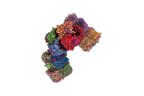 |
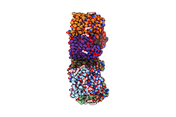
Crystal Structure Of The Ompk36 Clinical Isolate St258 From Klebsiella Pneumonia
Organism: Klebsiella pneumoniae
Method: X-RAY DIFFRACTION Resolution:3.23 Å Release Date: 2019-09-11 Classification: MEMBRANE PROTEIN |
Organism: Klebsiella pneumoniae
Method: X-RAY DIFFRACTION
Release Date: 2019-09-11
 |
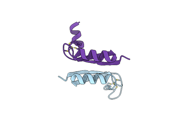
Crystal Structure Of The Wild Type Ompk36 From Klebsiella Pneumonia
Organism: Klebsiella pneumoniae
Method: X-RAY DIFFRACTION Resolution:1.98 Å Release Date: 2019-09-11 Classification: MEMBRANE PROTEIN Ligands: C8E, MG |
Organism: Klebsiella pneumoniae
Method: X-RAY DIFFRACTION
Release Date: 2019-09-11
Ligands: C8E, MG
 |
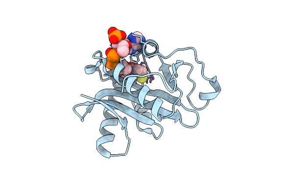
Crystal Structure Of Hpv E7 Cr3 Domain
Organism: Human papillomavirus type 1a
Method: X-RAY DIFFRACTION Resolution:1.60 Å Release Date: 2005-10-25 Classification: transcription, viral protein Ligands: ZN |
Organism: Human papillomavirus type 1a
Method: X-RAY DIFFRACTION
Release Date: 2005-10-25
Ligands: ZN
 |
Crystal Structure Of Tetrahymena Gcn5 With Bound Coenzyme A And A 19-Residue Histone H4 Peptide
Organism: Tetrahymena thermophila
Method: X-RAY DIFFRACTION Resolution:2.25 Å Release Date: 2004-08-03 Classification: TRANSFERASE/STRUCTURAL PROTEIN Ligands: COA |
Organism: Tetrahymena thermophila
Method: X-RAY DIFFRACTION
Release Date: 2004-08-03
Ligands: COA
 |
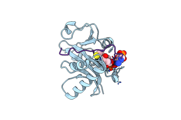
Structure And Autoregulation Of The Yeast Hst2 Homolog Of Sir2
Organism: Saccharomyces cerevisiae
Method: X-RAY DIFFRACTION Resolution:2.50 Å Release Date: 2003-09-30 Classification: HYDROLASE Ligands: ZN, CL |
Organism: Saccharomyces cerevisiae
Method: X-RAY DIFFRACTION
Release Date: 2003-09-30
Ligands: ZN, CL
 |
Crystal Structure Of Tetrahymena Gcn5 With Bound Coenzyme A And A 19-Residue Histone H3 Peptide
Organism: Tetrahymena thermophila
Method: X-RAY DIFFRACTION Resolution:2.30 Å Release Date: 2003-09-23 Classification: TRANSFERASE/STRUCTURAL PROTEIN Ligands: COA |
Organism: Tetrahymena thermophila
Method: X-RAY DIFFRACTION
Release Date: 2003-09-23
Ligands: COA
 |
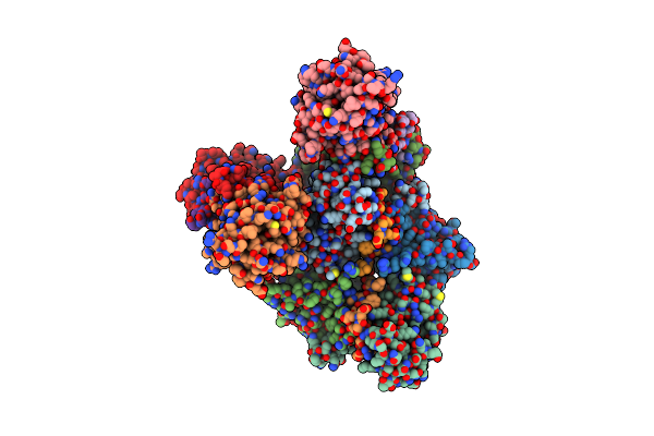
Crystal Structure Of Tetrahymena Gcn5 With Bound Coenzyme A And A Phosphorylated, 19-Residue Histone H3 Peptide
Organism: Tetrahymena thermophila
Method: X-RAY DIFFRACTION Resolution:2.30 Å Release Date: 2003-09-23 Classification: TRANSFERASE/STRUCTURAL PROTEIN Ligands: COA |
Organism: Tetrahymena thermophila
Method: X-RAY DIFFRACTION
Release Date: 2003-09-23
Ligands: COA
 |
Crystal Structure Of The Retinoblastoma Tumour Suppressor Protein Bound To E2F Peptide
Organism: Homo sapiens
Method: X-RAY DIFFRACTION Resolution:2.60 Å Release Date: 2003-03-06 Classification: APOPTOSIS |
Organism: Homo sapiens
Method: X-RAY DIFFRACTION
Release Date: 2003-03-06
 |
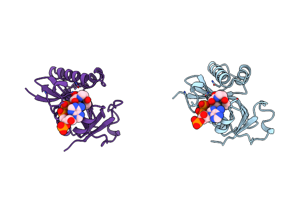
Crystal Structure Of The Mouse P53 Core Dna-Binding Domain At 2.7A Resolution
Organism: Mus musculus
Method: X-RAY DIFFRACTION Resolution:2.70 Å Release Date: 2001-07-04 Classification: DNA BINDING PROTEIN Ligands: ZN |
Organism: Mus musculus
Method: X-RAY DIFFRACTION
Release Date: 2001-07-04
Ligands: ZN
 |
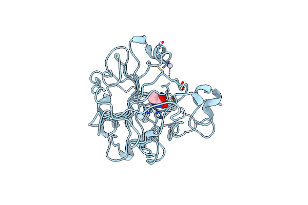
Crystal Structure Of The Pcaf/Coenzyme-A Complex
Organism: Homo sapiens
Method: X-RAY DIFFRACTION Resolution:2.30 Å Release Date: 1999-07-06 Classification: SIGNALING PROTEIN Ligands: COA |
Organism: Homo sapiens
Method: X-RAY DIFFRACTION
Release Date: 1999-07-06
Ligands: COA
 |
Carbonic Anhydrase Ii Inhibitor: Acetohydroxamate
Organism: Homo sapiens
Method: X-RAY DIFFRACTION Resolution:2.00 Å Release Date: 1998-06-24 Classification: HYDROLASE Ligands: HG, ZN, HAE |
Organism: Homo sapiens
Method: X-RAY DIFFRACTION
Release Date: 1998-06-24
Ligands: HG, ZN, HAE