

Search Count: 58
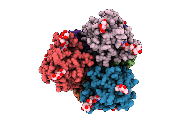 |
Structure Of The Machupo Virus Glycoprotein Complex
Organism: Mammarenavirus machupoense
Method: ELECTRON MICROSCOPY Release Date: 2025-07-16 Classification: VIRAL PROTEIN Ligands: NAG, ZN |
Organism: Mammarenavirus machupoense
Method: ELECTRON MICROSCOPY
Release Date: 2025-07-16
Ligands: NAG, ZN
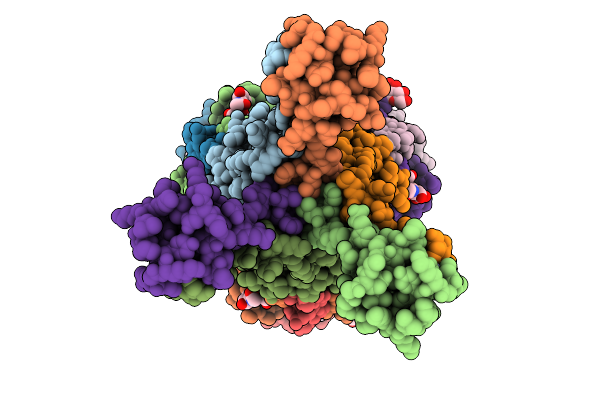 |
Structure Of The Junin Virus Glycoprotein Complex
Organism: Mammarenavirus juninense
Method: ELECTRON MICROSCOPY Release Date: 2025-07-16 Classification: VIRAL PROTEIN Ligands: NAG, ZN |
Organism: Mammarenavirus juninense
Method: ELECTRON MICROSCOPY
Release Date: 2025-07-16
Ligands: NAG, ZN
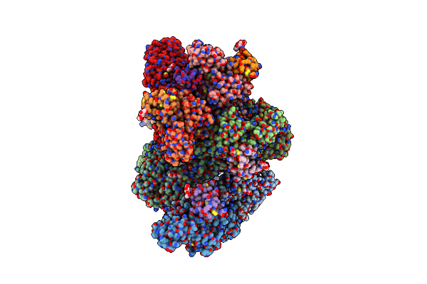 |
Structure Of Eastern Equine Encephalitis Virus Vlp In Complex With Vldlr La1
Organism: Eastern equine encephalitis virus, Homo sapiens
Method: ELECTRON MICROSCOPY Release Date: 2024-08-14 Classification: VIRUS LIKE PARTICLE Ligands: NAG, CA |
Organism: Eastern equine encephalitis virus, Homo sapiens
Method: ELECTRON MICROSCOPY
Release Date: 2024-08-14
Ligands: NAG, CA
 |
Structure Of Semliki Forest Virus Vlp In Complex With Vldlr La2
Organism: Semliki forest virus, Homo sapiens
Method: ELECTRON MICROSCOPY Release Date: 2024-08-14 Classification: VIRUS LIKE PARTICLE Ligands: NAG, CA |
Organism: Semliki forest virus, Homo sapiens
Method: ELECTRON MICROSCOPY
Release Date: 2024-08-14
Ligands: NAG, CA
 |
Structure Of Eastern Equine Encephalitis Virus Vlp Unliganded Quasi-Threefold Spike Protein
Organism: Eastern equine encephalitis virus
Method: ELECTRON MICROSCOPY Release Date: 2024-08-14 Classification: VIRUS LIKE PARTICLE Ligands: NAG |
Organism: Eastern equine encephalitis virus
Method: ELECTRON MICROSCOPY
Release Date: 2024-08-14
Ligands: NAG
 |
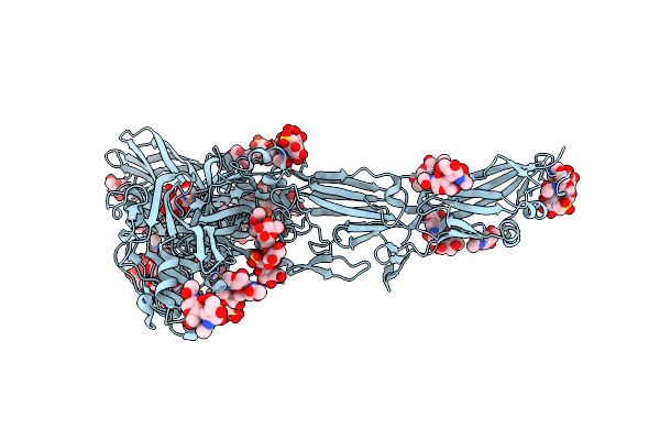
Two-Component Assembly Of Slaa And Slab S-Layer Proteins Of Sulfolobus Acidocaldarius
Organism: Sulfolobus acidocaldarius dsm 639
Method: ELECTRON MICROSCOPY Release Date: 2024-02-21 Classification: STRUCTURAL PROTEIN |
Organism: Sulfolobus acidocaldarius dsm 639
Method: ELECTRON MICROSCOPY
Release Date: 2024-02-21
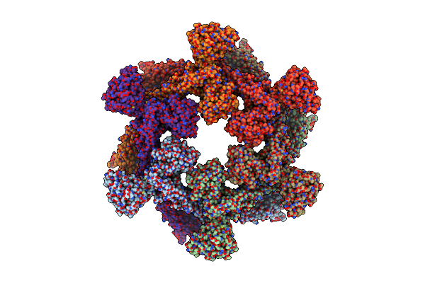 |
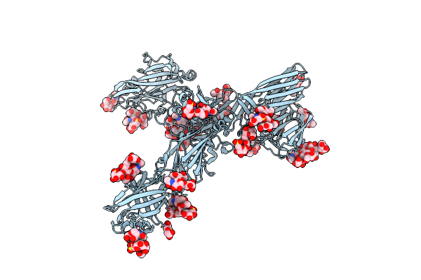
A Hexamer Pore In The S-Layer Of Sulfolobus Acidocaldarius Formed By Slaa Protein
Organism: Sulfolobus acidocaldarius dsm 639
Method: ELECTRON MICROSCOPY Release Date: 2024-02-21 Classification: STRUCTURAL PROTEIN |
Organism: Sulfolobus acidocaldarius dsm 639
Method: ELECTRON MICROSCOPY
Release Date: 2024-02-21
 |
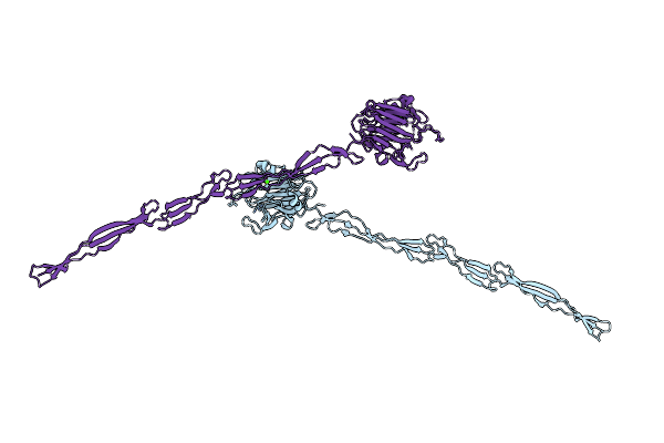
S-Layer Protein Slaa From Sulfolobus Acidocaldarius At Ph 10.0
Organism: Sulfolobus acidocaldarius dsm 639
Method: ELECTRON MICROSCOPY Resolution:3.20 Å Release Date: 2023-08-16 Classification: STRUCTURAL PROTEIN Ligands: NAG |
Organism: Sulfolobus acidocaldarius dsm 639
Method: ELECTRON MICROSCOPY
Release Date: 2023-08-16
Ligands: NAG
 |
S-Layer Protein Slaa From Sulfolobus Acidocaldarius At Ph 7.0
Organism: Sulfolobus acidocaldarius dsm 639
Method: ELECTRON MICROSCOPY Resolution:3.90 Å Release Date: 2023-08-16 Classification: STRUCTURAL PROTEIN Ligands: NAG |
Organism: Sulfolobus acidocaldarius dsm 639
Method: ELECTRON MICROSCOPY
Release Date: 2023-08-16
Ligands: NAG
 |
S-Layer Protein Slaa From Sulfolobus Acidocaldarius At Ph 4.0
Organism: Sulfolobus acidocaldarius
Method: ELECTRON MICROSCOPY Resolution:3.10 Å Release Date: 2023-06-14 Classification: STRUCTURAL PROTEIN Ligands: NAG, YZT |
Organism: Sulfolobus acidocaldarius
Method: ELECTRON MICROSCOPY
Release Date: 2023-06-14
Ligands: NAG, YZT
 |
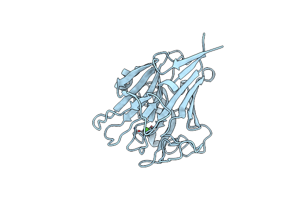
Structure Of Aap A Domain And B-Repeats (Residues 351-813) From Staphylococcus Epidermidis
Organism: Staphylococcus epidermidis rp62a
Method: X-RAY DIFFRACTION Resolution:2.30 Å Release Date: 2023-05-03 Classification: CELL ADHESION Ligands: CA, CL |
Organism: Staphylococcus epidermidis rp62a
Method: X-RAY DIFFRACTION
Release Date: 2023-05-03
Ligands: CA, CL
 |
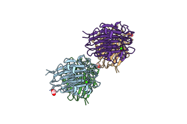
Structure Of Pls A-Domain (Residues 391-656; 513-518 Deletion Mutant) From Staphylococcus Aureus
Organism: Staphylococcus aureus
Method: X-RAY DIFFRACTION Resolution:2.75 Å Release Date: 2022-11-09 Classification: CELL ADHESION Ligands: CA |
Organism: Staphylococcus aureus
Method: X-RAY DIFFRACTION
Release Date: 2022-11-09
Ligands: CA
 |
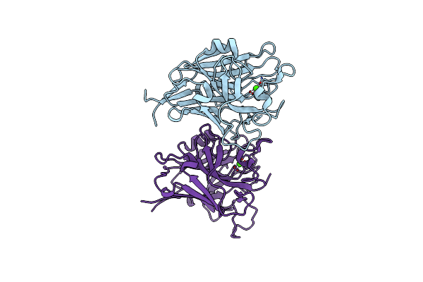
Structure Of Sasg A-Domain (Residues 163-419) From Staphylococcus Aureus
Organism: Staphylococcus aureus (strain nctc 8325 / ps 47)
Method: X-RAY DIFFRACTION Resolution:1.65 Å Release Date: 2022-11-02 Classification: CELL ADHESION Ligands: CA, EDO |
Organism: Staphylococcus aureus (strain nctc 8325 / ps 47)
Method: X-RAY DIFFRACTION
Release Date: 2022-11-02
Ligands: CA, EDO
 |
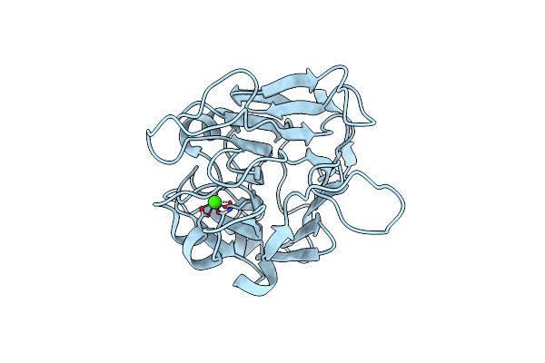
Structure Of Pls A-Domain (Residues 391-656) From Staphylococcus Aureus
Organism: Staphylococcus aureus subsp. aureus nctc 8325
Method: X-RAY DIFFRACTION Resolution:1.21 Å Release Date: 2022-10-26 Classification: CELL ADHESION Ligands: CA |
Organism: Staphylococcus aureus subsp. aureus nctc 8325
Method: X-RAY DIFFRACTION
Release Date: 2022-10-26
Ligands: CA
 |
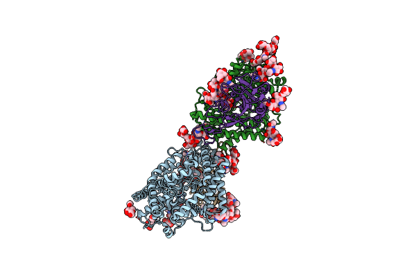
Structure Of Aap A-Domain (Residues 351-605) From Staphylococcus Epidermidis
Organism: Staphylococcus epidermidis (strain atcc 35984 / rp62a)
Method: X-RAY DIFFRACTION Resolution:1.30 Å Release Date: 2022-10-19 Classification: CELL ADHESION Ligands: CA, CL |
Organism: Staphylococcus epidermidis (strain atcc 35984 / rp62a)
Method: X-RAY DIFFRACTION
Release Date: 2022-10-19
Ligands: CA, CL
 |
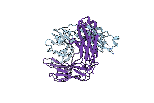
Crystal Structure Of Spike Protein Receptor Binding Domain Of Escape Mutant Sars-Cov-2 From Immunocompromised Patient (D146*) In Complex With Human Receptor Ace2
Organism: Homo sapiens, Severe acute respiratory syndrome coronavirus 2
Method: X-RAY DIFFRACTION Resolution:3.08 Å Release Date: 2021-12-08 Classification: VIRAL PROTEIN Ligands: ZN, CL, NAG |
Organism: Homo sapiens, Severe acute respiratory syndrome coronavirus 2
Method: X-RAY DIFFRACTION
Release Date: 2021-12-08
Ligands: ZN, CL, NAG
 |
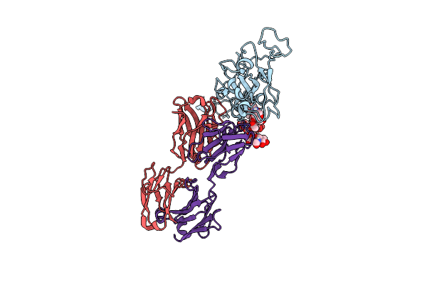
Structure Of Human Sars-Cov-2 Neutralizing Antibody C1C-A3 Fab
Organism: Homo sapiens
Method: X-RAY DIFFRACTION Resolution:1.47 Å Release Date: 2021-12-08 Classification: IMMUNE SYSTEM |
Organism: Homo sapiens
Method: X-RAY DIFFRACTION
Release Date: 2021-12-08
 |
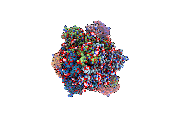
Structure Of Human Sars-Cov-2 Neutralizing Antibody C1C-A3 Fab
Organism: Severe acute respiratory syndrome coronavirus 2, Homo sapiens
Method: ELECTRON MICROSCOPY Release Date: 2021-12-08 Classification: IMMUNE SYSTEM |
Organism: Severe acute respiratory syndrome coronavirus 2, Homo sapiens
Method: ELECTRON MICROSCOPY
Release Date: 2021-12-08
 |
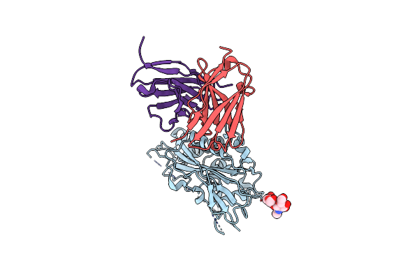
Structure Of Human Sars-Cov-2 Spike Glycoprotein Trimer Bound By Neutralizing Antibody C1C-A3 Fab (Variable Region)
Organism: Severe acute respiratory syndrome coronavirus 2, Homo sapiens
Method: ELECTRON MICROSCOPY Release Date: 2021-12-08 Classification: VIRAL PROTEIN/IMMUNE SYSTEM Ligands: NAG |
Organism: Severe acute respiratory syndrome coronavirus 2, Homo sapiens
Method: ELECTRON MICROSCOPY
Release Date: 2021-12-08
Ligands: NAG
 |
Wt Chicken Scap L1-L7 / Fab 4G10 Complex Focused Refinement
Organism: Gallus gallus, Mus musculus
Method: ELECTRON MICROSCOPY Release Date: 2021-06-30 Classification: LIPID BINDING PROTEIN |
Organism: Gallus gallus, Mus musculus
Method: ELECTRON MICROSCOPY
Release Date: 2021-06-30