

Search Count: 18
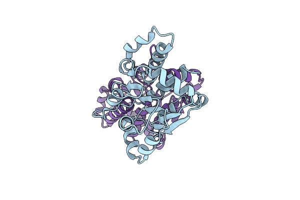 |
Crystal Structure Of The Cobh-Like Domain In Cobalt Regulator Coar
Organism: Synechocystis sp. (strain pcc 6803 / kazusa)
Method: X-RAY DIFFRACTION Resolution:3.20 Å Release Date: 2024-11-13 Classification: METAL BINDING PROTEIN Ligands: CL |
Organism: Synechocystis sp. (strain pcc 6803 / kazusa)
Method: X-RAY DIFFRACTION
Release Date: 2024-11-13
Ligands: CL
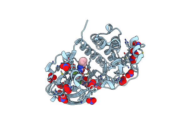 |
Crystal Structure Of Kit1 In Complex With Dp2976 Via Co-Crystallization
Organism: Homo sapiens
Method: X-RAY DIFFRACTION Resolution:1.80 Å Release Date: 2019-07-31 Classification: TRANSFERASE/TRANSFERASE INHIBITOR Ligands: NO3, JWY |
Organism: Homo sapiens
Method: X-RAY DIFFRACTION
Release Date: 2019-07-31
Ligands: NO3, JWY
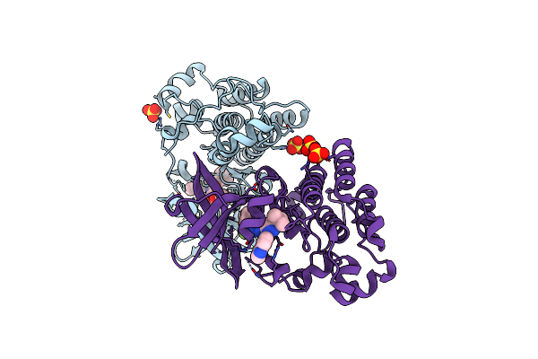 |
Crystal Structure Of Tie2 In Complex With Decipera Compound Dp1919
Organism: Homo sapiens
Method: X-RAY DIFFRACTION Resolution:2.05 Å Release Date: 2018-11-21 Classification: TRANSFERASE Ligands: SO4, 919 |
Organism: Homo sapiens
Method: X-RAY DIFFRACTION
Release Date: 2018-11-21
Ligands: SO4, 919
 |
Crystal Structure Of Human Pde1B Catalytic Domain In Complex With Inhibitor 16J (6-(4-Methoxybenzyl)-9-((Tetrahydro-2H-Pyran-4-Yl)Methyl)-8,9,10,11-Tetrahydropyrido[4',3':4,5]Thieno[3,2-E][1,2,4]Triazolo[1,5-C]Pyrimidin-5(6H)-One)
Organism: Homo sapiens
Method: X-RAY DIFFRACTION Resolution:1.82 Å Release Date: 2017-04-26 Classification: HYDROLASE/HYDROLASE INHIBITOR Ligands: ZN, MG, 8HM |
Organism: Homo sapiens
Method: X-RAY DIFFRACTION
Release Date: 2017-04-26
Ligands: ZN, MG, 8HM
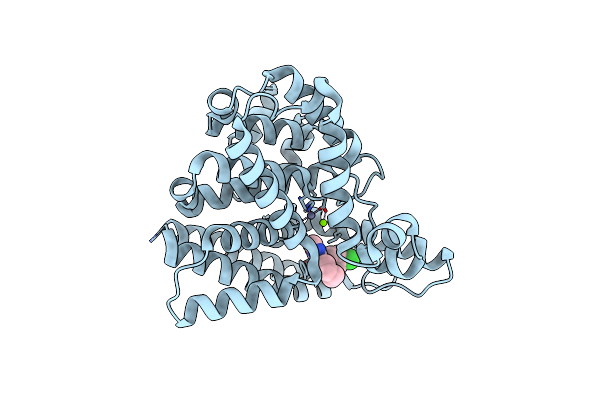 |
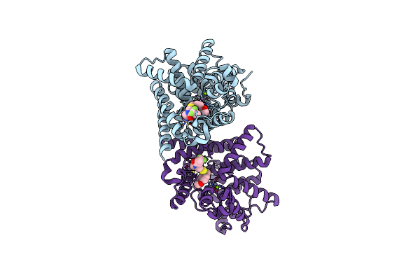
Crystal Structure Of Human Pde1B Catalytic Domain In Complex With Inhibitor 3 (6-(4-Chlorobenzyl)-8,9,10,11-Tetrahydrobenzo[4,5]Thieno[3,2-E][1,2,4]Triazolo[1,5-C]Pyrimidin-5(6H)-One)
Organism: Homo sapiens
Method: X-RAY DIFFRACTION Resolution:2.04 Å Release Date: 2017-04-26 Classification: HYDROLASE/HYDROLASE INHIBITOR Ligands: ZN, MG, 8HP |
Organism: Homo sapiens
Method: X-RAY DIFFRACTION
Release Date: 2017-04-26
Ligands: ZN, MG, 8HP
 |
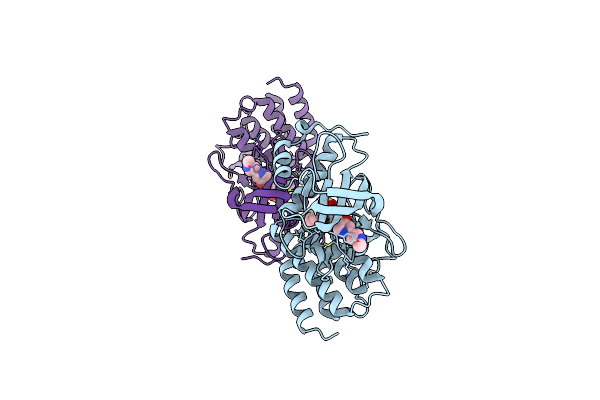
Crystal Structure Of Human Pde10A In Complex With Inhibitor 16D
Organism: Homo sapiens
Method: X-RAY DIFFRACTION Resolution:1.87 Å Release Date: 2017-04-26 Classification: Hydrolase/Hydrolase Inhibitor Ligands: 8Q7, ZN, MG |
Organism: Homo sapiens
Method: X-RAY DIFFRACTION
Release Date: 2017-04-26
Ligands: 8Q7, ZN, MG
 |
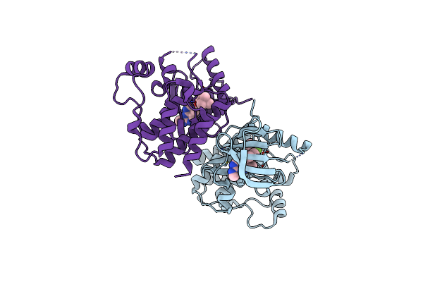
Crystal Structure Of The Tyrosine Kinase Domain Of The Hepatocyte Growth Factor Receptor C-Met In Complex With Altiratinib Analog Dp-4157
Organism: Homo sapiens
Method: X-RAY DIFFRACTION Resolution:2.60 Å Release Date: 2016-08-31 Classification: TRANSFERASE/TRANSFERASE INHIBITOR Ligands: 5B4 |
Organism: Homo sapiens
Method: X-RAY DIFFRACTION
Release Date: 2016-08-31
Ligands: 5B4
 |
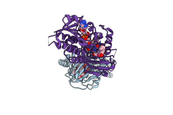
Crystal Structure Of Braf(V600E) In Complex With Ly3009120 Compnd
Organism: Homo sapiens
Method: X-RAY DIFFRACTION Resolution:2.70 Å Release Date: 2015-07-15 Classification: TRANSFERASE/TRANSFERASE INHIBITOR Ligands: 4Z5, CL |
Organism: Homo sapiens
Method: X-RAY DIFFRACTION
Release Date: 2015-07-15
Ligands: 4Z5, CL
 |
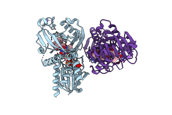
Co-Crystal Structure Of Compound 3 (4-Hydroxy-6-[2-(7-Hydroxy-2-Oxo-4-Phenyl-2H-Chromen-6-Yl)Ethyl]Pyridazin-3(2H)-One) And Fad Bound To Human Daao At 2.4A
Organism: Homo sapiens
Method: X-RAY DIFFRACTION Resolution:2.40 Å Release Date: 2014-07-16 Classification: oxidoreductase/oxidoreductase inhibitor Ligands: FAD, 31T, K |
Organism: Homo sapiens
Method: X-RAY DIFFRACTION
Release Date: 2014-07-16
Ligands: FAD, 31T, K
 |
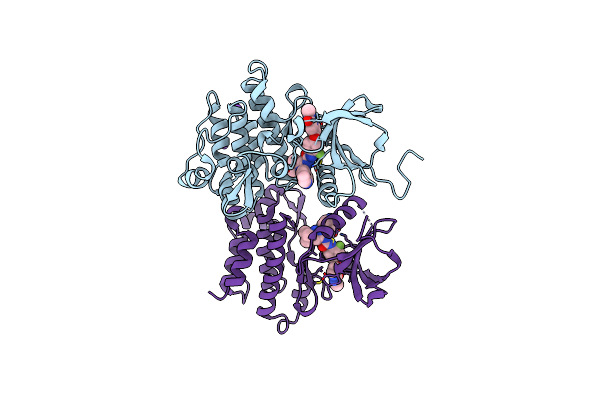
Co-Crystal Structure Of Compound 2 (3-(7-Hydroxy-2-Oxo-4-Phenyl-2H-Chromen-6-Yl)Propanoic Acid) And Fad Bound To Human Daao At 2.85A
Organism: Homo sapiens
Method: X-RAY DIFFRACTION Resolution:2.85 Å Release Date: 2014-07-16 Classification: oxidoreductase/oxidoreductase inhibitor |
Organism: Homo sapiens
Method: X-RAY DIFFRACTION
Release Date: 2014-07-16
 |
The Crystal Structure Of Human Abl1 Kinase Domain In Complex With Dcc-2036
Organism: Homo sapiens
Method: X-RAY DIFFRACTION Resolution:2.10 Å Release Date: 2011-06-01 Classification: TRANSFERASE/TRANSFERASE INHIBITOR |
Organism: Homo sapiens
Method: X-RAY DIFFRACTION
Release Date: 2011-06-01
 |
The Crystal Structure Of Human Abl1 Kinase Domain T315I Mutant In Complex With Dcc-2036
Organism: Homo sapiens
Method: X-RAY DIFFRACTION Resolution:1.82 Å Release Date: 2011-06-01 Classification: TRANSFERASE/TRANSFERASE INHIBITOR Ligands: 919 |
Organism: Homo sapiens
Method: X-RAY DIFFRACTION
Release Date: 2011-06-01
Ligands: 919
 |
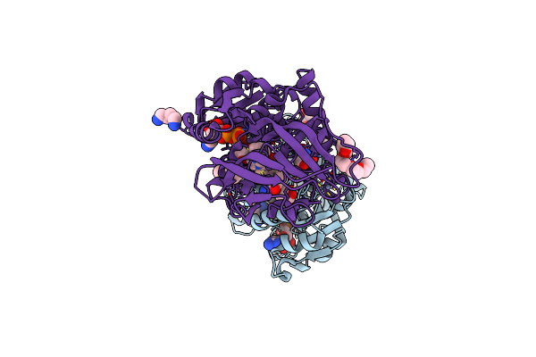
The Crystal Structure Of Human Abl1 Kinase Domain In Complex With Dp-987
Organism: Homo sapiens
Method: X-RAY DIFFRACTION Resolution:2.30 Å Release Date: 2011-06-01 Classification: TRANSFERASE/TRANSFERASE INHIBITOR Ligands: 9DP |
Organism: Homo sapiens
Method: X-RAY DIFFRACTION
Release Date: 2011-06-01
Ligands: 9DP
 |
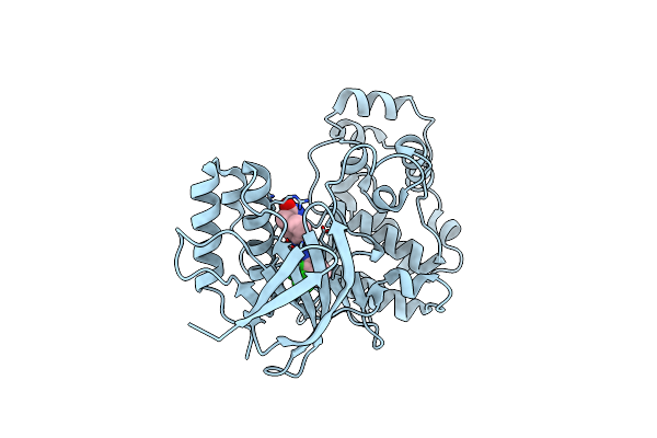
S-Nitrosoglutathione Reductase (Gsnor) In Complex With N6022
Organism: Homo sapiens
Method: X-RAY DIFFRACTION Resolution:1.90 Å Release Date: 2011-04-06 Classification: OXIDOREDUCTASE/OXIDOREDUCTASE INHIBITOR Ligands: ZN, K, NAD, 022, 1PE, N2P, TRS |
Organism: Homo sapiens
Method: X-RAY DIFFRACTION
Release Date: 2011-04-06
Ligands: ZN, K, NAD, 022, 1PE, N2P, TRS
 |
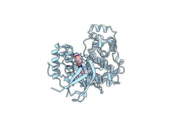
Crystal Structure Of P38 Alpha In Complex With Dp1376
Organism: Homo sapiens
Method: X-RAY DIFFRACTION Resolution:2.40 Å Release Date: 2010-09-15 Classification: TRANSFERASE Ligands: EDB |
Organism: Homo sapiens
Method: X-RAY DIFFRACTION
Release Date: 2010-09-15
Ligands: EDB
 |
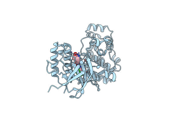
Crystal Structure Of P38 Alpha In Complex With Dp437
Organism: Homo sapiens
Method: X-RAY DIFFRACTION Resolution:2.10 Å Release Date: 2010-09-15 Classification: TRANSFERASE Ligands: 437 |
Organism: Homo sapiens
Method: X-RAY DIFFRACTION
Release Date: 2010-09-15
Ligands: 437
 |
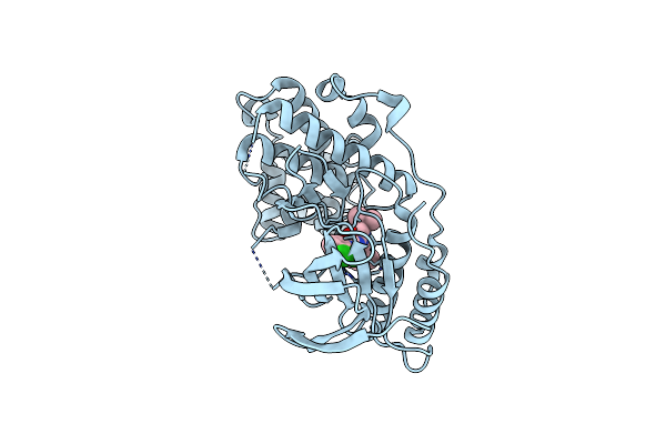
Crystal Structure Of P38 Alpha In Complex With Dp802
Organism: Homo sapiens
Method: X-RAY DIFFRACTION Resolution:1.89 Å Release Date: 2010-09-15 Classification: TRANSFERASE Ligands: EDD |
Organism: Homo sapiens
Method: X-RAY DIFFRACTION
Release Date: 2010-09-15
Ligands: EDD
 |
Crystal Structure Of Phosphorylated P38 Alpha In Complex With Dp802
Organism: Homo sapiens
Method: X-RAY DIFFRACTION Resolution:2.28 Å Release Date: 2010-09-15 Classification: TRANSFERASE Ligands: EDD |
Organism: Homo sapiens
Method: X-RAY DIFFRACTION
Release Date: 2010-09-15
Ligands: EDD