

Search Count: 32
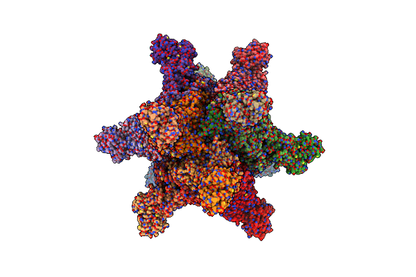 |
Cryoem Structure Of Helicobacter Pylori Urefd/Urease Complex
Organism: Helicobacter pylori 26695
Method: ELECTRON MICROSCOPY Release Date: 2023-05-03 Classification: HYDROLASE |
Organism: Helicobacter pylori 26695
Method: ELECTRON MICROSCOPY
Release Date: 2023-05-03
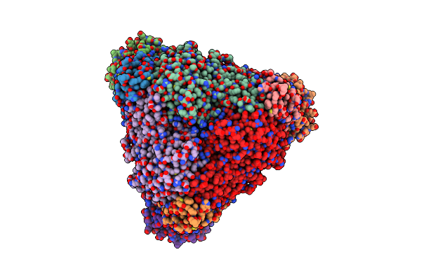 |
Cryoem Structure Of Klebsiella Pneumoniae Ured/Urease Complex
Organism: Klebsiella pneumoniae
Method: ELECTRON MICROSCOPY Release Date: 2023-05-03 Classification: HYDROLASE |
Organism: Klebsiella pneumoniae
Method: ELECTRON MICROSCOPY
Release Date: 2023-05-03
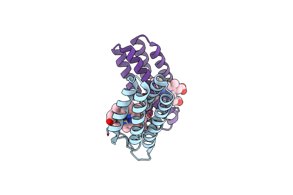 |
Ni(Ii)-Bound B2 Dimer (H60/H100/H104)
Organism: Escherichia coli
Method: X-RAY DIFFRACTION Resolution:1.89 Å Release Date: 2022-10-12 Classification: METAL BINDING PROTEIN Ligands: HEC, NI, MPD |
Organism: Escherichia coli
Method: X-RAY DIFFRACTION
Release Date: 2022-10-12
Ligands: HEC, NI, MPD
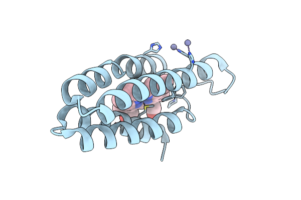 |
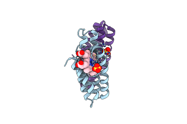
Zn(Ii)-Bound B2 Dimer (H60/H100/H104)
Organism: Escherichia coli
Method: X-RAY DIFFRACTION Resolution:1.70 Å Release Date: 2022-10-12 Classification: METAL BINDING PROTEIN Ligands: HEC, ZN |
Organism: Escherichia coli
Method: X-RAY DIFFRACTION
Release Date: 2022-10-12
Ligands: HEC, ZN
 |
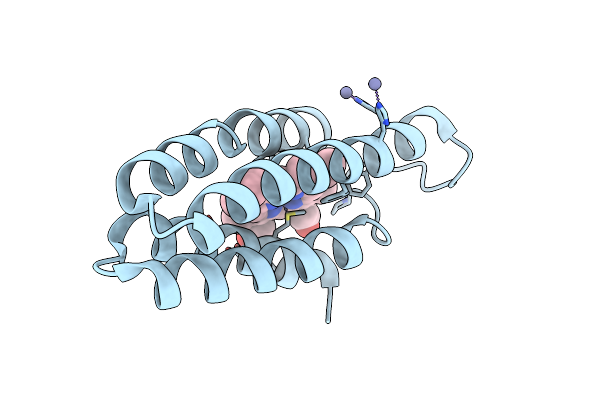
Apo B2 Dimer (H60/H100/H104) Formed In The Presence Of Cu(Ii)
Organism: Escherichia coli
Method: X-RAY DIFFRACTION Resolution:2.40 Å Release Date: 2022-10-12 Classification: METAL BINDING PROTEIN Ligands: HEC, SO4 |
Organism: Escherichia coli
Method: X-RAY DIFFRACTION
Release Date: 2022-10-12
Ligands: HEC, SO4
 |
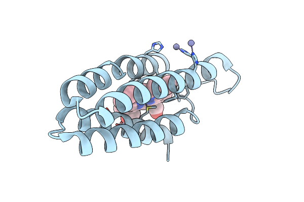
Zn(Ii)-Bound B2 Dimer (H60/H100/H104) Formed In Cu(Ii)//Zn(Ii) (M1 // M2) Condition
Organism: Escherichia coli
Method: X-RAY DIFFRACTION Resolution:1.65 Å Release Date: 2022-10-12 Classification: METAL BINDING PROTEIN Ligands: HEC, ZN |
Organism: Escherichia coli
Method: X-RAY DIFFRACTION
Release Date: 2022-10-12
Ligands: HEC, ZN
 |
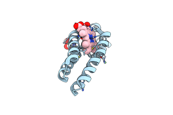
Zn(Ii)-Bound B2 Dimer (H60/H100/H104) Formed In Zn(Ii)//Cu(Ii) (M1 // M2) Condition
Organism: Escherichia coli
Method: X-RAY DIFFRACTION Resolution:1.55 Å Release Date: 2022-10-12 Classification: METAL BINDING PROTEIN Ligands: HEC, ZN |
Organism: Escherichia coli
Method: X-RAY DIFFRACTION
Release Date: 2022-10-12
Ligands: HEC, ZN
 |
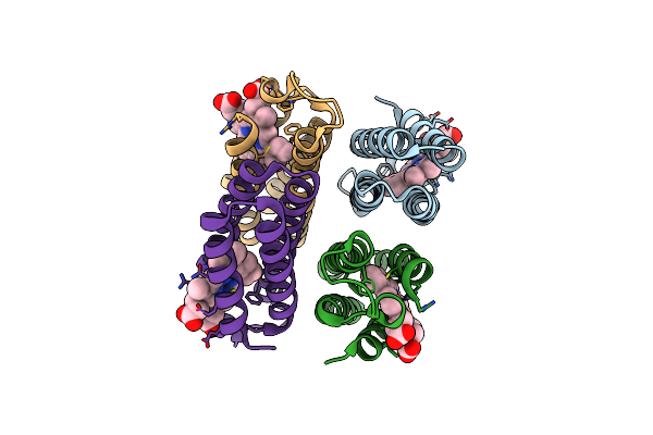
Crystal Structure Of Ni-Bound Ridc1 Variant In The Presence Of Reductant
Organism: Escherichia coli
Method: X-RAY DIFFRACTION Resolution:1.80 Å Release Date: 2022-06-29 Classification: METAL BINDING PROTEIN Ligands: NI, TRS, HEC, CL |
Organism: Escherichia coli
Method: X-RAY DIFFRACTION
Release Date: 2022-06-29
Ligands: NI, TRS, HEC, CL
 |
Crystal Structure Of A Metal-Free Ridc1 Variant
Organism: Escherichia coli
Method: X-RAY DIFFRACTION Resolution:2.20 Å Release Date: 2022-06-29 Classification: METAL BINDING PROTEIN Ligands: HEC |
Organism: Escherichia coli
Method: X-RAY DIFFRACTION
Release Date: 2022-06-29
Ligands: HEC
 |
Crystal Structure Of A Zn-Bound Ridc1 Variant
Organism: Escherichia coli
Method: X-RAY DIFFRACTION Resolution:1.70 Å Release Date: 2022-06-29 Classification: METAL BINDING PROTEIN Ligands: ZN, HEC, MPD, MRD, EPE |
Organism: Escherichia coli
Method: X-RAY DIFFRACTION
Release Date: 2022-06-29
Ligands: ZN, HEC, MPD, MRD, EPE
 |
Crystal Structure Of A Zn-Bound Ridc1 Variant In The Presence Of Reductant
Organism: Escherichia coli
Method: X-RAY DIFFRACTION Resolution:1.60 Å Release Date: 2022-06-29 Classification: METAL BINDING PROTEIN Ligands: ZN, CA, HEC |
Organism: Escherichia coli
Method: X-RAY DIFFRACTION
Release Date: 2022-06-29
Ligands: ZN, CA, HEC
 |
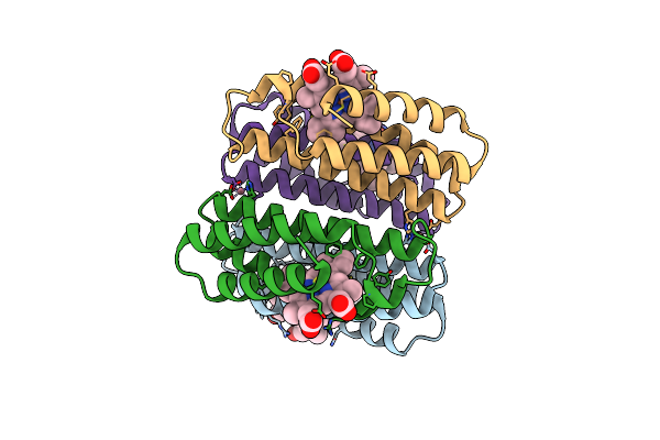
Crystal Structure Of A Fe-Bound Ridc1 Variant In The Presence Of Reductant
Organism: Escherichia coli
Method: X-RAY DIFFRACTION Resolution:2.20 Å Release Date: 2022-06-29 Classification: METAL BINDING PROTEIN Ligands: HEC, 1PE, FE, TRS |
Organism: Escherichia coli
Method: X-RAY DIFFRACTION
Release Date: 2022-06-29
Ligands: HEC, 1PE, FE, TRS
 |
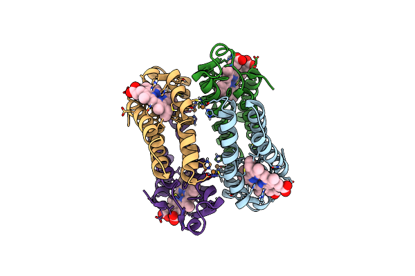
Crystal Structure Of A Co-Bound Ridc1 Variant
Organism: Escherichia coli
Method: X-RAY DIFFRACTION Resolution:2.00 Å Release Date: 2022-06-29 Classification: METAL BINDING PROTEIN Ligands: HEC, CO |
Organism: Escherichia coli
Method: X-RAY DIFFRACTION
Release Date: 2022-06-29
Ligands: HEC, CO
 |
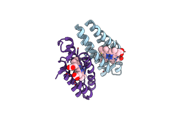
Crystal Structure Of A Cu-Bound Cytochrome Cb562 Variant In The Presence Of Reductant
Organism: Escherichia coli
Method: X-RAY DIFFRACTION Resolution:2.70 Å Release Date: 2022-06-29 Classification: METAL BINDING PROTEIN Ligands: HEC, CU |
Organism: Escherichia coli
Method: X-RAY DIFFRACTION
Release Date: 2022-06-29
Ligands: HEC, CU
 |
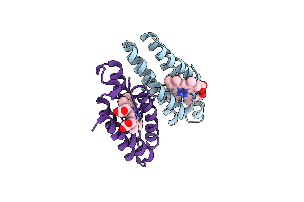
Ni-Bound Crystal Structure Of The Engineered Cyt Cb562 Variant, Ab2-H100A, Crystallized In The Presence Of Ni(Ii)
Organism: Escherichia coli
Method: X-RAY DIFFRACTION Resolution:1.80 Å Release Date: 2022-06-15 Classification: METAL BINDING PROTEIN Ligands: HEC, NI |
Organism: Escherichia coli
Method: X-RAY DIFFRACTION
Release Date: 2022-06-15
Ligands: HEC, NI
 |
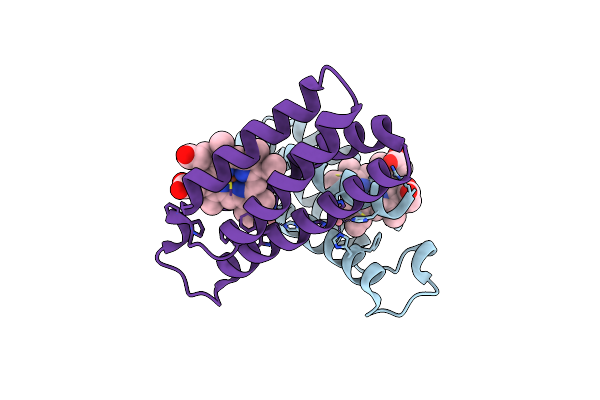
Co-Bound Crystal Structure Of The Engineered Cyt Cb562 Variant, Ab2-H100A, Crystallized In The Presence Of Co(Ii)
Organism: Escherichia coli
Method: X-RAY DIFFRACTION Resolution:1.93 Å Release Date: 2022-06-15 Classification: METAL BINDING PROTEIN Ligands: HEC, CO |
Organism: Escherichia coli
Method: X-RAY DIFFRACTION
Release Date: 2022-06-15
Ligands: HEC, CO
 |
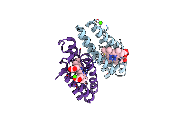
Co-Bound Crystal Structure Of The Engineered Cyt Cb562 Variant, Dicyt2
Organism: Escherichia coli
Method: X-RAY DIFFRACTION Resolution:1.27 Å Release Date: 2022-04-27 Classification: METAL BINDING PROTEIN Ligands: HEC, CO |
Organism: Escherichia coli
Method: X-RAY DIFFRACTION
Release Date: 2022-04-27
Ligands: HEC, CO
 |
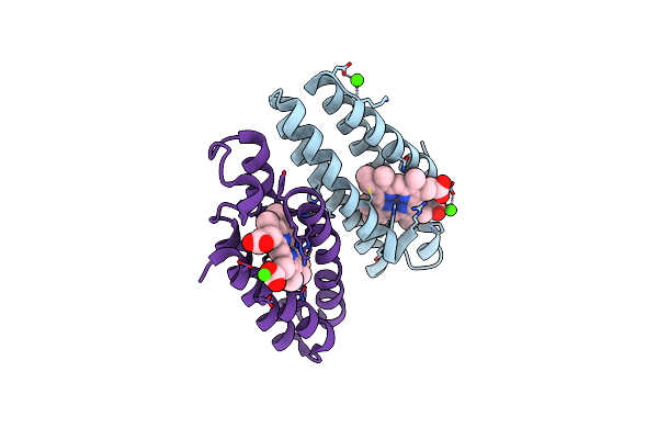
Cu-Bound Crystal Structure Of The Engineered Cyt Cb562 Variant, Dicyt2, Crystallized In The Presence Of Cu(Ii)
Organism: Escherichia coli
Method: X-RAY DIFFRACTION Resolution:1.91 Å Release Date: 2022-03-02 Classification: METAL BINDING PROTEIN Ligands: HEC, CA, CU |
Organism: Escherichia coli
Method: X-RAY DIFFRACTION
Release Date: 2022-03-02
Ligands: HEC, CA, CU
 |
Cu-Bound Crystal Structure Of The Engineered Cyt Cb562 Variant, Dicyt2 - H97A, Crystallized In The Presence Of Cu(Ii)
Organism: Escherichia coli
Method: X-RAY DIFFRACTION Resolution:1.99 Å Release Date: 2022-03-02 Classification: METAL BINDING PROTEIN Ligands: HEC, CA, CU |
Organism: Escherichia coli
Method: X-RAY DIFFRACTION
Release Date: 2022-03-02
Ligands: HEC, CA, CU
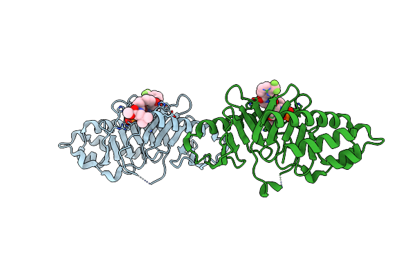 |
Crystal Structure Of Polo Box Domain In Complex With Cyclic Peptide Inhibitor
Organism: Homo sapiens, Synthetic construct
Method: X-RAY DIFFRACTION Resolution:1.85 Å Release Date: 2022-03-02 Classification: TRANSFERASE/Inhibitor |
Organism: Homo sapiens, Synthetic construct
Method: X-RAY DIFFRACTION
Release Date: 2022-03-02