
Search Count: 236
 |
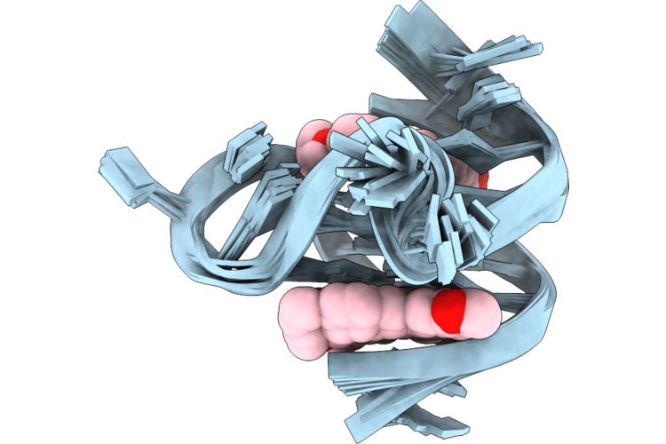 |
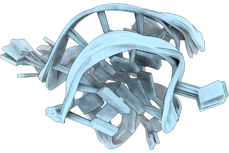
Organism: Homo sapiens
Method: SOLUTION NMR Release Date: 2026-06-03 Classification: DNA Ligands: BER |
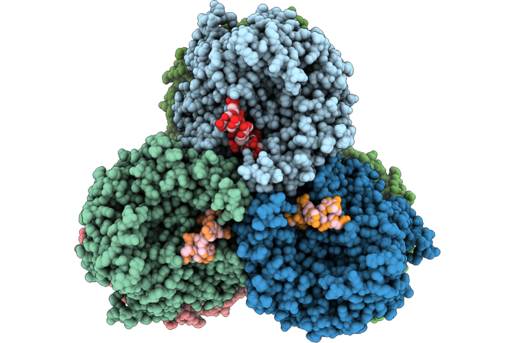 |
Organism: Enterobacteriaceae
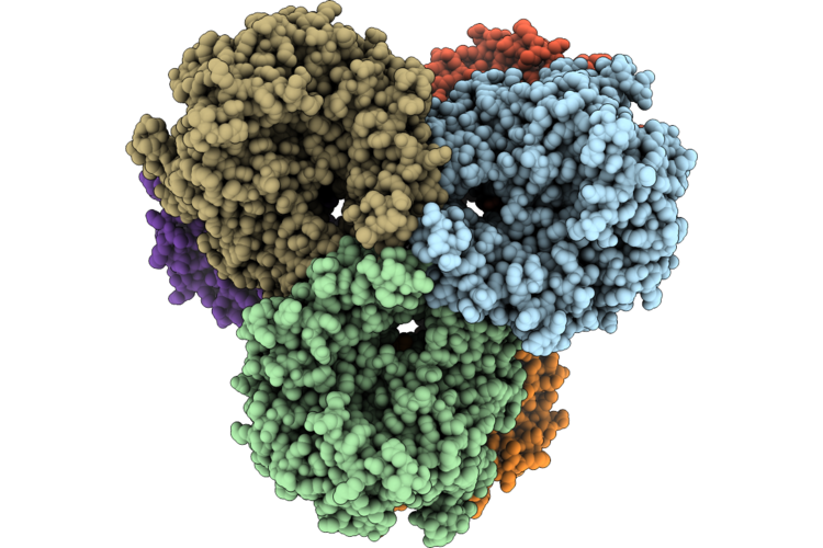
Method: ELECTRON MICROSCOPY Resolution:2.34 Å Release Date: 2026-05-27 Classification: RNA BINDING PROTEIN/DNA |
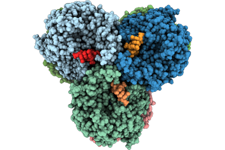 |
Organism: Enterobacteriaceae, Escherichia coli
Method: ELECTRON MICROSCOPY Resolution:2.68 Å Release Date: 2026-05-27 Classification: RNA BINDING PROTEIN/DNA Ligands: T8T, MG |
 |
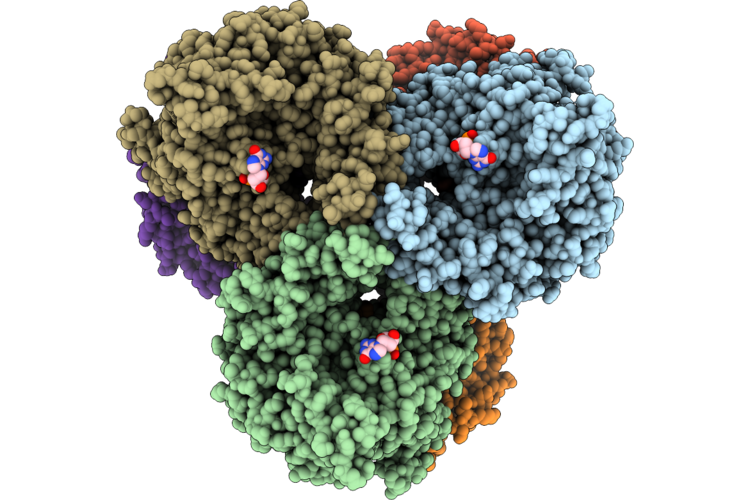
Organism: Enterobacteriaceae, Escherichia coli
Method: ELECTRON MICROSCOPY Resolution:2.84 Å Release Date: 2026-05-27 Classification: RNA BINDING PROTEIN/DNA Ligands: DTP, MG |
 |
Organism: Enterobacteriaceae
Method: ELECTRON MICROSCOPY Resolution:2.76 Å Release Date: 2026-05-27 Classification: RNA BINDING PROTEIN Ligands: T8T, MG, GMP |
 |
Organism: Enterobacteriaceae, Dna molecule
Method: ELECTRON MICROSCOPY Resolution:2.96 Å Release Date: 2026-05-27 Classification: RNA BINDING PROTEIN/DNA Ligands: DTP, MG |
 |
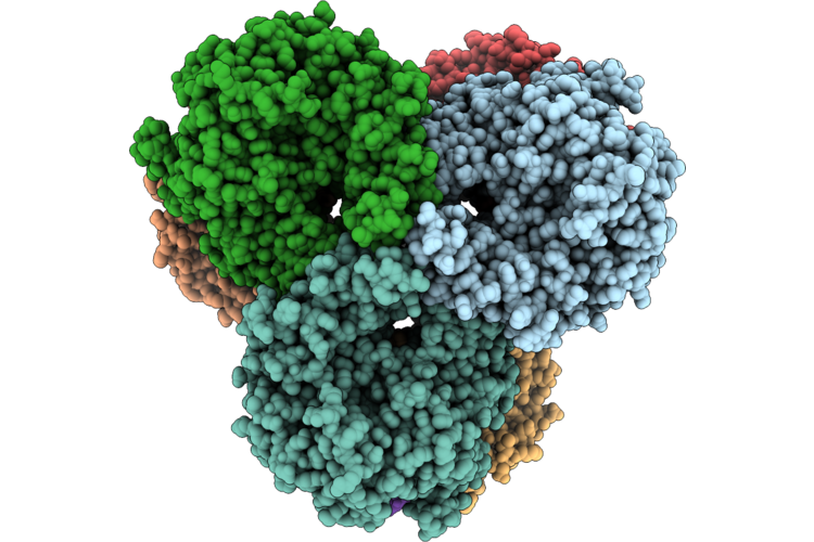
Organism: Enterobacteriaceae
Method: ELECTRON MICROSCOPY Resolution:2.80 Å Release Date: 2026-05-27 Classification: IMMUNE SYSTEM Ligands: DGT, MG |
 |
Organism: Enterobacteriaceae
Method: ELECTRON MICROSCOPY Resolution:2.54 Å Release Date: 2026-05-27 Classification: IMMUNE SYSTEM Ligands: DGT, MG, GMP |
 |
Organism: Enterobacteriaceae
Method: ELECTRON MICROSCOPY Resolution:2.22 Å Release Date: 2026-05-27 Classification: IMMUNE SYSTEM Ligands: DGT, MG, DGP, GMP |
 |
Organism: Enterobacteriaceae, Synthetic construct
Method: ELECTRON MICROSCOPY Resolution:2.68 Å Release Date: 2026-05-27 Classification: IMMUNE SYSTEM Ligands: DGT, MG |
 |
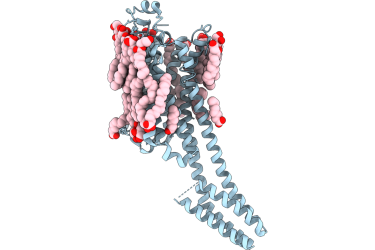
Organism: Homo sapiens, Escherichia coli
Method: X-RAY DIFFRACTION Resolution:2.49 Å Release Date: 2026-05-20 Classification: MEMBRANE PROTEIN |
 |
Organism: Homo sapiens, Escherichia coli
Method: X-RAY DIFFRACTION Resolution:2.43 Å Release Date: 2026-05-20 Classification: MEMBRANE PROTEIN |
 |
Organism: Homo sapiens, Escherichia coli
Method: X-RAY DIFFRACTION Resolution:2.66 Å Release Date: 2026-05-20 Classification: MEMBRANE PROTEIN |
 |
Organism: Homo sapiens, Escherichia coli
Method: X-RAY DIFFRACTION Resolution:2.66 Å Release Date: 2026-05-20 Classification: MEMBRANE PROTEIN |
 |
Organism: Homo sapiens, Escherichia coli
Method: X-RAY DIFFRACTION Resolution:2.49 Å Release Date: 2026-05-20 Classification: MEMBRANE PROTEIN |
 |
Organism: Homo sapiens, Escherichia coli
Method: X-RAY DIFFRACTION Resolution:2.52 Å Release Date: 2026-05-20 Classification: MEMBRANE PROTEIN |
 |
Organism: Homo sapiens, Escherichia coli
Method: X-RAY DIFFRACTION Resolution:2.43 Å Release Date: 2026-05-20 Classification: MEMBRANE PROTEIN |
 |
Organism: Homo sapiens, Escherichia coli
Method: X-RAY DIFFRACTION Resolution:2.52 Å Release Date: 2026-05-20 Classification: MEMBRANE PROTEIN |
 |
Organism: Homo sapiens, Escherichia coli
Method: X-RAY DIFFRACTION Resolution:2.54 Å Release Date: 2026-05-20 Classification: MEMBRANE PROTEIN |